Plants: The Inside Story
Plants: The Inside Story
Plants: The Inside Story

You also want an ePaper? Increase the reach of your titles
YUMPU automatically turns print PDFs into web optimized ePapers that Google loves.
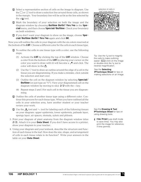
ì Select a representative section of cells on the image to diagram. Use<br />
the ¤ or › tool to draw a selection line around those cells, as shown<br />
in the margin. Your boundary line will be as fat as the line selected by<br />
the flÔ tool.<br />
ì Mark the boundary of your selection on both the image and the<br />
diagram window by choosing Outline Both? Yes/No in the Special<br />
menu, and then choose Special/Outline. Check your boundary<br />
on both windows.<br />
ì If you don’t want your diagram to show on the image, choose Special/Outline<br />
Both? Yes/No again and click OK.<br />
Now you will outline the cells in your diagram with the six colors reserved at<br />
the bottom of the LUT. Choose a different color for the cells in each tissue type.<br />
ì To outline the cells in one tissue type with a color, use the following<br />
steps:<br />
(1) Activate the LUT by clicking the top of the LUT window. Choose<br />
a color from the bottom of the LUT by placing your cursor on the<br />
color you want to draw with (it will become a Ï) and click. <strong>The</strong><br />
color will show in the ‰.<br />
(2) Use the fi tool to draw an outline around the edge of a cell in the<br />
tissue you are diagramming. If you make a mistake, click outside<br />
the selection and start over.<br />
(3) Outline the cell on the diagram window by selecting Special/<br />
Outline (or just type an “O”). View your diagrammed cell on the<br />
diagram window by moving to slice 2/2 with the > key.<br />
(4) Repeat steps 2 and 3 for each cell in the tissue you are diagramming.<br />
ì Outline the cells of another tissue type using a different color. Continue<br />
this process for each tissue type. When you have outlined all the<br />
cells in your selection area, have another student or your teacher<br />
review your work.<br />
ì Use the Ë tool and Ç tool for labeling each of the following tissues<br />
on your diagram: upper epidermis, lower epidermis, palisade layer,<br />
spongy layer, air spaces, stomata, xylem and phloem.<br />
3. Print your diagram of plant anatomy from the diagram window (slice<br />
2/2). Attach it to your Data Sheet. If you don’t have access to a printer,<br />
show your diagram to your teacher.<br />
4. Using your diagram and your texbook, describe the structure and function<br />
of each tissue in the leaf. How does the size, shape, and arrangement<br />
of cells in each tissue relate to its function? Write your answers in the<br />
table on your Data Sheet.<br />
selection of cells in<br />
leaf cross section<br />
Tip: Use the ÿ tool to magnify<br />
the cells to make outlining<br />
easier. æ-click on the image<br />
or double-click the ÿ tool to<br />
reverse magnification.<br />
See the Selecting<br />
IPTechnique Sheet for tips on<br />
making selections on an image.<br />
See the Drawing & Text<br />
IPTechnique Sheet for tips on<br />
using drawing tools.<br />
 File/Print (use draft mode<br />
to save time). You may also<br />
print the image for reference,<br />
if time permits.<br />
106 HIP BIOLOGY 1 © 1996 Arizona Board of Regents. Limited classroom reproduction rights are granted under the notice appearing earlier in this work.

![alumni e-mail addresses by classes - word[3]](https://img.yumpu.com/13381322/1/190x245/alumni-e-mail-addresses-by-classes-word3.jpg?quality=85)