Plants: The Inside Story
Plants: The Inside Story
Plants: The Inside Story

Create successful ePaper yourself
Turn your PDF publications into a flip-book with our unique Google optimized e-Paper software.
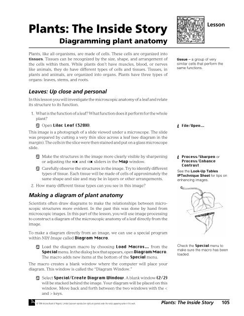
<strong>Plants</strong>: <strong>The</strong> <strong>Inside</strong> <strong>Story</strong><br />
Diagramming plant anatomy<br />
Lesson<br />
<strong>Plants</strong>, like all organisms, are made of cells. <strong>The</strong>se cells are organized into<br />
tissues. Tissues can be recognized by the size, shape, and arrangement of<br />
the cells within them. While plants don’t have muscles, blood, or nerves<br />
like animals, they do have different types of cells and tissues. Tissues, in<br />
plants and animals, are organized into organs. <strong>Plants</strong> have three types of<br />
organs: leaves, stems, and roots.<br />
tissue – a group of very<br />
similar cells that perform the<br />
same functions.<br />
Leaves: Up close and personal<br />
In this lesson you will investigate the microscopic anatomy of a leaf and relate<br />
its structure to its function.<br />
1. What is the function of a leaf? What function does it perform for the whole<br />
plant?<br />
ì Open Lilac Leaf (320X).<br />
This image is a photograph of a slide viewed under a microscope. <strong>The</strong> slide<br />
was prepared by cutting a very thin slice across a leaf (see diagram in the<br />
margin). <strong>The</strong> cells in the slice were then stained and put on a glass microscope<br />
slide.<br />
ì Make the structures in the image more clearly visible by sharpening<br />
or adjusting the ¬ and Æ sliders in the Map window.<br />
ì Carefully observe the structures in the image. Try to identify different<br />
types of tissue. Each tissue will be made of cells of approximately the<br />
same shape and size and may be in layers or other arrangements.<br />
2. How many different tissue types can you see in this image?<br />
 File/Open…<br />
 Process/Sharpen or<br />
Process/Enhance<br />
Contrast<br />
See the Look-Up Tables<br />
IPTechnique Sheet for tips on<br />
enhancing images.<br />
Making a diagram of plant anatomy<br />
Scientists often draw diagrams to make the relationships between microscopic<br />
structures more evident. In the past this was done by hand from<br />
microscopic images. In this part of the lesson, you will use image processing<br />
to construct a diagram of the microscopic anatomy of a leaf directly from the<br />
image.<br />
To make a diagram directly from an image, we can use a special program<br />
within NIH Image called Diagram Macro.<br />
ì Load the diagram macro by choosing Load Macros… from the<br />
Special menu. In the dialog box that appears, open Diagram Macro.<br />
<strong>The</strong> macro adds new items at the bottom of the Special menu.<br />
<strong>The</strong> macro creates a blank window where the computer will place your<br />
diagram. This window is called the “Diagram Window.”<br />
Check the Special menu to<br />
make sure the macro has been<br />
loaded.<br />
ì Select Special/Create Diagram Window. A blank window (2/2)<br />
will be stacked behind the image. Your diagram will be placed on this<br />
window. Move back and forth between the two windows with the <<br />
and > keys.<br />
© 1996 Arizona Board of Regents. Limited classroom reproduction rights are granted under the notice appearing earlier in this work.<br />
<strong>Plants</strong>: <strong>The</strong> <strong>Inside</strong> <strong>Story</strong> 105
ì Select a representative section of cells on the image to diagram. Use<br />
the ¤ or › tool to draw a selection line around those cells, as shown<br />
in the margin. Your boundary line will be as fat as the line selected by<br />
the flÔ tool.<br />
ì Mark the boundary of your selection on both the image and the<br />
diagram window by choosing Outline Both? Yes/No in the Special<br />
menu, and then choose Special/Outline. Check your boundary<br />
on both windows.<br />
ì If you don’t want your diagram to show on the image, choose Special/Outline<br />
Both? Yes/No again and click OK.<br />
Now you will outline the cells in your diagram with the six colors reserved at<br />
the bottom of the LUT. Choose a different color for the cells in each tissue type.<br />
ì To outline the cells in one tissue type with a color, use the following<br />
steps:<br />
(1) Activate the LUT by clicking the top of the LUT window. Choose<br />
a color from the bottom of the LUT by placing your cursor on the<br />
color you want to draw with (it will become a Ï) and click. <strong>The</strong><br />
color will show in the ‰.<br />
(2) Use the fi tool to draw an outline around the edge of a cell in the<br />
tissue you are diagramming. If you make a mistake, click outside<br />
the selection and start over.<br />
(3) Outline the cell on the diagram window by selecting Special/<br />
Outline (or just type an “O”). View your diagrammed cell on the<br />
diagram window by moving to slice 2/2 with the > key.<br />
(4) Repeat steps 2 and 3 for each cell in the tissue you are diagramming.<br />
ì Outline the cells of another tissue type using a different color. Continue<br />
this process for each tissue type. When you have outlined all the<br />
cells in your selection area, have another student or your teacher<br />
review your work.<br />
ì Use the Ë tool and Ç tool for labeling each of the following tissues<br />
on your diagram: upper epidermis, lower epidermis, palisade layer,<br />
spongy layer, air spaces, stomata, xylem and phloem.<br />
3. Print your diagram of plant anatomy from the diagram window (slice<br />
2/2). Attach it to your Data Sheet. If you don’t have access to a printer,<br />
show your diagram to your teacher.<br />
4. Using your diagram and your texbook, describe the structure and function<br />
of each tissue in the leaf. How does the size, shape, and arrangement<br />
of cells in each tissue relate to its function? Write your answers in the<br />
table on your Data Sheet.<br />
selection of cells in<br />
leaf cross section<br />
Tip: Use the ÿ tool to magnify<br />
the cells to make outlining<br />
easier. æ-click on the image<br />
or double-click the ÿ tool to<br />
reverse magnification.<br />
See the Selecting<br />
IPTechnique Sheet for tips on<br />
making selections on an image.<br />
See the Drawing & Text<br />
IPTechnique Sheet for tips on<br />
using drawing tools.<br />
 File/Print (use draft mode<br />
to save time). You may also<br />
print the image for reference,<br />
if time permits.<br />
106 HIP BIOLOGY 1 © 1996 Arizona Board of Regents. Limited classroom reproduction rights are granted under the notice appearing earlier in this work.
Further Exploration<br />
• Choose Name Groups… from the Special menu to name each of the<br />
tissues in your diagram. <strong>The</strong>n choose Special/Sort & Show Results<br />
to see measurements of area, perimeter, width, and height recorded for<br />
each cell. Average the values to define the average size of cells in each<br />
tissue.<br />
• Diagram a stem or a root. Open the Roots or Stems folder located in<br />
the Further Exploration folder. Choose an image to diagram using the<br />
techniques described for the leaf. Compare and contrast the similarities<br />
and differences between leaves, stems, and roots, or between monocots<br />
and dicots.<br />
selection of cells in<br />
root cross section<br />
stem<br />
roots<br />
• Look at the images in the Extra Color Images folder. <strong>The</strong>se images<br />
reveal the stains used to visualize the tissues under the light microscope.<br />
What features of the cells do the stains reveal?<br />
© 1996 Arizona Board of Regents. Limited classroom reproduction rights are granted under the notice appearing earlier in this work.<br />
<strong>Plants</strong>: <strong>The</strong> <strong>Inside</strong> <strong>Story</strong> 107
108 HIP BIOLOGY 1 © 1996 Arizona Board of Regents. Limited classroom reproduction rights are granted under the notice appearing earlier in this work.
<strong>Plants</strong>: <strong>The</strong> <strong>Inside</strong> <strong>Story</strong><br />
Name(s)<br />
Class<br />
Date<br />
Data<br />
Sheet<br />
1. What is the function of a leaf? What function does it perform for the whole plant?<br />
2. How many different tissue types can you see in this image?<br />
3. Print your diagram of plant anatomy from the diagram window (slice 2/2). Attach it to your Data Sheet. If<br />
you don’t have access to a printer, show your diagram to your teacher.<br />
4. Using your diagram and your texbook, describe the structure and function of each tissue in the leaf. How<br />
does the size, shape, and arrangement of cells in each tissue help it perform its function? Write your answers<br />
in the table below.<br />
Tissue<br />
Function<br />
How does shape and arrangement<br />
of cells contribute to function?<br />
upper<br />
epidermis<br />
palisade layer<br />
spongy layer<br />
lower<br />
epidermis<br />
© 1996 Arizona Board of Regents. Limited classroom reproduction rights are granted under the notice appearing earlier in this work.<br />
<strong>Plants</strong>: <strong>The</strong> <strong>Inside</strong> <strong>Story</strong> 109
110 HIP BIOLOGY 1 © 1996 Arizona Board of Regents. Limited classroom reproduction rights are granted under the notice appearing earlier in this work.


![alumni e-mail addresses by classes - word[3]](https://img.yumpu.com/13381322/1/190x245/alumni-e-mail-addresses-by-classes-word3.jpg?quality=85)