

TECHNICAL & POSTER EXHIBITION - ismrm
TECHNICAL & POSTER EXHIBITION - ismrm
TECHNICAL & POSTER EXHIBITION - ismrm

Create successful ePaper yourself
Turn your PDF publications into a flip-book with our unique Google optimized e-Paper software.
ISMRM<br />
BRIDGING THE GAP BETWEEN CLINICAL NEEDS<br />
AND TECHNOLOGICAL SOLUTIONS<br />
International Society for Magnetic Resonance in Medicine<br />
ISMRM 21 st ANNUAL MEETING & <strong>EXHIBITION</strong><br />
SMRT 22 nd ANNUAL MEETING<br />
ISMRM<br />
S<br />
alt<br />
21 st Annual<br />
Meeting & Exhibition<br />
Lake City, Utah, USA<br />
20–26 April 2013<br />
“Discovery, Innovation & Application –<br />
Advancing MR for Improved Health”<br />
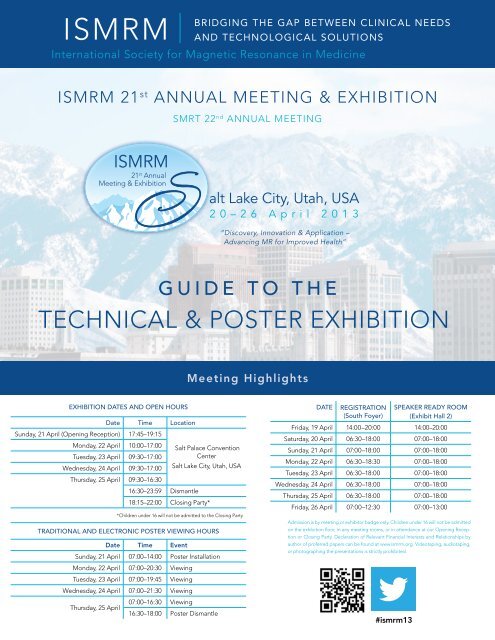
GUIDE TO THE<br />
<strong>TECHNICAL</strong> & <strong>POSTER</strong> <strong>EXHIBITION</strong><br />
Meeting Highlights<br />
<strong>EXHIBITION</strong> DATES AND OPEN HOURS<br />
Date Time Location<br />
Sunday, 21 April (Opening Reception) 17:45–19:15<br />
Monday, 22 April 10:00–17:00 Salt Palace Convention<br />
Tuesday, 23 April 09:30–17:00<br />
Center<br />
Wednesday, 24 April 09:30–17:00 Salt Lake City, Utah, USA<br />
Thursday, 25 April 09:30–16:30<br />
16:30–23:59 Dismantle<br />
18:15–22:00 Closing Party*<br />
*Children under 16 will not be admitted to the Closing Party<br />
TRADITIONAL AND ELECTRONIC <strong>POSTER</strong> VIEWING HOURS<br />
Date Time Event<br />
Sunday, 21 April 07:00–14:00 Poster Installation<br />
Monday, 22 April 07:00–20:30 Viewing<br />
Tuesday, 23 April 07:00–19:45 Viewing<br />
Wednesday, 24 April 07:00–21:30 Viewing<br />
Thursday, 25 April<br />
07:00–16:30 Viewing<br />
16:30–18:00 Poster Dismantle<br />
DATE<br />
REGISTRATION<br />
(South Foyer)<br />
SPEAKER READY ROOM<br />
(Exhibit Hall 2)<br />
Friday, 19 April 14:00–20:00 14:00–20:00<br />
Saturday, 20 April 06:30–18:00 07:00–18:00<br />
Sunday, 21 April 07:00–18:00 07:00–18:00<br />
Monday, 22 April 06:30–18:30 07:00–18:00<br />
Tuesday, 23 April 06:30–18:00 07:00–18:00<br />
Wednesday, 24 April 06:30–18:00 07:00–18:00<br />
Thursday, 25 April 06:30–18:00 07:00–18:00<br />
Friday, 26 April 07:00–12:30 07:00–13:00<br />
Admission is by meeting or exhibitor badge only. Children under 16 will not be admitted<br />
on the exhibition floor, in any meeting rooms, or in attendance at our Opening Reception<br />
or Closing Party. Declaration of Relevant Financial Interests and Relationships by<br />
author of proferred papers can be found at www.<strong>ismrm</strong>.org. Videotaping, audiotaping,<br />
or photographing the presentations is strictly prohibited.<br />
#<strong>ismrm</strong>13
Acknowledgements to Our Corporate Members<br />
ISMRM COMMUNITY<br />
FOR CLINICIANS<br />
AND SCIENTISTS<br />
Thank You<br />
to our corporate members<br />
Gold Corporate Members:<br />
GE Healthcare<br />
Philips Healthcare<br />
Siemens<br />
Silver Corporate Members:<br />
Bayer HealthCare<br />
Bruker<br />
Bronze Corporate Members:<br />
Bracco<br />
Hitachi<br />
Shanghai United Imaging Healthcare Co., Ltd.<br />
Toshiba<br />
Associate Corporate Members:<br />
Nova Medical, Inc.<br />
ZMT Zurich MedTech AG<br />
2
MESSAGE FROM THE PROGRAM CHAIR<br />
Garry Gold, M.D.<br />
Chair, Annual Meeting Program Committee<br />
Our 21 st Annual Meeting & Exhibition will be a celebration<br />
of over 20 years of innovation in MRI—innovation<br />
that has transformed imaging and medicine.<br />
The theme this year is “Discovery, Innovation and<br />
Application – Advancing MR for Improved Health.”<br />
In our plenary lectures, you will learn about the rich<br />
history of innovation in magnetic resonance. Past Gold<br />
medal winners of the ISMRM will<br />
describe their moments of innovation<br />
that led to their biggest<br />
the Intersection of Engineering and Biology.” The<br />
NIBIB New Horizons Lecture, will be given by Scott<br />
Reeder, M.D., Ph.D., of the University of Wisconsin<br />
at Madison, entitled “Frontiers in Body MRI: from<br />
Qualitative to Quantitative.” The Education program<br />
is spread throughout the week and is targeted towards<br />
all levels of expertise. Courses will be clinical,<br />
technical and mixed in nature—a<br />
reflection of our diverse society.<br />
The setting in Salt Lake City is<br />
discoveries. Experts will discuss<br />
“ON BEHALF OF<br />
spectacular; it is a clean and<br />
major advances in breast cancer<br />
accessible city surrounded by<br />
using MRI and the application THE ISMRM ANNUAL<br />
snow-capped mountains. The<br />
of MRI to clinical trials. You will<br />
MEETING PROGRAM convention center is large with a<br />
learn about the power of high<br />
great format for our meeting. A<br />
field brain imaging for assessing<br />
COMMITTEE, WELCOME host of excellent shops, bars and<br />
the microstructure and con-<br />
restaurants are situated within<br />
nections within the brain, and TO A celebration<br />
walking distance of the convention<br />
how the landscape of MRI may<br />
of twenty years<br />
center, providing an ideal<br />
be transformed when it is combined<br />
with Positron Emission of innovation and<br />
setting for breaks during and<br />
after the day’s meeting.<br />
Tomography (PET).<br />
advances in MRI.” On behalf of the Annual Meeting<br />
Klaas Pruessmann, Ph.D., ETH,<br />
Zurich, will deliver the Lauterbur<br />
lecture entitled “Beyond<br />
Fourier Encoding: The Need,<br />
the Challenges, and the Rewards of Breaking Out<br />
of K-Space.” Richard Ehman, M.D., Professor of<br />
Radiology at the Mayo Clinic and former President<br />
of the ISMRM, will deliver the Mansfield lecture,<br />
Program Committee, I welcome<br />
you to Salt Lake City, Utah, USA,<br />
for the 21 st Annual Meeting &<br />
Exhibition of the ISMRM. Be part of the celebration<br />
of over 20 years of innovation and advances in MRI.<br />
The setting and the venue are spectacular, the people<br />
are friendly and the Rocky Mountains await you after<br />
the meeting.<br />
“MRI and Mechanobiology: Emerging Science at<br />
Garry Gold, M.D.<br />
Chair, ISMRM Annual Meeting Program Committee<br />
“Discovery, Innovation & Application – Advancing MR for Improved Health”<br />
3
GE Healthcare<br />
CAN YOU PLEASE KEEP<br />
IT DOWN UP THERE?<br />
INTRODUCING SILENT SCAN<br />
Humanizing MR isn’t just our philosophy. It’s our promise.<br />
Our promise to change how patients feel, see and hear MR<br />
for the better. However, now is the time to break the silent<br />
barrier and change the way patients hear MR forever.<br />
Introducing Silent Scan * . Using a unique combination of<br />
breakthrough technologies, we’ve made MR as silent as a<br />
whisper. The day when your patients can undergo an MR<br />
scan without the added anxiety of loud noise is here. And<br />
we’ve accomplished this while still providing the excellent<br />
image quality you need to make a confident diagnosis.<br />
It’s time to hear the difference.<br />
© 2013 General Electric Company. All rights reserved.<br />
GE Healthcare, a division of General Electric Company.<br />
GE and GE monogram are trademarks of General Electric Company.<br />
* Trademark of General Electric Company
The first-ever digital broadband MR is changing expectations,<br />
and lives. That’s the power of Philips Imaging 2.0.<br />
Thanks to Philips Imaging 2.0, a revolutionary new imaging approach, the Philips Ingenia 1.5T and 3.0T MR<br />
systems set a new standard in clarity, speed and expandability. Ingenia captures and digitizes the signal closest<br />
to the patient to improve SNR by up to 40%. Easier coil handling and improved patient comfort help increase<br />
productivity by up to 30%. And, Ingenia is designed to meet the growing<br />
needs in oncology imaging. Discover the revolution in MR technology at<br />
www.philips.com/ISMRM or visit us at booth 274.
A91MR-9257-A3-7600<br />
www.siemens.com/prisma<br />
MAGNETOM Prisma *<br />
Understanding functional processes and the most threatening diseases.<br />
MAGNETOM Prisma, our upcoming and powerful 3T MRI<br />
system, is built to tackle the most demanding research<br />
challenges of today and tomorrow. It delivers maximum<br />
performance under prolonged high-strain conditions<br />
opening new possibilities for imaging functional<br />
processes and understanding the most threatening<br />
diseases. Only one of many high performance features is<br />
the new gradient system. With its higher gradient<br />
amplitude it delivers significantly higher signal-to-noise<br />
ratio, enhancing for example physiological imaging or<br />
morphometric measurements. With higher spatial and<br />
temporal resolution you can see excellent anatomical<br />
detail, for example displaying functional and structural<br />
brain connectivity. MAGNETOM Prisma delivers<br />
benchmark 3T magnet homogeneity – the basis for<br />
superior quantitative evaluations. Our new, powerful 3T<br />
system helps you enter new areas of research and<br />
strengthen your leadership in MRI.<br />
* MAGNETOM Prisma is currently under development; it is not for sale<br />
in the U.S. and other countries. Its future availability cannot be<br />
guaranteed.<br />
Answers for life.
ISMRM<br />
BRIDGING THE GAP BETWEEN CLINICAL NEEDS<br />
AND TECHNOLOGICAL SOLUTIONS<br />
International Society for Magnetic Resonance in Medicine<br />
THE POWER OF SCIENCE,<br />
TECHNOLOGY<br />
& PRACTICE<br />
Visit us in<br />
Booth #342<br />
WHAT<br />
WE DO:<br />
• We bridge the gap between the<br />
clinical and scientific communities.<br />
• We foster research and development<br />
in basic and clinical MR science and its<br />
application to healthcare.<br />
• We provide international forums for MR<br />
science in medicine, biology and other<br />
industry hot topics.<br />
• We promote communication and<br />
understanding about cutting-edge<br />
MR developments.<br />
• We provide educational channels and<br />
other opportunities for continuing<br />
medical education credits.<br />
• We publish two journals as well as<br />
proceedings and syllabi from premier<br />
scientific and clinical events.<br />
AND WE IMPROVE HUMAN HEALTH<br />
As a member of ISMRM, you become a part<br />
of a community of your peers. You contribute<br />
to the development of MR techniques and<br />
technologies. And you help improve the<br />
health of people around the globe.<br />
BECOME A MEMBER TODAY!<br />
WHOJOINS?<br />
• Basic or clinical scientists<br />
who are developing MR<br />
techniques and applications<br />
• Technologists who want to<br />
improve their understanding<br />
and utilization of MR<br />
• Clinicians who are interested<br />
in MR clinical education<br />
• Students, including postdocs,<br />
residents and fellows, engaged<br />
full-time in an academic or<br />
training program<br />
BENEFITS<br />
THAT<br />
RESONATE:<br />
LEARN. Benefits of membership include<br />
a full year’s subscription to either Magnetic<br />
Resonance in Medicine or Journal<br />
of Magnetic Resonance Imaging, the<br />
journals of the International Society for<br />
Magnetic Resonance in Medicine. Members<br />
may opt to select both journals.<br />
The journal subscription is optional for<br />
technologists and students.<br />
DISCOVER. Stay current with clinical and<br />
scientific developments by attending<br />
conferences, scientific workshops,<br />
educational courses, annual meetings and<br />
chapter meetings. Also access over 6,000<br />
oral presentations online.<br />
CONNECT. Meet other MR professionals<br />
and make valuable connections. Tap into<br />
the ISMRM online membership directory<br />
to connect with peers.<br />
ENGAGE. Volunteer for abstract review,<br />
committee, study group and/or chapter<br />
membership and leadership.<br />
DEVELOP. Access an ever-growing listing<br />
of available positions through the ISMRM<br />
Career Center, the largest MR community<br />
online resource for posting your position<br />
and/or your CV.<br />
SAVE. Reduce the registration fees for the<br />
Annual Meeting and other programming<br />
throughout the year. Receive a 25% discount<br />
on any items purchased through the ISMRM<br />
bookstore.<br />
ACCESS. As a member, you have access to<br />
educational stipends, seed grants and other<br />
funding opportunities.<br />
to find out more about membership levels, study groups and many other benefits of ISMRM membership<br />
Visit us at www.<strong>ismrm</strong>.org or call +1 510 841 1899<br />
International society for magnetic resonance in medicine • 2030 addison street • suite 700 • berkeley • california 94704 usa
ISMRM Program-At-A-Glance<br />
SATURday,<br />
20 APRIL 2013<br />
Educational<br />
Course:<br />
Innovation in<br />
Body MRI<br />
Program-At-A-Glance • EDUCATIONAL COURSES<br />
Educational<br />
Course:<br />
Pre-Clinical MR<br />
of Cancer<br />
Educational<br />
Course:<br />
Perfusion<br />
Imaging:<br />
ASL, DCE & DSC<br />
(morning)<br />
08:00–12:35<br />
Educational<br />
Course:<br />
fMRI: From Basic to<br />
Intermediate Brain<br />
Connectivity, Part 1<br />
(afternoon)<br />
Educational<br />
Course:<br />
Single-Subject<br />
Neuroimaging<br />
(morning)<br />
08:30-12:45<br />
Educational<br />
Course:<br />
Diffusion<br />
Goes Mad<br />
(afternoon)<br />
Educational<br />
Course:<br />
Challenges in<br />
Musculoskeletal<br />
Imaging<br />
Educational<br />
Course:<br />
Advanced<br />
Neuroimaging 1:<br />
Brain & Spinal Cord<br />
Educational<br />
Course:<br />
MR Systems<br />
Engineering<br />
Educational<br />
Course:<br />
MR Physics for<br />
Physicists<br />
08:30–17:15 08:30–17:15 13:30–18:15 14:00–18:15 08:30–16:45 09:00–17:15 08:30–17:15 08:30–18:15<br />
Room<br />
150 AG<br />
Room<br />
155 EF<br />
Room<br />
155 BC<br />
Room<br />
151 AG<br />
Room<br />
255 EF<br />
Room<br />
255 BC<br />
Room<br />
250 BCEF<br />
Room<br />
251 BCEF<br />
SMRT 22 nd Annual Meeting<br />
“Changing the World through MR Education and Innovation”<br />
(Technologist/Radiographer Program)<br />
Day 1 • 07:45–17:05 • Salt Palace Convention Center, Room 355 ABCDEF<br />
ISMRM Program-At-A-Glance<br />
SUNday,<br />
21 APRIL 2013<br />
Educational<br />
Course:<br />
Molecular &<br />
Cellular Imaging:<br />
From Bench to the<br />
Bed<br />
Educational<br />
Course:<br />
Clinical Cancer<br />
MRI—Case-Based<br />
Teaching<br />
Educational<br />
Course:<br />
Recent Innovation<br />
in Cardiac MR<br />
Educational<br />
Course:<br />
Everything You<br />
Wanted to Know<br />
about MR-PET<br />
(morning)<br />
08:00–12:15<br />
Educational<br />
Course:<br />
A Practical Guide<br />
to MR Safety<br />
(afternoon)<br />
Educational<br />
Course:<br />
Advanced<br />
Diffusion<br />
Acquisition:<br />
Targeted Methods<br />
(morning)<br />
08:00-12:45<br />
Educational<br />
Course:<br />
fMRI: From Basic to<br />
Intermediate Brain<br />
Connectivity, Part 2<br />
(afternoon)<br />
Educational<br />
Course:<br />
Advanced<br />
Neuroimaging 2:<br />
Across the Lifespan<br />
Educational<br />
Course:<br />
RF Engineering—<br />
Coils<br />
Educational<br />
Course:<br />
Imaging<br />
Acquisition &<br />
Reconstruction<br />
08:30–16:45 08:30–17:15 08:00–18:00 13:30–17:45 13:30–18:15 09:00–17:15 08:30–16:45 08:30–17:45<br />
Room<br />
155 EF<br />
Room<br />
150 AG<br />
Room<br />
155 BC<br />
Room<br />
151 AG<br />
12:30–13:30 Silver Corporate Symposium • Bayer HealthCare (no CME credit) • Plenary Hall<br />
Room<br />
255 EF<br />
Room<br />
255 BC<br />
Room<br />
250 BCEF<br />
Room<br />
251 BCEF<br />
SMRT 22 nd Annual Meeting<br />
“Changing the World through MR Education and Innovation”<br />
(Technologist/Radiographer Program)<br />
Day 2 • 07:45–16:45 • Salt Palace Convention Center, Room 355 ABCDEF<br />
Opening Reception in Technical Exhibition • Exhibit Hall • 17:45–19:15<br />
8
SMRT 22 nd ANNUAL MEETING: CHANGING THE WORLD THROUGH MR EDUCATION & INNOVATION<br />
20–21 April 2013, Salt Lake City, Utah, USA • ROOM 355 ABCDEF • Please visit www.<strong>ismrm</strong>.org/smrt/13 for program updates.<br />
PROGRAM<br />
SMRT Poster Walking Tour & Reception – Friday, 19 April 2013, 18:00 – 20:00<br />
Time<br />
Saturday, 20 April 2013, 07:45–17:05<br />
(8.0 Category A CE)<br />
Time<br />
Sunday, 21 April 2013 • 07:45–16:45<br />
(7.0 Category A CE)<br />
07:45<br />
Welcome & Announcements<br />
Vera Kimbrell, B.S., R.T. (R)(MR), SMRT President 2012–2013<br />
G. Barry Southers, M.Ed., R.T. (R)(MR), SMRT Program Chair 2013<br />
07:45<br />
Welcome & Announcements<br />
Ben Kennedy, B.App.Sc., MMRT, SMRT President 2013-2014<br />
G. Barry Southers, M.Ed., R.T. (R)(MR), SMRT Program Chair 2013<br />
Forum 1: History & Future of MR<br />
Moderator: Anne Marie Sawyer, B.S., R.T. (R)(MR), FSMRT<br />
Forum 6: Musculoskeletal MR<br />
Moderator: Vanessa Louise Orchard, DCR (R), PGDip.(NM)<br />
08:00<br />
Localized Origins<br />
Paul Arthur Bottomley, Ph.D.<br />
08:00<br />
Post-Operative Imaging of the Rotator Cuff<br />
Lynne S. Steinbach, M.D.<br />
08:50<br />
MRI: From Science to Society<br />
Vivian S. Lee, M.D., Ph.D., M.B.A<br />
08:50<br />
GE Healthcare - Corporate Meeting Sponsor<br />
Lloyd Estkowski, R.T. (R)(MR)<br />
09:40 Break<br />
09:55<br />
Forum 2: Cardiovascular MR<br />
Moderator: Maureen N. Hood, Ph.D., R.N., R.T. (MR), FSMRT<br />
Cardiac MR: Getting the Numbers Right<br />
Wendy Strugnell, B.App.Sc. (MIT), FSMRT<br />
09:00<br />
09:50 Break<br />
Optimization of Musculoskeletal MRI<br />
William Morrison, M.D.<br />
Forum 7: Whole Body MR<br />
Moderator: Rhonda Walcarius, B.Sc., R.T. (R)(MR)<br />
10:25<br />
Philips Healthcare – Corporate Meeting Sponsor<br />
Gerard O’ Leary, B.App.Sci.<br />
10:05<br />
MR Elastography Abdomen/Liver<br />
Scott B. Reeder, M.D., Ph.D.<br />
10:35<br />
MRI Cardiac Perfusion<br />
James C. Carr, M.D.<br />
10:35<br />
MR Enterography<br />
Kristan Harrington, M.B.A., R.T. (R)(MR)<br />
11:05<br />
Non-contrast CMR<br />
Debiao Li, Ph.D.<br />
11:05<br />
Whole Body DWI<br />
Winfried A. Willinek, M.D.<br />
11:35 SMRT Annual Business Meeting<br />
12:05 Lunch<br />
12:50<br />
13:10<br />
13:20<br />
13:30<br />
13:40<br />
14:30<br />
15:20 Break<br />
15:35<br />
16:05<br />
16:35<br />
Forum 3: Proffered Papers –<br />
Originations of MR Innovation: Research Focus<br />
Moderator: Kendra Huber, B.S., R.T. (R)(M)(CT)(MR)<br />
President’s Award: Whole Brain Tractography Mapping Reveals Abnormal<br />
Structural Connections in Neuronal Heterotopia<br />
– Shawna Farquharson, B.Sc., M.Sc.<br />
1 st Place Research Focus Award: Discrimination of Various Calcium Compounds<br />
Using Phase Images of Magnetic Resonance Imaging<br />
– Tomoka Doi, B.Sc., R.T.<br />
2 nd Place Research Focus Award: 2D T1-weighted TSE vs. 3D Merge in<br />
Carotid Artery Wall Imaging – Sandra van den Berg, R.T. (MR)<br />
3 rd Place Research Focus Award: Investigation of Spin-Echo T1 Contrast at 3T<br />
Using 32-Channel Coil – Renee Hill, R.T.<br />
Forum 4: Pediatric MR<br />
Moderator: Glenn Cahoon, B.App.Sc., Dip.Ed., MApp.Sc.<br />
Stroke in the Pediatric Environment<br />
Michael Kean, R.T., FSMRT<br />
MRI Assessment of Inflammatory Bowel Disease<br />
Shreyas Vasanawala, M.D., Ph.D.<br />
Forum 5: MR Physics & Technology<br />
Moderator: Scott Dunn, R.T. (MR)<br />
RF Coil & MR Hardware<br />
Robert V. Mulkern, Jr., Ph.D.<br />
Reduction of Metal Artifacts<br />
Brian A. Hargreaves, Ph.D.<br />
MR Protocol Optimization<br />
William Faulkner, B.S., R.T. (R)(MR)(CT), FSMRT<br />
17:05 Announcements/Close<br />
19:00 SMRT Reception – The Grand America Hotel<br />
11:35 Lunch<br />
12:20<br />
12:30<br />
12:40<br />
Forum 8: Proffered Papers –<br />
Originations of MR Innovation: Clinical Focus<br />
Moderator: Sheryl Foster, MHSc. (MRI)<br />
1st Place Clinical Focus Award: Dixon Imaging of the Bone Marrow in<br />
Whole Body MRI – Ian Simcock, B.Sc. (Hons.)<br />
2 nd Place Clinical Focus Award: Susceptibility Weighted Imaging: Clinical<br />
Significance and Limitations - Kimberley Krueger, B.Sc., R.T. (MR)<br />
3 rd Place Clinical Focus Award: The Electronic Medical Record: An<br />
Innovative Approach to Ensuring MRI Safety<br />
– Amanda Golsch, B.S., R.T. (R)(MR)<br />
12:50 SMRT Awards Presentation<br />
13:30<br />
14:00<br />
14:30 Break<br />
14:45<br />
15:15<br />
15:45<br />
16:15<br />
Forum 9: Breast MR<br />
Moderator: Rosemary Fisher, R.T. (R)(CT)(MR)<br />
Clinical Use of Breast MRI<br />
Christiane K. Kuhl, M.D., Ph.D.<br />
MR Mammography: How Do They Do It?<br />
Carolyn Kaut Roth, R.T. (R)(MR)(CT)(M)(CV), FSMRT<br />
Forum 10: Recent Innovations in MR<br />
Moderator: Carol Lee, B.S., R.T. (R)(CT)(MR)<br />
Companion Animal Imaging<br />
James J. Stuppino, B.S., R.T. (R)(MR)<br />
UTE Imaging<br />
Emily McWalter, Ph.D., M.A.Sc.<br />
In Vivo Magnetic Resonance Spectroscopy to Characterize Changes<br />
in the Brain Chemistry Associated with Altitude<br />
Perry F. Renshaw, M.D., Ph.D., M.B.A.<br />
Perception in Medical Imaging<br />
Richard L. Ehman, M.D.<br />
16:45 Announcements/Close<br />
9
ISMRM Program-At-A-Glance<br />
Monday,<br />
22 APRIL 2013<br />
Time Room Plenary Session Presenter(s)<br />
Program-At-A-Glance • Scientific Programs<br />
07:30 Plenary Hall Welcome & Awards Thomas M. Grist, M.D., F.A.C.R., 2012–13 ISMRM President<br />
08:20 Lauterbur Lecture: Beyond Fourier Encoding: The Need, the Challenges & the<br />
Rewards of Breaking Out of K-Space,<br />
Klaas P. Pruessmann, Ph.D.<br />
Plenary Session:<br />
Panning for Gold: 20 Years of Innovation in MRI<br />
Organizers: Garry E. Gold, M.D. & Thomas M. Grist, M.D., F.A.C.R.<br />
09:05 Plenary Hall Surface Coils Joseph J. H. Ackerman, Ph.D.<br />
09:20 Inversion Recovery & Early Contrast Studies in the Brain: A Brief History Ian R. Young, Ph.D.<br />
09:35 Contrast MR Angiography Martin R. Prince, M.D., Ph.D.<br />
09:50 Excitation K-Space & uTE MRI John M. Pauly, Ph.D.<br />
10:05 Fast Spin Echo Jürgen K. Hennig, Ph.D.<br />
10:20 Adjourn<br />
10:20–10:45 Break<br />
10:45–12:45<br />
Traditional<br />
Poster<br />
Session:<br />
(no CME credit)<br />
Electronic<br />
Poster<br />
Sessions:<br />
(no CME credit)<br />
Neuro A<br />
Study<br />
Group<br />
Sessions:<br />
(no CME credit)<br />
Cardiovascular<br />
Interventional<br />
MRI<br />
Study<br />
Group<br />
Sessions:<br />
(no CME credit)<br />
Hyperpolarized<br />
Media MR<br />
Young<br />
Investigator<br />
Award<br />
Presentations<br />
Extreme<br />
Encoding<br />
Methods<br />
Renal MRI<br />
fMRI Connectivity:<br />
Mechanisms<br />
&<br />
Analysis<br />
Advanced<br />
MRI in<br />
Multiple<br />
Sclerosis<br />
Diffusion<br />
Acquisition<br />
Educational<br />
Course:<br />
MR<br />
Physics for<br />
Clinicians<br />
Educational<br />
Course:<br />
MRI of<br />
Musculoskeletal<br />
Impingement<br />
Syndromes<br />
Educational<br />
Course:<br />
Integrated<br />
Comprehensive<br />
Approach<br />
to the<br />
Brain<br />
Tumor<br />
Patient:<br />
A Case<br />
Study<br />
Exhibition<br />
Hall<br />
Exhibition<br />
Hall<br />
Room<br />
155 ABC<br />
Room<br />
254 ABC<br />
Room<br />
150 AG<br />
Room<br />
151 AG<br />
Room<br />
155 EF<br />
Room<br />
255 EF<br />
Room<br />
355 BC<br />
Room<br />
355 EF<br />
Room<br />
250 BCEF<br />
Room<br />
255 BC<br />
Room<br />
251 BCEF<br />
12:45–14:00 LUNCH<br />
13:00–14:00 Gold Corporate Symposium • GE Healthcare (no CME credit) • Plenary Hall<br />
14:15–16:15<br />
Traditional<br />
Poster<br />
Session:<br />
(no CME credit)<br />
Young<br />
Investigator<br />
Award<br />
Presentations<br />
Electronic<br />
Poster<br />
Sessions:<br />
(no CME credit)<br />
Diffusion &<br />
Perfusion<br />
Study<br />
Group<br />
Sessions:<br />
(no CME credit)<br />
MR<br />
Engineering;<br />
MR Safety<br />
Study<br />
Group<br />
Sessions:<br />
(no CME credit)<br />
Dynamic<br />
NMR<br />
Spectroscopy<br />
Flow<br />
Quantification<br />
RF Pulse<br />
Design<br />
Animal<br />
Models 1<br />
Prostate:<br />
Clinical<br />
Arterial<br />
Spin<br />
Labeling<br />
Educational<br />
Course:<br />
Imaging<br />
Metabolism<br />
with<br />
Hyperpolarized<br />
Nuclei<br />
Educational<br />
Course:<br />
ISMRM/<br />
SMRT<br />
Forum:<br />
Safe &<br />
Ethical<br />
Imaging of<br />
Patients &<br />
Research<br />
Subjects<br />
Special<br />
Session:<br />
(no CME credit)<br />
Mock<br />
Grant<br />
Review<br />
Educational<br />
Course:<br />
New<br />
Advances<br />
in<br />
Neurodegenerative<br />
Disease<br />
Exhibition<br />
Hall<br />
Exhibition<br />
Hall<br />
Room<br />
155 ABC<br />
Room<br />
254 ABC<br />
Room<br />
150 AG<br />
Room<br />
151 AG<br />
Room<br />
155 EF<br />
Room<br />
355 BC<br />
Room<br />
355 EF<br />
Room<br />
250 BCEF<br />
Room<br />
255 BC<br />
Room<br />
255 EF<br />
Room<br />
251 BCEF<br />
16:15–16:30 Break<br />
16:30–18:30<br />
Traditional<br />
Poster<br />
Session:<br />
(no CME credit)<br />
Diffusion &<br />
Perfusion<br />
Electronic<br />
Poster<br />
Sessions:<br />
(no CME credit)<br />
Functional<br />
MRI<br />
(Neuro)<br />
Study<br />
Group<br />
Sessions:<br />
(no CME credit)<br />
High Field<br />
Systems &<br />
Applications<br />
Study<br />
Group<br />
Sessions:<br />
(no CME credit)<br />
Cardiac<br />
MR<br />
MRS:<br />
Normal<br />
Metabolism<br />
& Systems<br />
Under<br />
Stress<br />
New<br />
Systems &<br />
Probes<br />
Bone,<br />
Tendon &<br />
Menisci:<br />
State of<br />
the Art<br />
Novel<br />
Contrast<br />
Agents &<br />
Reporters<br />
Image<br />
Reconstruction<br />
Translational<br />
Scientific<br />
Session:<br />
Susceptibility<br />
Image in<br />
the Brain<br />
Advanced<br />
Fetal &<br />
Pediatric<br />
CNS<br />
Imaging<br />
Educational<br />
Course:<br />
Female<br />
Pelvis<br />
Educational<br />
Course:<br />
Added<br />
Value<br />
of DWI<br />
for Your<br />
Clinical<br />
Practice<br />
Exhibition<br />
Hall<br />
Exhibition<br />
Hall<br />
Room<br />
155 BC<br />
Room<br />
254 ABC<br />
Room<br />
150 AG<br />
Room<br />
151 AG<br />
Room<br />
155 EF<br />
Room<br />
255 BC<br />
Room<br />
255 EF<br />
Room<br />
355 BC<br />
Room<br />
355 EF<br />
Room<br />
250 BCEF<br />
Room<br />
251 BCEF<br />
10
Young Investigator Awards Finalists Presentations<br />
Monday,<br />
22 APRIL 2013<br />
The winners of the Young Investigator Awards will be presented on Thursday, 25 April 2013, at 08:00 in the Plenary Hall.<br />
Finalist Poster Topic Presentation Date Time Room<br />
Kun Qing 06<br />
Regional Mapping of Gas Uptake by Red Blood<br />
Cells & Tissue in the Human Lung Using Hyperpolarized<br />
Xenon-129 MRI<br />
Oral<br />
Presentation<br />
Poster<br />
Presentation<br />
Monday,<br />
22 April<br />
10:45 150 AG<br />
14:15 Exhibition Hall<br />
Christopher Roy 07<br />
Dynamic Imaging of the Fetal Heart Using Metric<br />
Optimized Gating<br />
Oral<br />
Presentation<br />
Poster<br />
Presentation<br />
Monday,<br />
22 April<br />
11:05 150 AG<br />
14:35 Exhibition Hall<br />
Susanne Schnell 08<br />
3D Hemodynamics in Intracranial Aneurysms:<br />
Influence of Size & Morphology<br />
Oral<br />
Presentation<br />
Poster<br />
Presentation<br />
Monday,<br />
22 April<br />
11:25 150 AG<br />
14:55 Exhibition Hall<br />
Adrienne Campbell-<br />
Washburn<br />
09<br />
Multi-Slice Cardiac Arterial Spin Labeling using<br />
Improved Myocardial Perfusion Quantification<br />
with Simultaneously Measured Blood Pool Input<br />
Function<br />
Oral<br />
Presentation<br />
Poster<br />
Presentation<br />
Monday,<br />
22 April<br />
11:45 150 AG<br />
15:15 Exhibition Hall<br />
Jeremy Gordon 10<br />
Joint K-T Reconstruction & Oversampled Spirals for<br />
Single-Shot 2D Spatial/1D Spectral Imaging of 13 C<br />
Dynamics<br />
Oral<br />
Presentation<br />
Poster<br />
Presentation<br />
Monday,<br />
22 April<br />
12:05 150 AG<br />
15:35 Exhibition Hall<br />
Chad T. Harris 11<br />
A New Approach to Shimming: The Dynamically<br />
Controlled Adaptive Current Network<br />
Oral<br />
Presentation<br />
Poster<br />
Presentation<br />
Monday,<br />
22 April<br />
12:25 150 AG<br />
15:55 Exhibition Hall<br />
ISMRM BUSINESS MEETING OPEN TO ALL MEMBERS<br />
ISMRM COMMUNITY<br />
FOR CLINICIANS<br />
AND SCIENTISTS<br />
All ISMRM members are invited to attend the Annual ISMRM Business Meeting:<br />
Wednesday, 24 April 18:15–19:15 in room 150 AG.<br />
Salute outgoing officers, meet incoming officers and central office staff, receive<br />
updates on society business, discover volunteer opportunities, make your voice<br />
heard and network with colleagues.<br />
11
ISMRM Program-At-A-Glance<br />
TUESday,<br />
23 APRIL 2013<br />
Time Room Sunrise Educational Courses<br />
07:00–<br />
07:50<br />
250 BCEF Hot Topics in Body MRI<br />
150 AG MRS<br />
155 A Cardiac MR Today & Tomorrow<br />
251 BCEF Emerging Clinical Techniques<br />
255 BC Practical Quantitative Imaging<br />
255 EF Translational Pathways & Validation<br />
151 AG Absolute Beginner’s Guide to Neuroimaging Methods<br />
155 EF Advanced MSK MRI Techniques with Clinical Applications<br />
355 BC From Pulse Sequence to Clinical Applications in the Brain<br />
355 EF Nuts & Bolts of Advanced Imaging<br />
09:30 –10:00 BREAK<br />
Time Room Plenary Session Presenter(s)<br />
Plenary Session:<br />
MRI in Cancer: Promises, Controversies & Technical Innovation in Breast MRI<br />
Organizers: Brian A. Hargreaves, Ph.D. & Elizabeth A. Morris, M.D., F.A.C.R.<br />
08:15 Plenary<br />
Hall<br />
How Clinical Research<br />
Trials Changed the Use<br />
of Breast MRI<br />
08:40 Innovations in Breast<br />
MRI<br />
09:05 Screening for Breast<br />
Cancer with MRI<br />
09:30 Adjourn<br />
Constance D. Lehman, M.D., Ph.D.<br />
Donald B. Plewes, Ph.D.<br />
Christiane K. Kuhl, M.D., Ph.D.<br />
10:00–12:00<br />
Traditional<br />
Poster<br />
Session:<br />
(no CME credit)<br />
Neuro A<br />
Electronic<br />
Poster<br />
Sessions:<br />
(no CME credit)<br />
Musculoskeletal;<br />
Cancer<br />
Study<br />
Group<br />
Sessions:<br />
(no CME credit)<br />
Psychiatric<br />
MR<br />
Spectroscopy<br />
& Imaging<br />
Translational<br />
Scientific<br />
Session:<br />
Fast<br />
Cardiac<br />
Imaging<br />
Breast MRI:<br />
Clinical &<br />
Technical<br />
Advanced<br />
Stroke<br />
Imaging<br />
MRS:<br />
Cancer &<br />
Aberrant<br />
Metabolism<br />
Thermotherapy<br />
&<br />
Thermometry<br />
Targeted<br />
Molecular<br />
Imaging<br />
Agents<br />
Sequences<br />
& Applications<br />
Educational<br />
Course:<br />
Bowel<br />
Educational<br />
Course:<br />
Imaging<br />
Bone<br />
Architecture<br />
& Composition<br />
Exhibition<br />
Hall<br />
Exhibition<br />
Hall<br />
Room<br />
254 ABC<br />
Room<br />
150 AG<br />
Room<br />
151 AG<br />
Room<br />
155 EF<br />
Room<br />
255 BC<br />
Room<br />
255 EF<br />
Room<br />
355 BC<br />
Room<br />
355 EF<br />
Room<br />
250 BCEF<br />
Room<br />
251 BCEF<br />
12:00–13:30 LUNCH<br />
12:15–13:15 Gold Corporate Symposium • Philips Healthcare (no CME credit) • Plenary Hall<br />
13:30–15:30<br />
Traditional<br />
Poster<br />
Session:<br />
(no CME credit)<br />
Pulse<br />
Sequences<br />
& Reconstruction<br />
A<br />
Electronic<br />
Poster<br />
Sessions:<br />
(no CME credit)<br />
Neuro B<br />
Study<br />
Group<br />
Sessions:<br />
(no CME credit)<br />
Molecular<br />
& Cellular<br />
Imaging<br />
Study<br />
Group<br />
Sessions:<br />
(no CME credit)<br />
MR Flow<br />
& Motion<br />
Quantitation<br />
Tissue<br />
Characterization<br />
of the<br />
Myocardium:<br />
Different<br />
Insights<br />
High<br />
Resolution<br />
Brain<br />
Morphometry<br />
Hepatobiliary/<br />
Pancreas<br />
Transmit<br />
Arrays &<br />
RF Safety<br />
High<br />
Resolution<br />
fMRI<br />
Applications<br />
to<br />
Neuroscience<br />
Motion<br />
Artifact<br />
Correction<br />
Educational<br />
Course:<br />
Bringing<br />
Radiation<br />
Therapy to<br />
the Next<br />
Level:<br />
Technical<br />
Concepts<br />
& Clinical<br />
Applications<br />
Educational<br />
Course:<br />
Imaging<br />
Muscle<br />
Structure<br />
& Function<br />
Educational<br />
Course:<br />
Cerebrovascular<br />
Disease:<br />
From<br />
Acute to<br />
Chronic<br />
Exhibition<br />
Hall<br />
Exhibition<br />
Hall<br />
Room<br />
155 ABC<br />
Room<br />
254 ABC<br />
Room<br />
150 AG<br />
Room<br />
151 AG<br />
Room<br />
155 EF<br />
Room<br />
255 EF<br />
Room<br />
355 BC<br />
Room<br />
355 EF<br />
Room<br />
250 BCEF<br />
Room<br />
255 BC<br />
Room<br />
251 BCEF<br />
15:30 –16:00 Break<br />
16:00–18:00<br />
Traditional<br />
Poster<br />
Session:<br />
(no CME credit)<br />
Body;<br />
Molecular<br />
Imaging<br />
Electronic<br />
Poster<br />
Sessions:<br />
(no CME credit)<br />
Pulse<br />
Sequences<br />
& Reconstruction<br />
A<br />
Study<br />
Group<br />
Sessions:<br />
(no CME credit)<br />
MR of<br />
Cancer<br />
Study<br />
Group<br />
Sessions:<br />
(no CME credit)<br />
Perfusion<br />
Myocardial<br />
Perfusion:<br />
Technical<br />
Development<br />
&<br />
Clinical<br />
Needs<br />
fMRI Connectivity:<br />
Applications<br />
Animal<br />
Models 2<br />
Spine &<br />
Spinal<br />
Cord<br />
Muscle:<br />
Physiology<br />
& Function<br />
B 1<br />
Mapping<br />
& Corrections<br />
Educational<br />
Course:<br />
Cardiovascular<br />
MR<br />
Imaging:<br />
Pushing<br />
the<br />
Limits—<br />
Part 1:<br />
CMR in<br />
Cardiac<br />
Arrhythmias<br />
Educational<br />
Course:<br />
MR<br />
Physics &<br />
Techniques<br />
for<br />
Clinicians<br />
Educational<br />
Course:<br />
Revenge:<br />
Game<br />
Show<br />
12<br />
Exhibition<br />
Hall<br />
Exhibition<br />
Hall<br />
Room<br />
155 ABC<br />
Room<br />
254 ABC<br />
Room<br />
150 AG<br />
Room<br />
151 AG<br />
18:30 –20:30 Bronze Corporate Symposium • Bracco (no CME credit) • 255 EF<br />
Room<br />
155 EF<br />
Room<br />
255 EF<br />
Room<br />
355 BC<br />
Room<br />
355 EF<br />
Room<br />
255 BC<br />
Room<br />
250 BCEF<br />
Room<br />
251 BCEF
ISMRM Program-At-A-Glance<br />
WEDNESday,<br />
24 APRIL 2013<br />
Time Room Sunrise Educational Courses<br />
07:00–<br />
07:50<br />
250 BCEF Hot Topics in Body MRI<br />
150 AG MRS<br />
155 A Cardiac MR Today & Tomorrow<br />
251 BCEF Emerging Clinical Techniques<br />
255 BC Practical Quantitative Imaging<br />
255 EF Translational Pathways & Validation<br />
151 AG Absolute Beginner’s Guide to Neuroimaging Methods<br />
155 EF Advanced MSK MRI Techniques with Clinical Applications<br />
355 BC From Pulse Sequence to Clinical Applications in the Brain<br />
355 EF Nuts & Bolts of Advanced Imaging<br />
09:30 –10:00 BREAK<br />
Time Room Plenary Session Presenter(s)<br />
Plenary Session:<br />
Standardization of MR-Based Biomarkers for Evidence Based Medicine Across Institutions<br />
Organizers: Robert E. Lenkinski, Ph.D. & Keith R. Thulborn, M.D., Ph.D.<br />
08:10 Plenary<br />
Hall<br />
The Evolution in the MR-Based<br />
Biomarker<br />
08:30 The European Experience with<br />
Multi-Center Breast MR Trials<br />
08:50 The American College of Radiology<br />
Imaging Network (ACRIN): Successes<br />
& Failures<br />
09:10 NIBIB New Horizons: Frontiers<br />
in Body MRI: From Qualitative to<br />
Quantitative<br />
09:30 Adjourn<br />
Neil M. Rofsky, M.D.<br />
Francesco Sardanelli,<br />
M.D.<br />
Mitchell D. Schnall,<br />
M.D., Ph.D.<br />
Scott B. Reeder, M.D.,<br />
Ph.D.<br />
10:00–12:00<br />
Traditional<br />
Poster<br />
Session:<br />
(no CME credit)<br />
Pulse Sequences<br />
&<br />
Reconstruction<br />
B<br />
Electronic<br />
Poster<br />
Sessions:<br />
(no CME credit)<br />
Molecular<br />
Imaging;<br />
Study<br />
Group<br />
Sessions:<br />
(no CME credit)<br />
White<br />
Matter<br />
MR Spectroscopy<br />
Neurodegenerative:<br />
Clinical<br />
RF Engineering:<br />
Far Fields<br />
& High<br />
Dielectrics<br />
Translational<br />
Scientific<br />
Session:<br />
Fat-Water<br />
Imaging-<br />
Translational<br />
Applications<br />
Novel fMRI<br />
Acquisition<br />
Methods<br />
& Contrast<br />
Mechanisms<br />
Saturation<br />
Transfer:<br />
New<br />
Frontiers &<br />
Applications<br />
Cartilage<br />
& Basic<br />
Science:<br />
Emerging<br />
Techniques<br />
Preclinical<br />
Cancer<br />
Imaging:<br />
Molecular &<br />
Traditional<br />
Educational<br />
Course:<br />
Lung<br />
Educational<br />
Course:<br />
Motion<br />
Artifacts &<br />
Practical<br />
Solutions<br />
Exhibition<br />
Hall<br />
Exhibition<br />
Hall<br />
Room<br />
155 ABC<br />
Room<br />
150 AG<br />
Room<br />
151 AG<br />
Room<br />
155 EF<br />
Room<br />
255 BC<br />
Room<br />
255 EF<br />
Room<br />
355 BC<br />
Room<br />
355 EF<br />
Room<br />
250 BCEF<br />
Room<br />
251 BCEF<br />
10:00–<br />
12:00<br />
Hands-On Workshop 1 • GE Healthcare (no CME credit) • Room 155 D<br />
Hands-On Workshop 1 • Philips Healthcare • Neuro/MSK (no CME credit) • Room 255 D<br />
12:00–13:30 LUNCH<br />
Hands-On Workshop 1 • Siemens • MR Angiography Techniques, Protocol Optimization & Post-Processing (no CME credit) • Room 355 D<br />
12:15–13:15 Gold Corporate Symposium • Siemens (no CME credit) • Plenary Hall<br />
13:30–15:30<br />
Traditional<br />
Poster<br />
Session:<br />
(no CME credit)<br />
Musculoskeletal;<br />
Engineering<br />
Electronic<br />
Poster<br />
Sessions:<br />
(no CME credit)<br />
Body<br />
Study<br />
Group<br />
Sessions:<br />
(no CME credit)<br />
Current<br />
Issues<br />
in Brain<br />
Function<br />
Study<br />
Group<br />
Sessions:<br />
(no CME credit)<br />
MR in<br />
Drug<br />
Research<br />
Novel<br />
Neuroimaging<br />
Methods<br />
Relaxometry<br />
&<br />
Parameter<br />
Mapping<br />
Of<br />
Catheters,<br />
Guidewires<br />
&<br />
Needles:<br />
MR-<br />
Guided<br />
Interventions<br />
Cardiac<br />
Microstructure<br />
&<br />
Function<br />
Diffusion<br />
Biophysics<br />
&<br />
Modeling<br />
Tumor<br />
Therapy<br />
Response:<br />
Clinical &<br />
Preclinical<br />
Educational<br />
Course:<br />
MR<br />
Cardiovascular<br />
MR<br />
Imaging:<br />
Pushing the<br />
Limits—<br />
Part 2:<br />
Case-<br />
Based<br />
Studies in<br />
CMR<br />
Special<br />
Session:<br />
Women in<br />
MRI—<br />
Networking<br />
&<br />
Panel<br />
Discussion<br />
(no CME credit)<br />
Educational<br />
Course:<br />
Multiple<br />
Sclerosis:<br />
from<br />
Pathology<br />
to<br />
Patients’<br />
Monitoring<br />
Exhibition<br />
Hall<br />
Exhibition<br />
Hall<br />
Room<br />
155 ABC<br />
Room<br />
254 ABC<br />
Room<br />
150 AG<br />
Room<br />
151 AG<br />
Room<br />
155 EF<br />
Room<br />
255 EF<br />
Room<br />
355 BC<br />
Room<br />
355 EF<br />
Room<br />
250 BCEF<br />
Room<br />
255 BC<br />
Room<br />
251 BCEF<br />
13:30–15:30 Hands-On Workshop 2 • GE Healthcare (no CME credit) • Room 155 D<br />
Hands-On Workshop 2 • Philips Healthcare • Body/Cardiovascular (no CME credit) • Room 255 D<br />
Hands-On Workshop 2 • Siemens • Conditional Metal Implant Imaging & MapIT, Protocol Optimization & Post-Processing (no CME credit) • Room 355 D<br />
15:30–16:00 BREAK<br />
16:00–18:00<br />
Traditional<br />
Poster<br />
Session:<br />
(no CME credit)<br />
Cancer;<br />
Interventional<br />
Electronic<br />
Poster<br />
Sessions:<br />
(no CME credit)<br />
Pulse<br />
Sequences<br />
& Reconstruction<br />
B<br />
Study<br />
Group<br />
Sessions:<br />
(no CME credit)<br />
Susceptibility<br />
Weighted<br />
Imaging<br />
Study<br />
Group<br />
Sessions:<br />
(no CME credit)<br />
Musculoskeletal<br />
MR<br />
Human<br />
Brain<br />
Tumors:<br />
Diagnosis<br />
&<br />
Response<br />
fMRI in<br />
Brain<br />
Disorders<br />
MRS:<br />
Methods,<br />
Physiologic<br />
&<br />
MR Parameters<br />
MRA:<br />
Still Worth<br />
Mining<br />
Correction<br />
for Eddy<br />
Currents &<br />
Off-Resonance<br />
Acquisition<br />
&<br />
Detection<br />
Strategies<br />
in<br />
Molecular<br />
Imaging<br />
Educational<br />
Course:<br />
Body MR<br />
Artifacts:<br />
A Game<br />
Show!<br />
- Case-<br />
Based<br />
Teaching<br />
Educational<br />
Course:<br />
MR Physics<br />
& Techniques<br />
for<br />
Clinicians<br />
Educational<br />
Course:<br />
Emerging<br />
Technologies<br />
for<br />
Clinical<br />
Neuroimaging<br />
Exhibition<br />
Hall<br />
Exhibition<br />
Hall<br />
Room<br />
155 ABC<br />
Room<br />
254 ABC<br />
Room<br />
150 AG<br />
18:15 –19:15 ISMRM Business Meeting (no CME credit) • Room 150 AG<br />
Room<br />
151 AG<br />
Room<br />
155 EF<br />
Room<br />
255 EF<br />
Room<br />
355 BC<br />
Room<br />
355 EF<br />
Room<br />
250 BCEF<br />
Room<br />
251 BCEF<br />
Room<br />
255 BC<br />
13
ISMRM Program-At-A-Glance<br />
thurSday,<br />
25 APRIL 2013<br />
Time Room Sunrise Educational Courses<br />
07:00–<br />
07:50<br />
250 BCEF Hot Topics in Body MRI<br />
150 AG MRS<br />
155 A Cardiac MR Today & Tomorrow<br />
251 BCEF Emerging Clinical Techniques<br />
255 BC Practical Quantitative Imaging<br />
255 EF Translational Pathways & Validation<br />
151 AG Absolute Beginner’s Guide to Neuroimaging Methods<br />
155 EF Advanced MSK MRI Techniques with Clinical Applications<br />
355 BC From Pulse Sequence to Clinical Applications in the Brain<br />
355 EF Nuts & Bolts of Advanced Imaging<br />
10:00–10:30 BREAK<br />
Time Room Plenary Session Presenter(s)<br />
08:00<br />
08:15<br />
09:00<br />
09:20<br />
Plenary<br />
Hall<br />
Young Investigator Awards<br />
Presentation<br />
Mansfield Lecture:<br />
MRI & Mechanobiology: Emerging<br />
Science at the Intersection of<br />
Engineering & Medicine<br />
Peter Jezzard, Ph.D.,<br />
2013-14 ISMRM President<br />
Richard L. Ehman, M.D.<br />
Plenary Session:<br />
MR-PET<br />
Organizers: Roland Bammer, Ph.D., Marco Essig, M.D., Ph.D. & Garry E. Gold, M.D.<br />
Plenary<br />
Hall<br />
Next Generation of Integrated<br />
Diagnostics<br />
MR-PET Instrumentation & the<br />
Gains for Both Modalities<br />
Sanjiv S. Gambhir, M.D., Ph.D.<br />
Bernd J. Pichler, Ph.D.<br />
09:40 A Clinical Tool for Radiologists? Ciprian Catana, M.D., Ph.D.<br />
10:00 Adjourn<br />
10:30–12:30<br />
Traditional<br />
Poster<br />
Session:<br />
(no CME credit)<br />
Functional<br />
MRI<br />
Electronic<br />
Poster<br />
Sessions:<br />
(no CME credit)<br />
Study<br />
Group<br />
Sessions:<br />
(no CME credit)<br />
Diffusion<br />
Study<br />
Group<br />
Sessions:<br />
(no CME credit)<br />
Detection<br />
&<br />
Correction<br />
of Motion<br />
in MRI &<br />
MRS<br />
MR<br />
Spectroscopy;<br />
Interventional;<br />
Engineering<br />
Cutting-<br />
Edge<br />
Cardiac<br />
MRI<br />
Perfusion<br />
& Permeability<br />
Measured<br />
with<br />
Contrast<br />
Agents<br />
Emerging<br />
Body MR<br />
Techniques<br />
Compressed<br />
Sensing:<br />
Novel<br />
Methods &<br />
Applications<br />
Advanced<br />
Imaging<br />
for<br />
Dementia<br />
Advanced<br />
Neurovascular<br />
MRA<br />
Cartilage:<br />
Clinical &<br />
Translational<br />
Pulmonary<br />
Imaging:<br />
From<br />
Mouse to<br />
Man<br />
Special<br />
Session:<br />
(no CME credit)<br />
Emerging<br />
Techniques:<br />
Meet the<br />
Experts<br />
Exhibition<br />
Hall<br />
Exhibition<br />
Hall<br />
Room<br />
155 ABC<br />
Room<br />
254 ABC<br />
Room<br />
150 AG<br />
Room<br />
151 AG<br />
Room<br />
155 EF<br />
Room<br />
250 BCEF<br />
Room<br />
251 BCEF<br />
Room<br />
255 EF<br />
Room<br />
355 BC<br />
Room<br />
355 EF<br />
Room<br />
255 BC<br />
10:30–12:30 Hands-On Workshop 3 • GE Healthcare (no CME credit) • Room 155 D<br />
12:30–13:30 LUNCH<br />
Hands-On Workshop 3 • Philips Healthcare • Neuro/MSK (no CME credit) • Room 255 D<br />
Hands-On Workshop 3 • Siemens • fMRI & DTI: Acquisition Protocols & Post-Processing (no CME credit) • Room 355 D<br />
13:30 - 15:30<br />
Traditional<br />
Poster<br />
Session:<br />
(no CME credit)<br />
Neuro B<br />
Electronic<br />
Poster<br />
Sessions:<br />
(no CME credit)<br />
Cardiovascular<br />
Hyperpolarized<br />
13 C<br />
Gradients,<br />
Shims & Field<br />
Monitoring<br />
Diabetes,<br />
Nutrition &<br />
Gastrointestinal<br />
Translational<br />
Scientific<br />
Session:<br />
Musculoskeletal<br />
Translational<br />
Imaging<br />
Advances<br />
in Image<br />
Analysis<br />
Microstructure<br />
By All<br />
Means<br />
Perfusion &<br />
Permeability:<br />
Applications<br />
Educational<br />
Course:<br />
Cardiovascular<br />
MR Imaging:<br />
Pushing the<br />
Limits - Part 3:<br />
Accelerated<br />
Cardiovascular<br />
Imaging:<br />
Technique &<br />
Clinical Applications<br />
Educational<br />
Course:<br />
Off-<br />
Mainstream<br />
Techniques<br />
Exhibition<br />
Hall<br />
Exhibition<br />
Hall<br />
Room<br />
150 AG<br />
Room<br />
151 AG<br />
Room<br />
155 EF<br />
Room<br />
255 BC<br />
Room<br />
255 EF<br />
Room<br />
355 BC<br />
Room<br />
355 EF<br />
Room<br />
250 BCEF<br />
Room<br />
251 BCEF<br />
13:30–15:30 Hands-On Workshop 4 • GE Healthcare (no CME credit) • Room 155 D<br />
Hands-On Workshop 4 • Philips Healthcare • Body/Cardiovascular (no CME credit) • Room 255 D<br />
Hands-On Workshop 4 • Siemens • Biograph mMR, Tissue4D & Onco-Treat (no CME credit) • Room 355 D<br />
15:30–16:00 BREAK<br />
16:00–18:00<br />
RF Circuits &<br />
Concepts<br />
Imaging<br />
Biomarkers<br />
in Psychiatric<br />
Diseases<br />
Body Perfusion<br />
& Contrast<br />
Agents<br />
fMRI with<br />
Simultaneous<br />
[Insert Modality<br />
Here]<br />
UTE: Methods &<br />
Applications<br />
Fibers &<br />
Tractography<br />
Brain Diffusion<br />
Imaging: Clinical<br />
Applications<br />
Across the<br />
Lifespan<br />
Educational<br />
Course:<br />
MR Physics &<br />
Techniques for<br />
Clinicians<br />
Educational<br />
Course:<br />
Game Show:<br />
Lower-Higher:<br />
The Transition<br />
from Low Field<br />
to High Field<br />
—Case-Based<br />
Teaching<br />
Room<br />
150 AG<br />
Room<br />
151 AG<br />
Room<br />
155 EF<br />
Room<br />
255 BC<br />
Room<br />
255 EF<br />
Room<br />
355 BC<br />
Room<br />
355 EF<br />
Room<br />
251 BCEF<br />
Room<br />
250 BCEF<br />
14<br />
CLOSING Party • Hall AB • 18:15–22:00
ISMRM Program-At-A-Glance<br />
FRIday,<br />
26 APRIL 2013<br />
Time Room Plenary Session Presenter(s)<br />
Plenary Session:<br />
Connectomics: A New Frontier in Neuroscience<br />
Organizers: Xiaoping P. Hu, Ph.D., Derek K. Jones, Ph.D. & Karla L. Miller, Ph.D.<br />
08:15 Plenary Hall Establishing the Brain’s Connections: How Connectomics Will Change Basic & Clinical Neuroscience Klaas E. Stephan, M.D.<br />
08:40 State of the Art in Hardware, Acquisition & Analysis for In-Vivo Connectivity Lawrence L. Wald, Ph.D.<br />
09:05 Innovations in Multi-Modal Imaging for Mapping a Comprehensive Human Connectome Kamil Ugurbil, Ph.D.<br />
09:30 Adjourn<br />
09:30–10:30 BREAK<br />
10:30–12:30<br />
Normal<br />
Developing<br />
Brain<br />
Preclinical<br />
Cancer<br />
Spectroscopy<br />
Hyperpolarized<br />
Gases: The Lung<br />
& Beyond<br />
Hybrid<br />
Systems<br />
Cortex,<br />
Connections &<br />
Connectomes<br />
Probing Brain<br />
Physiology &<br />
Metabolism with<br />
fMRI<br />
MRS of the Brain<br />
Contrast<br />
Generation &<br />
Elastography<br />
Vessel Wall<br />
Imaging<br />
Room<br />
150 AG<br />
Room<br />
151 AG<br />
Room<br />
155 EF<br />
Room<br />
250 BCEF<br />
Room<br />
251 BCEF<br />
Room<br />
255 BC<br />
Room<br />
255 EF<br />
Room<br />
355 BC<br />
Room<br />
355 EF<br />
MARK YOUR CALENDAR FOR OUR NEXT MEETING<br />
JOINT ANNUAL MEETING ISMRM-ESMRMB<br />
10–16 MAY 2014<br />
SMRT 23 rd ANNUAL MEETING • 10-11 May 2014<br />
Joint Annual Meeting<br />
ISMRM–ESMRMB<br />
Fashioning MR to Improve<br />
Global Healthcare<br />
Milan ITALY<br />
10–16 MAY 2014<br />
ABSTRACT DEADLINE: 13 NOVEMBER 2013<br />
15
Corporate SymposiA<br />
Presenter Date Time Room<br />
Gold Corporate Symposium (no CME credit)<br />
GE Healthcare Monday, 22 April 13:00–14:00 Plenary Hall<br />
Philips Healthcare Tuesday, 23 April 12:15–13:15 Plenary Hall<br />
Siemens Wednesday, 24 April 12:15–13:15 Plenary Hall<br />
Silver Corporate Symposium (no CME credit)<br />
Bayer HealthCare Sunday, 21 April 12:30– 13:30 Plenary Hall<br />
Bronze Corporate Symposium (no CME credit)<br />
Bracco Tuesday, 23 April 18:30–20:30 Room 255 EF<br />
GROW WITH US<br />
ISMRM + YOUR COMPANY<br />
ENJOY THE BENEFITS OF ISMRM CORPORATE MEMBERSHIP.<br />
CONTACT ROBERTA KRAVITZ, ISMRM EXECUTIVE DIRECTOR<br />
ROBERTA@ISMRM.ORG<br />
+1 510 841 1899<br />
16
meeting details and accreditation<br />
TO RECEIVE CREDIT for the ISMRM Meeting<br />
If you wish to receive credit and/or a credit certificate, you must:<br />
1. Complete and submit evaluation forms online.<br />
(Evaluation is entirely online; there are no paper forms.)<br />
2. Complete the CE LOG section on the evaluation form.<br />
ACCREDITATION<br />
The International Society for Magnetic Resonance in Medicine is<br />
accredited by the Accreditation Council for Continuing Medical<br />
Education to provide continuing medical education for physicians.<br />
ISMRM DESIGNATION OF CREDIT<br />
The International Society for Magnetic Resonance in Medicine<br />
designates this live activity for a maximum of 52 AMA PRA<br />
Category 1 Credits TM . Physicians should claim only the credit<br />
commensurate with the extent of their participation in the activity.<br />
SMRT ANNUAL MEETING<br />
North America:<br />
8.0 Category A CE credits for Saturday, 20 April; 7.0 Category A<br />
CE credits for Sunday, 21 April; 15.0 Total Category A CE credits<br />
for SMRT Annual Meeting. Monday ISMRM/SMRT Joint Forum,<br />
2 hours Category A CE and selected ISMRM Annual Meeting<br />
sessions.<br />
Australia:<br />
Australia Institute of Radiology (AIR), CPD Activity is approved for<br />
the SMRT Annual Meeting and selected ISMRM Annual Meeting<br />
sessions.<br />
United Kingdom:<br />
College of Radiographers (UK) has approved the SMRT Annual<br />
Meeting for CPD Credits and selected ISMRM Annual Meeting<br />
sessions.<br />
SPEAKER READY ROOM (Audiovisual Preview)<br />
Located in Exhibit Hall 2 of the Salt Palace Convention Center, an<br />
audio-visual technician will be on duty in the Speaker Ready Room<br />
throughout the meeting to assist oral presenters and e-poster<br />
presenters with their materials.<br />
The Speaker Ready Room will be open during the following hours:<br />
Friday, 19 April • 14:00–20:00<br />
Saturday–Thursday, 20–25 April • 07:00-18:00<br />
Friday, 26 April • 07:00–13:00<br />
SESSION ROOM ETIQUETTE<br />
The Annual Meeting Program Committee requests your<br />
cooperation in observing the following guidelines for etiquette in<br />
session rooms. Please respect your colleagues and follow the rules!<br />
• Videotaping or photographing the presentations is strictly<br />
prohibited.<br />
• Mobile phones and pagers and other devices generating sound<br />
must be turned off in the session room.<br />
• Attendees using laptop computers, personal digital assistants, or<br />
other electronic devices generating light must sit in the back half<br />
of the room to avoid disturbing fellow attendees.<br />
• Admission to the Educational Programs, the Scientific Sessions<br />
and the Technical Exhibition is restricted to individuals wearing<br />
name badges. Please wear your name badge at all times.<br />
Remember that children under 16 are not allowed in any meeting<br />
sessions or evening events (no exceptions)!<br />
MONDAY–FRIDAY COURSES<br />
Scientific Meeting & additional courses: up to 35.75 AMA PRA<br />
Category 1 Credits TM (study group meetings, lunchtime programs,<br />
poster sessions, and hands-on workshops are not certified for<br />
credit).<br />
ISMRM CERTIFICATES:<br />
After the meeting participants who submitted evaluation forms<br />
online with completed CE logs will be able to print certificates<br />
showing number of credits earned. Certificates may be printed for<br />
60 days after the meeting by going to the meeting website.<br />
MEETING EVALUATION ONLINE ONLY<br />
While in the convention center, use one of the free computer<br />
evaluation stations. Outside the convention center, you can access<br />
the ISMRM website at any time with your own computer. The online<br />
evaluation pages will be available for two weeks after the meeting.<br />
There is a separate form for each weekend course, plus a form for<br />
each course Monday through Friday. Please use the link from the<br />
main meeting page (www.<strong>ismrm</strong>.org/13), then click on the forms.<br />
OUTSTANDING TEACHER AWARDS<br />
To recognize outstanding educational contributions to the ISMRM<br />
Annual Meeting, the Annual Meeting Program Committee will<br />
acknowledge the highest rated speakers in weekend and Monday–<br />
Friday educational courses. Recipients of these awards will be<br />
determined by the evaluation scores which attendees give to<br />
speakers. Recipients will be recognized in MR Pulse and on the<br />
ISMRM website, in addition to receiving certificates of appreciation.<br />
We encourage our attendees to let us know about the outstanding<br />
teachers in our educational courses. Please fill out your evaluation<br />
forms completely.<br />
MEET THE TEACHER BREAKS<br />
“Meet the Teacher” breaks will follow each weekend session.<br />
Speakers will stay into the following break, and be available for<br />
one-to-one contact with attendees, providing an opportunity for<br />
informal questions and discussion.<br />
17
ISMRM Officers • Meeting Committees<br />
ISMRM OFFICERS<br />
Thomas M. Grist, M.D.,<br />
F.A.C.R., President<br />
Debiao Li, Ph.D.,<br />
Past President<br />
Peter Jezzard, Ph.D.,<br />
F.A.C.R., Vice President<br />
Jeffrey Joseph Neil, M.D.,<br />
Ph.D., Vice President-Elect<br />
Margaret A. Hall-Craggs, M.D.,<br />
Secretary<br />
David A. Bluemke, M.D., Ph.D.,<br />
Treasurer<br />
Garry E. Gold, M.D.,<br />
Program Chair<br />
ISMRM BOARD OF TRUSTEES<br />
Thomas M. Grist, M.D.,<br />
F.A.C.R., President<br />
Debiao Li, Ph.D.,<br />
Past President<br />
Peter Jezzard, Ph.D.,<br />
Vice President<br />
Jeffrey Joseph Neil, M.D.,<br />
Ph.D., Vice President-Elect<br />
Margaret A. Hall-Craggs,M.D.,<br />
Secretary<br />
David A. Bluemke, M.D.,<br />
Ph.D., Treasurer<br />
Garry E. Gold, M.D.,<br />
Program Chair<br />
Matt A. Bernstein, Ph.D.,<br />
Ex Officio<br />
Fernando Calamante, Ph.D.<br />
Christine Chung, M.D.<br />
John A. Detre, M.D.<br />
Xiaoping P. Hu, Ph.D.<br />
Petra S. Hüppi, M.D.<br />
Shahid M. Hussain, M.D., Ph.D.<br />
Derek K. Jones, Ph.D.<br />
Vera K. Kimbrell, B.S., R.T.<br />
Mark E. Ladd, Ph.D.<br />
Pia C. Maly Sundgren, M.D.,<br />
Ph.D.<br />
Karla L. Miller, Ph.D.<br />
Dennis L. Parker, Ph.D.<br />
C. Leon Partain, M.D., Ph.D.,<br />
Ex Officio<br />
Peter Seres, M.Sc.E.,<br />
Ex Officio<br />
Clare Tempany-Afdhal, M.D.<br />
Kaori Togashi, M.D., Ph.D.<br />
Steven M. Wright, Ph.D.<br />
ANNUAL PROGRAM COMMITTEE<br />
Garry E. Gold, M.D., Chair<br />
Derek K. Jones, Ph.D.,<br />
Vice Chair<br />
Christine Chung, M.D.,<br />
Vice Chair-Elect<br />
James G. Pipe, Ph.D.,<br />
Past Chair<br />
Thomas M. Grist, M.D.,<br />
F.A.C.R., ex officio<br />
Peter Jezzard, Ph.D.,<br />
F.A.C.R., ex officio<br />
Joseph J. H. Ackerman, Ph.D.<br />
Andrew L. Alexander, Ph.D.<br />
Adam W. Anderson, Ph.D.<br />
Neal K. Bangerter, Ph.D.<br />
Kevin M. Bennett, Ph.D.<br />
Michael Bock, Ph.D.<br />
Richard W. Bowtell, Ph.D., M.A.<br />
David L. Buckley, Ph.D.<br />
Christopher M. Collins, Ph.D.<br />
Nicola F. De Zanche, Ph.D.<br />
E. Jim Delikatny, Ph.D.<br />
Daniel B. Ennis, Ph.D.<br />
Victor A. Ferrari, M.D.<br />
Chris A. Flask, Ph.D.<br />
James C. Gee, Ph.D.<br />
Kristine Glunde, Ph.D.<br />
Martin J. Graves, Ph.D.<br />
David B. Hackney, M.D.<br />
Masoom A. Haider, M.D.,<br />
F.R.C.P.C.<br />
Brian A. Hargreaves, Ph.D.<br />
Xiaoping P. Hu, Ph.D.<br />
Richard Kijowski, M.D.<br />
Harald Kramer, M.D.<br />
Kagayaki Kuroda, Ph.D.<br />
Meng Law, M.D., M.B.B.S.,<br />
F.R.A.C.R.<br />
Robert E. Lenkinski, Ph.D.<br />
Zhi-Pei Liang, Ph.D.<br />
Ian Marshall, Ph.D.<br />
Craig H. Meyer, Ph.D.<br />
Elizabeth A. Morris, M.D.,<br />
F.A.C.R.<br />
William B. Morrison, M.D.<br />
John P. Mugler, III, Ph.D.<br />
Pratik Mukherjee, M.D., Ph.D.<br />
Jeffrey Joseph Neil, M.D., Ph.D.<br />
Mark D. Pagel, Ph.D.<br />
Robia G. Pautler, Ph.D.<br />
Ivan Pedrosa, M.D.<br />
James J. Pekar, Ph.D.<br />
James G. Pipe, Ph.D.<br />
Jonathan R. Polimeni, Ph.D.<br />
Scott B. Reeder, M.D., Ph.D.<br />
Ravinder R. Regatte, Ph.D.<br />
Maria A. Rocca, M.D.<br />
Howard A. Rowley, M.D.<br />
Alexey Samsonov, Ph.D.<br />
Jeanette Schulz-Menger, M.D.<br />
Nicole E. Seiberlich, Ph.D.<br />
G. Barry Southers, M.Ed., R.T.(R)<br />
(MR)<br />
Daniel M. Spielman, Ph.D.<br />
Toshiaki Taoka, M.D., Ph.D.<br />
Keith R. Thulborn, M.D., Ph.D.<br />
Jeffrey Tsao, Ph.D., M.B.A.<br />
Kamil Uludag, Ph.D.<br />
Shreyas S. Vasanawala, M.D.,<br />
Ph.D.<br />
Claudia A. Wheeler-Kingshott,<br />
Ph.D.<br />
Eric C. Wong, M.D., Ph.D.<br />
Greg Zaharchuk, M.D., Ph.D.<br />
Maxim Zaitsev, Ph.D.<br />
Xiaohong Joe Zhou, Ph.D.,<br />
D.A.B.R.<br />
Junior Fellow, Observers:<br />
Myriam M. Chaumeil, Ph.D.<br />
Kayvan R. Keshari, Ph.D.<br />
Wei Li, Ph.D.<br />
Deqiang Qiu, Ph.D.<br />
Hans F. Wehrl, Dipl.-Phys.<br />
Andrew L. Wentland, M.S.<br />
ISMRM CENTRAL OFFICE STAFF<br />
Roberta A. Kravitz, Executive Director<br />
Jennifer Olson, Associate Executive Director<br />
Jacob Coverstone, Director of Education • Stephanie M. Haaf, Education Coordinator<br />
Sandra Daudlin, Director of Meetings • Melisa Martinez, Meetings Coordinator<br />
Mary Keydash, Director of Publications<br />
Mariam Barzin, Director of Finance • Julia White, Accounting Coordinator<br />
Kristina King, Registrar<br />
Sally Moran, Director of Electronic Communications • Allison Barbour, Electronic Communications Coordinator<br />
Liz Tharpe, Membership & Study Group Coordinator<br />
Linda O-Brown, SMRT Coordinator<br />
Mary Day, Office Manager • John Celio, Administrative Assistant<br />
18
ISMRM<br />
GET INVOLVED!<br />
JOIN US ON FACEBOOK, TWITTER, LINKEDIN<br />
• Track the latest events and upcoming deadlines<br />
• Join the discussion on the Standards in Quantitative MR List Serve<br />
• Follow Members in the News<br />
• Follow breaking news on Twitter and Facebook<br />
ISMRM<br />
#ISMRM<br />
ISMRM<br />
Connect with ISMRM today!<br />
www.<strong>ismrm</strong>.org<br />
twitter.com/<strong>ismrm</strong><br />
www.facebook.com/<strong>ismrm</strong>
EXHIBITOR INFORMATION & BOOTH number (alphabetical)<br />
A<br />
Agilent Technologies, Inc..................................104<br />
L<br />
Liquids Research................................................181<br />
Aspect Imaging..................................................131<br />
LMT Medical Systems GmbH...........................110<br />
Aspect Imaging..................................................349<br />
M<br />
MagResource.....................................................183<br />
Avotec, Inc..........................................................132<br />
Metrasens, Ltd...................................................127<br />
B<br />
Bayer HealthCare...............................................114<br />
Miltenyi Biotec...................................................307<br />
BIOPAC Systems, Inc.........................................167<br />
MR Instruments, Inc...........................................164<br />
Bracco.................................................................258<br />
MR Solutions, Ltd..............................................242<br />
Brain Products....................................................169<br />
MRI Tec/MR:comp GmbH.................................183<br />
Brain Vision LLC.................................................169<br />
MRIpad...............................................................305<br />
Bruker.................................................................328<br />
N<br />
Nata Technologies.............................................174<br />
C<br />
Cambridge Research Systems, Ltd..................359<br />
Neoptix Fiber Optic Sensors, Inc.....................128<br />
Cedrus Corporation..........................................204<br />
NordicNeuroLab AS..........................................320<br />
The Coil Company.............................................200<br />
Nova Medical, Inc..............................................260<br />
Communication Power Corporation................129<br />
NUKEM Isotopes GmbH...................................111<br />
Compumedics USA, Inc....................................302<br />
O<br />
Olea Medical......................................................120<br />
Conaptic Limited...............................................377<br />
Opsens, Inc........................................................352<br />
CST of America, Inc...........................................182<br />
P<br />
PearlTec AG........................................................306<br />
D<br />
Doty Scientific, Inc.............................................180<br />
Pepric..................................................................207<br />
E<br />
Electrical Geodesics, Inc. (EGI).........................206<br />
The Phantom Laboratory..................................166<br />
Elsevier, Inc.........................................................176<br />
Philips Healthcare..............................................274<br />
EM Software & Systems - FEKO.......................379<br />
Pure Devices......................................................376<br />
Ergospect GmbH...............................................168<br />
R<br />
RAPID Biomedical GmbH.................................121<br />
ESMRMB.............................................................203<br />
Remcom.............................................................170<br />
ETS-Lindgren.....................................................205<br />
Resonance Technology, Inc...............................358<br />
F<br />
Fiera Milano Congressi.....................................202<br />
Rockland Technimed.........................................111<br />
FUS Instruments.................................................378<br />
S<br />
SA Instruments, Inc............................................175<br />
G<br />
GE Healthcare....................................................148<br />
ScanMed............................................................112<br />
GMW Associates...............................................165<br />
Shelley Medical Imaging Technologies...........113<br />
Guerbet LLC.......................................................364<br />
Siemens AG Healthcare Sector........................310<br />
H<br />
Hitachi Medical Systems America, Inc.............158<br />
St. Jude Children’s Research Hospital.............303<br />
I<br />
International Electric Company........................126<br />
T<br />
Tesla Engineering Ltd........................................354<br />
ISMRM Central...................................................342<br />
Toshiba America Medical Systems...................220<br />
ISMRM Product Theater....................................336<br />
V<br />
Visualization Sciences Group............................304<br />
Invivo...................................................................236<br />
John Wiley & Sons LTD.....................................353<br />
K<br />
KinetiCor............................................................201<br />
W<br />
World Molecular Imaging Society....................355<br />
Kopp Development...........................................361<br />
Z<br />
ZMT Zurich MedTech AG..................................348<br />
20
Exhibitor Information & Booth Numbers<br />
BOOTH 104<br />
Agilent Technologies, Inc. is one of the<br />
world’s leading suppliers of research MRI<br />
and NMR systems, with an installed base<br />
of several thousand systems worldwide.<br />
Agilent systems are used in life sciences,<br />
drug development, agricultural and<br />
materials research. Our unique approach<br />
Agilent Technologies, Inc.<br />
5301 Stevens Creek Blvd. • Santa Clara, CA 95051 USA<br />
Phone: +1 800 227 9770 • Fax: +1 302 633 8944<br />
www.agilent.com<br />
to magnetic resonance instrument design<br />
offers high-performance, configurable,<br />
and integrated systems for pre-clinical<br />
and human imaging, micro imaging, and<br />
NMR spectroscopy. Agilent MRI Systems<br />
offer a comprehensive library of applications<br />
for routine use and powerful, flexible<br />
programming tools for rapid development<br />
of advanced techniques. Installed<br />
imaging systems range in field from 2<br />
Tesla to 16 Tesla and in bore size from 5<br />
cm to 90 cm.<br />
BOOTH 131<br />
Aspect Imaging<br />
522 University Ave., Suite 1003 • Toronto, ON M5G 1W7 Canada<br />
Phone: +1 416 274 8166 • Email: rsandler@aspectimaging.com<br />
www.aspectimaging.com<br />
Aspect Imaging’s world-class M2<br />
compact high-performance MRI imaging<br />
system enables unique applications of MR<br />
imaging in markets where conventional<br />
MRI is too expensive, too complex to<br />
install and operate, and limited due to<br />
its need for a shielded facility. Aspect<br />
Imaging is pleased to showcase a<br />
complete line of four compact, highperformance<br />
magnets at ISMRM 2013<br />
showing the breadth of self-shielded,<br />
compact high-performance systems<br />
available for pre-clinical and advanced<br />
industrial applications. As with all of<br />
Aspect Imaging compact MRI systems,<br />
the magnets have the common benefits<br />
of self-shielding (no dedicated facilities<br />
or shielded rooms required), silent operation,<br />
no special cooling or power requirements,<br />
and having no eddy current and<br />
virtually no external fringe field—which<br />
allow the systems the flexibility to be<br />
placed in many locations in a hospital,<br />
lab or industrial setting. Please also visit<br />
Aspect Imaging’s sister booth (#349) to<br />
see the application of our technology<br />
for the clinical market with a new M2<br />
compact MRI system for clinical imaging<br />
of the wrist.<br />
BOOTH 349<br />
Aspect Imaging<br />
522 University Ave., Suite 1003 • Toronto, ON M5G 1W7 Canada<br />
Phone: +1 416 274 8166 • Email: rsandler@aspectimaging.com<br />
www.aspectimaging.com<br />
Aspect Imaging’s world-class M2<br />
compact high-performance MRI imaging<br />
system enables unique applications of MR<br />
imaging in markets where conventional<br />
MRI is too expensive, too complex to<br />
install and operate, and limited due to<br />
its need for a shielded facility. Aspect’s<br />
clinical program is introducing a new<br />
compact MRI system for clinical extremity<br />
imaging that will change the way MRI<br />
is perceived, used and incorporated in<br />
routine imaging of the wrist. The system<br />
enables high-throughput wrist imaging<br />
in a comfortable, stress-free environment<br />
and requires no shielding, specialty power<br />
or cooling. In the pre-clinical market,<br />
Aspect’s high-performance permanent<br />
magnet and gradients are used in Bruker’s<br />
industry-leading compact ICON<br />
system for pre-clinical research as well<br />
as Mediso’s nanoScan ® integrated wholebody<br />
pre-clinical PET-MR and SPECT-MR<br />
systems. In addition, the M2 compact<br />
high-performance MRI imaging system<br />
is also being marketed for 3D MR-based<br />
histology applications enabling exquisite<br />
results, affordability, portability and easeof-use.<br />
For advanced industrial markets,<br />
Aspect’s FlowScan platform provides<br />
unmatched real-time quantitative results<br />
for industrial rheological applications.<br />
Aspect also produces a state-of-the-art<br />
NMR platform for inline and bench-top<br />
measurement and real-time analysis of<br />
liquid composition in crude oil, FCC and<br />
petrochemical production.<br />
BOOTH 132<br />
Avotec, Inc.<br />
603 N. Buck Hendry Way • Stuart, FL 34994 USA<br />
Phone: +1 772 692 0750 • Fax: +1 772 692 0788 • Email: Sales@avotecinc.com<br />
www.avotecinc.com<br />
For more than 20 years, Avotec has led<br />
the way with accessories for the rapidly<br />
changing MR industry. Silent Scan,<br />
Silent Vision and Real Eye systems<br />
are synonymous with innovation and<br />
reliability. Reliability that is proven every<br />
day, around the world, in clinical and<br />
research settings. Innovation that gives<br />
you the tools you need for today, as well<br />
as for tomorrow.<br />
Safety and reliability are ensured by<br />
Avotec’s In-bore Inert design philosophy.<br />
In-bore Inert means all components<br />
inside the bore are made of MR-inert glass<br />
and plastic. In-bore inert materials, used<br />
with exacting RF shielding standards,<br />
result in the unique Avotec guarantee:<br />
Avotec systems are indistinguishable from<br />
background noise. Any magnet. Any time.<br />
Avotec’s passion for customer support is<br />
as strong as our commitment to product<br />
excellence. Purchase a Silent Scan audio<br />
system, Silent Vision visual system, or Real<br />
Eye eye monitoring/tracking system. Get<br />
the cost-effective system you need, along<br />
with the committed support you expect.<br />
21
Exhibitor Information & Booth Numbers<br />
BOOTH 114<br />
ISMRM SILVER<br />
CORPORATE<br />
MEMBER<br />
Bayer Radiology and Interventional (R&I)<br />
is a world leading diagnostic imaging<br />
and therapeutic solutions provider established<br />
in January 2012. Mobilizing the<br />
combined power of Bayer’s Medrad and<br />
Diagnostic Imaging unit integration,<br />
R&I transforms insight into innovation<br />
Bayer HealthCare<br />
100 Global View Drive • Warrendale, PA 15086<br />
Phone: +1 724 947 6800 • Fax: +1 412 767 2898<br />
www.ri.bayer.com<br />
to enable improved patient care and<br />
productivity in CT, MRI, PET and interventional<br />
studies. The comprehensive and<br />
continually evolving R&I portfolio includes<br />
medical devices; contrast and radiationdose<br />
management software; contrast<br />
media; and equipment service. Bayer’s<br />
R&I business tailor customer solutions<br />
that can help health care teams perform<br />
their work with greater confidence and<br />
satisfaction.<br />
This year, Bayer is celebrating 25 years<br />
of contrast in MRI. Please stop by and<br />
celebrate with us!<br />
BOOTH 167<br />
BIOPAC Systems, Inc.<br />
42 Aero Camino • Goleta, CA 93117 USA<br />
Phone: + 1 805 685 0066 • Fax: +1 805 685 0067 • Email: info@biopac.com<br />
www.biopac.com<br />
MRI Solutions for Human and Animal<br />
Studies<br />
BIOPAC provides physiological data<br />
acquisition and analysis systems specifically<br />
for human and small animal MRI life<br />
science research applications.<br />
NEW! Remote Monitor lets you view<br />
subject data on another machine –<br />
bedside monitor display. Track the welfare<br />
of the subject with alarms to warn when<br />
signals fall out of range.<br />
AcqKnowledge Software includes<br />
automation & specialized MRI tools, and is<br />
available with BIOPAC Basic Scripting for<br />
complete software customization.<br />
NEW! MRI Smart Amplifiers incorporate<br />
advanced signal processing circuitry<br />
which removes spurious MRI artifact from<br />
the source physiological data.<br />
NEW! Gating System complete gating<br />
solutions for human and animal research<br />
applications. The dual channel gating<br />
system sends cardiac trigger pulses to<br />
the MRI when a respiration signal is in the<br />
quiet phase. Pre-processing filters and<br />
gain controls further refine the quality of<br />
the signal and ensure reliable triggering.<br />
BIOPAC also provides Human-safe<br />
Isolated RF Cable/Filter Systems transducers,<br />
stimulus options, electrodes and<br />
leads with advanced software tools for<br />
safe data collection, subject monitoring<br />
and clean physiological signals in the MRI<br />
environment.<br />
Visit the BIOPAC booth to discuss your<br />
MRI research requirements…see why our<br />
systems are used in thousands of labs and<br />
cited in thousands of publications.<br />
BOOTH 258<br />
ISMRM BRONZE<br />
CORPORATE<br />
MEMBER<br />
Bracco<br />
259 Prospect Plains Rd. • Monroe Township, NJ 08831 USA<br />
Phone: +1 609 514 2200 • Fax: +1 609 514 2514<br />
usa.braccoimaging.com<br />
Bracco Imaging S.p.A. is one of the<br />
world’s leading companies in the diagnostic<br />
imaging business. Bracco Imaging<br />
develops, manufactures and markets<br />
diagnostic imaging agents and solutions<br />
that meet medical needs and facilitate<br />
clinical solutions. Headquartered in<br />
Milan, Italy, Bracco Imaging operates<br />
in over 90 markets worldwide, either<br />
directly or indirectly, through subsidiaries,<br />
joint ventures, licenses and distribution<br />
partnership agreements.<br />
Bracco Imaging is a subsidiary of Bracco<br />
S.p.A., the holding company of the Bracco<br />
Group which also markets Ethical and<br />
Over the Counter (OTC) pharmaceutical<br />
products in Italy as well as Medical<br />
Devices and Advanced injection systems<br />
for contrast imaging products worldwide.<br />
Furthermore, the Bracco Group offers<br />
diagnosis services through the Milanbased<br />
Centro Diagnostico Italiano (Italian<br />
Diagnostic Center).<br />
22
Bracco. The Contrast Imaging Specialists.<br />
Solutions for Your Practice<br />
and Your Patients<br />
Portfolio<br />
Science<br />
Services<br />
Come to the<br />
Bracco booth,<br />
no. 258!<br />
• Bracco is focused on contrast agents for medical<br />
imaging and provides a wide portfolio of products<br />
and services for your practice and your patients<br />
• Our offering can be tailored to fi t the unique needs<br />
of your practice in CT, Cardiac Cath, Ultrasound, MRI<br />
and Interventional/Diagnostic Radiology
Exhibitor Information & Booth Numbers<br />
BOOTH 169<br />
Brain Products – Brain Vision<br />
Zeppelinstrasse 7 • Gliching, 82205 Germany<br />
Phone: +49 0 8105 73384 0 • Fax: +49 0 8105 73384 505 • Email: sales@brainproducts.com<br />
www.brainproducts.com • www.brainvision.com<br />
Brain Products GmbH is a leading<br />
manufacturer of soft- and hardware for<br />
neurophysiological research. Brain Vision<br />
LLC is its exclusive distributor in the US<br />
and Canada.<br />
For EEG/fMRI co-registrations, we offer:<br />
- Electrode caps (BrainCap MR) with up<br />
to 256 channels;<br />
- EEG amplifiers (BrainAmp MR Series)<br />
with up to 256 channels;<br />
- Software solutions for signal recording,<br />
MR artefact correction and on-/offline<br />
signal analysis (BrainVision Recorder,<br />
Analyzer and RecView);<br />
- Various sensors to record ECG, EMG,<br />
GSR, acceleration, respiration, etc.<br />
synchronously with EEG/fMRI;<br />
- Consumables (like MR pillows, electrode<br />
gels, etc.);<br />
- And, last but not least, our long-time<br />
experience in the EEG/fMRI field proven<br />
by more than 300 PubMed-listed user<br />
publications (www.brainproducts.com/<br />
references.php).<br />
Our EEG/fMRI experts are looking forward<br />
to meeting you at our booth!<br />
BOOTH 328<br />
ISMRM SILVER<br />
CORPORATE<br />
MEMBER<br />
Bruker<br />
15 Fortune Drive • Billerica, NJ 01821 USA<br />
Phone: +1 978 667 9580 • Fax: +1 978 667 0985 • Email: info@bruker-biospin.com<br />
www.bruker.com/imaging<br />
Bruker is the worldwide technology and<br />
market leader in preclinical MRI, providing<br />
advanced solutions for small animal<br />
MRI in preclinical research and molecular<br />
imaging. Our products enable the latest<br />
imaging and spectroscopy applications<br />
for in vivo investigation of small animals.<br />
The Icon is an easy-to-use 1 Tesla<br />
desktop MRI scanner for small rodents<br />
that combines simplicity with compact<br />
dimensions, bringing magnetic resonance<br />
imaging (MRI) within everyone’s reach.<br />
The BioSpec ® is a multipurpose system<br />
for biomedical research designed for<br />
maximum flexibility in implementing<br />
the latest developments in imaging and<br />
spectroscopy.<br />
The PharmaScan ® has been designed<br />
for routine, dedicated applications in<br />
molecular imaging and pharmaceutical<br />
research.<br />
The ClinScan ® is designed to further facilitate<br />
translational research from “mice to<br />
men” in the field of preclinical MRI.<br />
The software package ParaVision ®<br />
provides “ease of use” for the routine<br />
user, and yet retains the flexible and<br />
powerful programming features required<br />
by the expert user.<br />
Bruker designs, manufactures and distributes<br />
life science and analytical research<br />
tools based on magnetic resonance core<br />
technology. Our technologies include<br />
NMR, EPR, MRI as well as superconducting<br />
magnets.<br />
BOOTH 359<br />
Cambridge Research Systems, Ltd.<br />
80 Riverside Estate • Rochester • Kent, ME24BH, UK<br />
Phone: +1 866 846 2929 • Fax:+ 011 44 1634 720 719 • Email: sales@crsltd.com<br />
www.toolsforfmri.com<br />
At Cambridge Research Systems, our<br />
reputation is founded on values of scientific<br />
rigour and integrity. You can be confident<br />
that our tools provide the precision<br />
and control needed for scientific applications,<br />
while remaining practical and<br />
affordable – the engineering philosophy<br />
which we have held since our incorporation<br />
in 1989.<br />
Our culture is to collaborate openly<br />
with academic partners and other likeminded<br />
companies, enabling us to deliver<br />
integrated, single-source solutions from<br />
a broad range of specially selected, high<br />
quality equipment. We offer you the flexibility<br />
and choice you need to advance our<br />
understanding of the brain.<br />
Our flagship BOLDscreen MRI-compatible<br />
LCD monitor range includes high brightness,<br />
high resolution, stereo and dichoptic<br />
presentation options. Our products are<br />
market leaders, our people, committed<br />
and knowledgeable. Our ambition is to<br />
continue setting standards in the neuroscience<br />
community, of which we are proud<br />
to be a part.<br />
24
Exhibitor Information & Booth Numbers<br />
BOOTH 204<br />
Cedrus Corporation<br />
PO Box 6309 • San Pedro, CA 90734 USA<br />
Phone: +1 310 548 9595 • Fax: +1 310 548 9537 • Email: torres@cedrus.com<br />
www.cedrus.com<br />
Cedrus Corporation offers robust, fiber<br />
optic response pads which are compatible<br />
with all applications including<br />
SuperLab, E-Prime, and Presentation.<br />
Please stop by our booth to see the<br />
newly introduced Lumina controller and<br />
response pads.<br />
BOOTH 200<br />
The Coil Company<br />
4744 Nighthorse Court • Parker, CO 80134-4526 USA<br />
Phone: +1 303 919 3822 • Fax: +1 303 997 6669 • Email: info@thecoilcompany.com<br />
www.thecoilcompany.com<br />
The Coil Company has been developing<br />
prototype cryogenically cooled coils<br />
for both animal and clinical diagnostic<br />
and treatment monitoring purposes<br />
and for medical research applications.<br />
These are simple in design, simple to<br />
make and will improve image quality<br />
by a factor of 2-3 fold. This improvement<br />
can make MRI exams significantly<br />
faster and/or improve image quality<br />
and resolution. The impact of this on<br />
biomedical research and health care<br />
should be significant. This is especially<br />
the case for lower fields (7T and below)<br />
and at clinically relevant fields of 1.0T<br />
and 1.5T- where we have demonstrated<br />
SNR gains of > than 3 at 1.5T using<br />
phased arrays. TCC provides custom<br />
research coils, preamplifiers and<br />
similar accessories for high throughput<br />
imaging and enhanced SNR – we are<br />
currently involved in R&D on coils for<br />
improved array imaging. Clients include<br />
the FDA, Pfizer, and the Univ. of New<br />
Mexico. Contact us at info@thecoilcompany.com<br />
or call Ray Nunnally directly<br />
at +1 303 919 3822.<br />
BOOTH 129<br />
Communication Power Corporation CPC<br />
80 Davids Drive Suite 3 • Hauppauge, NY 11788 USA<br />
Phone: +1 631 434 7306 • Fax: +1 631 434 7026 • Email: info@cpcamps.com<br />
www.cpcamps.com<br />
Communication Power Corporation (CPC,<br />
founded in 1994) is a leading amplifier<br />
supplier to the MRI, MRS, HFMRI, NMR,<br />
EPR and NQR markets. Our products<br />
are designed to provide the best performance<br />
for each customer whether the end<br />
user application is OEM, clinical, research<br />
institution or industrial/analytical. CPCs<br />
amplifiers cover all MRI applications<br />
(head, extremity, whole body) at all field<br />
strengths (now up to 10.5T whole body)<br />
and have a various amplifier products<br />
for parallel transmit (B 1<br />
shimming) applications.<br />
BOOTH 302<br />
Compumedics USA, Inc.<br />
6605 West W.T. Harris Blvd., Suite F • Charlotte, NC 28269<br />
Phone: +1 704 749 3200 • Fax: +1 704 749 3299<br />
www.compumedicsusa.com<br />
Compumedics Neuroscan is dedicated to<br />
expanding knowledge and understanding<br />
of the human brain and central nervous<br />
system through advanced technology.<br />
Neuroscan, founded in 1985, is the world’s<br />
leading provider of technologies for highdensity<br />
EEG recordings, electro-magnetic<br />
source localization, multi-modal neuroimaging<br />
and enhancements to functional<br />
MRI. Compumedics Neuroscan, products<br />
are in use at over 1500 universities, corporate<br />
laboratories and national research<br />
institutes in more than 40 countries.<br />
Through its world-leading technology,<br />
Compumedics Neuroscan and its parent<br />
company Compumedics Ltd., provides<br />
products for a full range of neuroscience<br />
research and neurodiagnostic applications.<br />
Compumedics Neuroscan was<br />
first to offer a commercially available<br />
system for the simultaneous acquisition<br />
of EEG and fMRI data. This technology<br />
has evolved into the integrated Curry/<br />
MicroMagLink system, with the capacity to<br />
record high quality EEG/fMRI data using<br />
sampling rates from 500-20,000 Hz, with<br />
gradient and ballistocardiogram artifact<br />
suppression in real time. This system has<br />
been used for applications in both cognitive<br />
and clinical neuroscience research,<br />
including the first ECoG recordings during<br />
fMRI. The seamless integration of our<br />
research systems with our clinical systems<br />
provides the opportunity to select one<br />
platform to satisfy all applications, in the<br />
laboratory or in the clinic.<br />
25
Exhibitor Information & Booth Numbers<br />
BOOTH 377<br />
Conaptic Limited<br />
Didcot Business Centre, Market Place • Didcot, Oxon OX117RE UK<br />
Phone: +44 845 6808459 • Fax: +44 870 622 0845 • Email: paul@conaptic.com<br />
www.conaptic.com<br />
Conaptic produces Calpendo, an intelligent<br />
booking system designed to<br />
manage bookings and projects for shared<br />
equipment. Originally designed for a busy<br />
MR laboratory at the University of Oxford,<br />
Calpendo is now in use in universities<br />
across North America, Europe and Asia.<br />
You can choose the rules that control who<br />
can make what bookings and Calpendo<br />
enforces your rules for you.<br />
Calpendo takes the hassle out of scheduling<br />
to minimise mistakes and stress, and<br />
maximise efficient use of your scanner.<br />
Calpendo will save you time and money.<br />
You won’t want to change your booking<br />
system again.<br />
Conaptic also produces Exprodo,<br />
web-based database software that can<br />
be used by a project to store all of its<br />
non-imaging data and control exactly who<br />
has what access to your data. Exprodo<br />
can also keep track of all your project<br />
tasks which is especially useful for those<br />
projects that need to see subjects multiple<br />
times.<br />
BOOTH 182<br />
CST of America, Inc.<br />
492 Old Connecticut Path, Suite 500 • Framingham, MA 01701 USA<br />
Phone: +1 508 665 4412 • Fax: +1 508 665 4401 • Email: debra.gasser@cst.com<br />
www.cst.com<br />
CST develops and markets CST STUDIO<br />
SUITE, a comprehensive electromagnetic<br />
and circuit simulator that have many applications<br />
in optimizing MRI systems. A large<br />
number of available biological models<br />
allow virtual simulation of biological<br />
effects and safety considerations.<br />
Magnetic Resonance Imaging (MRI)<br />
systems rely on a complex interaction<br />
of different physical domains and CST<br />
STUDIO SUITE is able to model and<br />
optimize many parts of the overall system<br />
including the RF coils in order to improve<br />
the homogeneity of the underlying RF<br />
fields. RF circuit matching and tuning is<br />
included.<br />
A new CST MRI-toolbox helps to directly<br />
evaluate the essential quantities such<br />
as the B1+ and B1- fields, their statistical<br />
properties, but also safety relevant<br />
quantities such as general averaged SAR<br />
results, “worst case SAR” of multi-channel<br />
systems or “total SAR per material.”.<br />
Transient thermal heating based on the<br />
bioheat equation can also be monitored.<br />
BOOTH 180<br />
Doty Scientific, Inc.<br />
700 Clemson Road • Columbia, SC 29229 USA<br />
Phone: +1 803 788 6497 • Fax: +1 803 736 5495 • Email: judy@dotynmr.com<br />
www.dotynmr.com<br />
Doty Scientific specializes in coils for<br />
small animal and pre-clinical imaging.<br />
Doty makes RF volume coils and surface<br />
coils as well as microscopy probes - which<br />
include gradients and RF. Doty’s patented<br />
simple-tune Litz and Litzcage RF coils are<br />
easy to use, yet provide extraordinary<br />
homogeneity and unmatched S/N. Litz<br />
small animal imaging platforms come<br />
in standard coil sizes, allow maximum<br />
flexibility for animal handling, and can<br />
be single or dual frequency. Litz imaging<br />
modules may have dimensions customized<br />
to your specifications, and can also<br />
be single or dual frequency.<br />
Vertical bore microscopy probes with 5 to<br />
12 mm diameter samples can be tuned<br />
up to 900 MHz. With 350 G/cm pulsed<br />
gradient strength and Litz RF coils, the<br />
S/N is unmatched.<br />
Hundreds of numerically optimized<br />
surface coils for up to twice the S/N are<br />
available as transmit-receive, receive only,<br />
passively detuned, or dual frequency, for<br />
many applications.<br />
26
Exhibitor Information & Booth Numbers<br />
BOOTH 206<br />
Electrical Geodesics, Inc. EGI<br />
1600 Millrace Drive, Suite 307 • Eugene, OR 97403 USA<br />
Phone: +1 541 687 7962 • Fax: +1 541 687 7963 • Email: info@egi.com<br />
www.egi.com<br />
EGI is the sole provider of the Geodesic<br />
EEG Platform for advanced brain research.<br />
Geodesic EEG offers the advantages of<br />
whole head coverage, dense array EEG for<br />
high spatial resolution, and the HydroCel<br />
Geodesic Sensor Net electrode placement<br />
system for exceptional comfort and<br />
ease of use. Geodesic EEG Systems come<br />
with 32, 64, 128, or 256 channels, and<br />
include the HydroCel Geodesic Sensor<br />
Net; amplifiers for up to 256 channels;<br />
and Net Station software for acquisition,<br />
review, and analysis. MetaFile Format<br />
facilitates interoperation with third party<br />
analysis and signal processing routines.<br />
Systems can be easily upgraded for<br />
compatible with fMRI. EGI also offers an<br />
integrated source estimation and optical<br />
sensor localization system, experimental<br />
control software, integrated eye tracking<br />
systems, and polygraphic input<br />
boxes. Excellence in customer support<br />
is provided with all products. Stop by the<br />
EGI booth for a demo!<br />
BOOTH 176<br />
Elsevier, Inc<br />
1600 JFK Blvd., Suite 1800 • Philadelphia, PA 19103 USA<br />
Phone: +1 215 239 3491 • Fax: +1 215 239 3494<br />
www.elsevierhealth.com<br />
ELSEVIER is a leading publisher of health<br />
science publications, advancing medicine<br />
by delivering superior reference information<br />
and decision support tools to<br />
doctors, nurses, health practitioners<br />
and students. With an extensive media<br />
spectrum — print, online and handheld,<br />
we are able to supply the information<br />
you need in the most convenient format.<br />
BOOTH 379<br />
EM Software & Systems - FEKO<br />
100 Exploration Way, Suite 310-B • Hampton, VA 23666 USA<br />
Phone: +1 757 224 9128 • Fax: +1 757 282 5897 • Email: feko.admin@emssusa.com<br />
www.feko.info<br />
FEKO (www.feko.info) is a powerful tool<br />
with Method of Moments (MoM) and<br />
Finite Element Method (FEM) solvers.<br />
FEKO has been successfully used in a<br />
number of MRI radio frequency (RF) coil<br />
designs and magnetic resonance (MR)<br />
RF safety analyses. RF coil design in<br />
FEKO allows for the accurate prediction<br />
of signal-to-noise ratio (SNR), field<br />
homogeneity and coil safety. Accurate<br />
prediction of coil performance not only<br />
helps to optimize the coil design itself, but<br />
also reduces the time and cost involved in<br />
developing a superior RF coil for a specific<br />
application in MR. FEKO modeling and<br />
simulation greatly assist in the development<br />
of surface coils, volume coils and<br />
array coils. For RF safety, FEKO calculates<br />
very reliably volume averaged SAR<br />
(Specific Absorption Rate) in 1-g, 10-g<br />
and whole body of interest for a given<br />
power input to coil element/s. The full<br />
unrestricted version of FEKO is available<br />
for a free trial for 45 days by registering<br />
at the FEKO website.<br />
BOOTH 168<br />
Ergospect GmbH<br />
Oppolzerstrasse 6 • Innsbruck, Tirol, 6020 Austria<br />
Phone: +1 00 43 699 1602 0200 • Fax: +1 00 43 5128 4598 • Email: info@ergospect.com<br />
www.ergospect.com<br />
Ergospect is specialized in the development<br />
and production of “Diagnostic<br />
Pedals” for the examination of different<br />
muscle groups, the myocardium and<br />
the musculoskeletal system. The main<br />
diagnostic focus is on the evaluation of<br />
peripheral arterial disease besides supervision<br />
of training success in professional<br />
sports. By objective evaluation of blood<br />
flow, blood circulation and differential<br />
diagnosis of orthopedic and neurological<br />
indications the muscle physiology<br />
and energy metabolism in the extremities<br />
can be assessed accurately. With<br />
advanced MRI techniques pathological<br />
alteration, which may be masked and<br />
therefore be underdiagnosed during rest,<br />
can be examined during stress or physical<br />
exercise.<br />
The pedals are compatible with all<br />
MRI-systems (up to 7 Tesla) and consist<br />
of a basic platform to be combined with<br />
different modules.<br />
On request, we can also develop and<br />
deliver individual solutions according to<br />
customer specifications.<br />
27
Exhibitor Information & Booth Numbers<br />
BOOTH 203<br />
European Society for Magnetic Resonance in Medicine and Biology (ESMRMB)<br />
Neutorgasse 9 • Vienna, 1010 Austria<br />
Phone: 0043 1 5351306 • Fax: 0043 1 5357041 • Email: office@esmrmb.org<br />
www.esmrmb.org<br />
Founded in 1984 as a platform for clinicians,<br />
physicists and basic scientists with<br />
an interest in the field of MR, ESMRMB<br />
has around 1,200 active members.<br />
In 1994, MAGMA was introduced as<br />
the official journal (which is included in<br />
the membership) and has become wellestablished<br />
since then with a remarkably<br />
high impact factor.<br />
ESMRMB runs several successful educational<br />
programmes: The School of<br />
MRI, which offers a variety of advanced<br />
clinical courses (14 courses in 2013),<br />
and the Lectures on MR programme,<br />
which provides teaching courses for<br />
MR physicists and basic scientists (6-8<br />
courses in 2013). The Hands-On MRI<br />
course programme, launched in 2009, is<br />
designed for radiographers and physicians<br />
with 50% lectures and 50% hands-on<br />
sessions on the scanner.<br />
The ESMRMB is furthermore proud to<br />
be holding its 30th Annual Scientific<br />
Meeting from October 3-5, 2013 in<br />
Toulouse offering several highlights of<br />
an equally balanced programme forming<br />
the European Forum for MR research and<br />
clinical practice.<br />
For further information please refer to our<br />
website www.esmrmb.org or contact us<br />
directly at the ESMRMB Office at office@<br />
esmrmb.org<br />
BOOTH 205<br />
ETS-Lindgren<br />
1301 Arrow Point Drive • Cedar Park, TX 78613 USA<br />
Phone: +1 512 531 6400 • Fax: +1 512 531 6500<br />
www.ets-lindgren.com<br />
ETS-Lindgren’s MRI shielding systems<br />
feature Clear Choice products: ferromagnetic<br />
detection systems to enhance<br />
safety and energy efficient LED lighting.<br />
Global support and services include<br />
installation, testing and site surveys.<br />
The company’s RF engineering and<br />
consulting division offers acoustic,<br />
vibration and magnetic design expertise<br />
resulting from decades of MRI<br />
shielding R&D.<br />
BOOTH 202<br />
Fiero Milano Congressi<br />
P.le Carlo Magno, 1 • Milano, 20149 Italy<br />
Phone: +39 02 49977694 • Fax: +39 02 49977695<br />
www.fieramilanocongressi.it/Home_en.html<br />
Fiera Milano Congressi, part of the Fiera<br />
Milano Group, has been Italy’s leading<br />
conference management operator since<br />
1994.<br />
Specialized in managing event venues,<br />
today we run two conference centres<br />
and an executive business suite with a<br />
total capacity of over 20,000 seats: MiCo -<br />
Milano Congressi, Europe’s largest; Stella<br />
Polare, the futuristic conference centre<br />
in the new Milan tradeshow complex;<br />
MoMeC, the executive business suite in<br />
the heart of Rome.<br />
An innovative management model<br />
designed to meet all your demands:<br />
luxurious meeting rooms and state-ofthe-art<br />
audiovisual equipment; huge<br />
multi-purpose spaces and innovative<br />
telecommunications; flexible conference<br />
halls and top class catering. Countless<br />
services, just one contractor: what could<br />
be easier than that?<br />
We manage over 500 events a year<br />
ranging from conventions and congresses<br />
to gala dinners and product launches<br />
– and we provide each with the same<br />
service: Excellence with a capital E.<br />
The secret of our success? People.<br />
Our criteria for selecting Fiera Milano<br />
Congressi team members are enthusiasm,<br />
professional skills and experience. In<br />
every department, from Sales to Admin,<br />
from Project Management to Marketing,<br />
we guarantee you our tireless support<br />
because we work with a single objective:<br />
to deliver an unforgettable event!<br />
BOOTH 378<br />
FUS Instruments<br />
415-170 The Donway West • Toronto, ON M3C2G3 Canada<br />
Phone: +1 416 480 6142 • Email: rronen@fusinstruments.com<br />
www.fusinstruments.com<br />
FUS Instruments, formed in 2009, was<br />
spun-out of research conducted at<br />
Sunnybrook Research Institute in Toronto,<br />
where scientists and engineers have<br />
developed focused ultrasound technology<br />
for over two decades. FUS Instruments<br />
develops image-guided focused ultrasound<br />
systems for preclinical research.<br />
Our flagship product, the RK-100 is a<br />
focused ultrasound system that is fully<br />
compatible with MRI, CT and diagnostic<br />
ultrasound imaging. The company’s<br />
mission is to lower the technology barrier<br />
that exists in investigating applications of<br />
focused ultrasound by inventing turnkey<br />
solutions to enable research in this exciting<br />
field.<br />
28
Exhibitor Information & Booth Numbers<br />
BOOTH 148<br />
ISMRM GOLD<br />
CORPORATE<br />
MEMBER<br />
GE Healthcare<br />
3200 N. Grandview Blvd. • Waukesha, WI 53188<br />
Phone: +1 262 951 9320 • Email: cory.stahl@ge.com<br />
www.gehealthcare.com<br />
GE Healthcare provides transformational<br />
medical technologies and services<br />
that are shaping a new age of patient<br />
care. Our broad expertise in medical<br />
imaging and information technologies,<br />
medical diagnostics, patient monitoring<br />
systems, drug discovery, biopharmaceutical<br />
manufacturing technologies,<br />
performance improvement and<br />
performance solutions services help<br />
our customers to deliver better care<br />
to more people around the world at a<br />
lower cost. In addition, we partner with<br />
healthcare leaders, striving to leverage<br />
the global policy change necessary to<br />
implement a successful shift to sustainable<br />
healthcare systems.<br />
BOOTH 165<br />
GMW Associates<br />
955 Industrial Road • San Carlos, CA 94070 USA<br />
Phone: +1 650 802 8292 • Fax: +1 650 802 8298 • Email: ian@gmw.com<br />
www.gmw.com<br />
GMW will be showing Instrumentation<br />
for magnetic field and electric current<br />
measurement including:<br />
Bartington Three-Component Fluxgate<br />
Magnetic Field Sensors with frequency<br />
response from dc to 3kHz for high<br />
resolution mapping or active cancellation<br />
of fringe magnetic fields. Metrolab<br />
Three Component Fluxgate and Hall<br />
Teslameters with USB Interface with<br />
Probes covering the field range from 10nT<br />
to 20T for mapping fringe and magnet<br />
fields for safety and equipment placement<br />
requirements. Metrolab NMR Teslameters<br />
and Probe Arrays for B0 magnet mapping<br />
and shimming. PEM Flux Probes for ac<br />
and pulse fields with single or differential<br />
coils and frequency response bands<br />
that can be specified between
Exhibitor Information & Booth Numbers<br />
BOOTH 158<br />
ISMRM BRONZE<br />
CORPORATE<br />
MEMBER<br />
Hitachi Medical Systems America, Inc.<br />
1959 Summit Commerce Park • Twinsburg, OH 44087 USA<br />
Phone: +1 330 425 1313 • Fax: +1 330 405 8173 • Email: johnstonm@hitachimed.com<br />
www.hitachimed.com<br />
Hitachi Medical Systems America<br />
(HMSA) located in Twinsburg, Ohio,<br />
a wholly owned subsidiary of Hitachi<br />
Medical Corporation offers a broad<br />
range of diagnostic imaging equipment<br />
including MRI, CT and Ultrasound.<br />
Our innovations in diagnostic imaging<br />
provide technology that drives clinical<br />
solutions to deliver diagnostic confidence,<br />
improve workflow efficiency and<br />
provide a better patient experience.<br />
In addition to exceptional equipment,<br />
HMSA is known for its comprehensive<br />
customer support programs to maximize<br />
the lifecycle value of equipment through<br />
responsive service maintenance, significant<br />
software upgrades and on-going<br />
applications support.<br />
BOOTH 126<br />
International Electric Co. (IECO), established<br />
in 1974, designs and manufactures<br />
precision power electronics, MRI gradient<br />
amplifiers, bipolar/unipolar magnet<br />
power supplies, and precision temperature<br />
controllers for MRI and other applications.<br />
IECO introduced its first gradient amplifier<br />
in 1994. This revolutionary PWM amplifier<br />
enabled excellent image quality in<br />
open MRI systems. Simultaneously IECO<br />
also launched the first D-class magnet<br />
International Electric Company<br />
Sahaajankatu 48 • Helsinki, FI-00880 Finland<br />
Phone: +358 09 759 4470 • Fax: +358 09 759 447 57 • Email: kimmo.alho@ieco.fi<br />
www.ieco.fi<br />
power supply delivering new efficiency<br />
levels with 0,1ppm accuracy. IECO’s<br />
expertise has recently been utilized in<br />
the development of the industry’s first<br />
High Temperature Superconductive (HTS)<br />
MRI magnets.<br />
IECO gradient amplifiers and bipolar<br />
magnet power supplies have modular<br />
design so they can be flexibly matched<br />
to a wide range of coils. Compact amplifier<br />
units can be connected in series or<br />
in parallel in Master/Slave operation to<br />
gain output voltages up to 1100V and<br />
output currents over 1500A. Amplifiers are<br />
utilized in resistive, superconductive and<br />
permanent magnet MRI systems, both in<br />
human and in research scanning systems.<br />
IECO bipolar magnet power supplies are<br />
ideal for e.g. pulsed magnet applications.<br />
IECO has ISO 9001 and ISO 13485 certified<br />
quality system and is headquartered<br />
in Helsinki, Finland.<br />
BOOTH 342<br />
International Society for Magnetic Resonance in Medicine<br />
2030 Addison Street, Suite 700, Berkeley, CA 94704 USA<br />
Phone: +1 510 841 1899 • Fax: +1 510 841 2300 • Email: info@<strong>ismrm</strong>.org<br />
www.<strong>ismrm</strong>.org<br />
On 1 January 1994, the Society of<br />
Magnetic Resonance in Medicine and<br />
the Society of Magnetic Resonance<br />
Imaging merged to form the Society<br />
of Magnetic Resonance (now named<br />
the International Society for Magnetic<br />
Resonance in Medicine). The first annual<br />
meeting of the merged Society was held<br />
in Dallas, Texas, USA, in March 1994. The<br />
most recent annual meeting was held in<br />
Stockholm, Sweden in 2010.<br />
The merged International Society for<br />
Magnetic Resonance in Medicine is<br />
an international, nonprofit, scientific<br />
association whose purpose is to promote<br />
communication, research, development,<br />
and applications in the field of magnetic<br />
resonance in medicine and biology and<br />
other related topics and to develop<br />
and provide channels and facilities for<br />
continuing education in the field. Its multidisciplinary<br />
membership of over 6,000<br />
consists of clinicians, physicists, engineers,<br />
biochemists, and technologists.<br />
In addition to its large scientific meetings,<br />
the Society holds workshops and publishes<br />
two journals, Magnetic Resonance in<br />
Medicine and the Journal of Magnetic<br />
Resonance Imaging, and a newsletter,<br />
MR Pulse. It also sponsors study groups<br />
on specific areas of scientific interest and<br />
chapters based on geographical location.<br />
BOOTH 236<br />
Invivo<br />
3630 SW 47th Avenue • Gainesville, FL 32608 USA<br />
Phone: +1 877 468 4861 • Email: info@invivo.com<br />
www.invivocorp.com<br />
30<br />
Invivo is proud to deliver innovative high<br />
performance RF coils and advanced<br />
clinical MRI solutions, including visualization,<br />
analysis, MRI targeting and<br />
reporting systems for breast, prostate,<br />
neuro, orthopedic, and fMRI applications.<br />
System solutions streamline MR<br />
image viewing, provide automatic analysis<br />
processing, simplified review and<br />
interventional planning tools for rapid,<br />
repeatable results and improved diagnostic<br />
confidence. Invivo is also the world<br />
leader in MRI wireless patient monitoring,<br />
providing accurate and high performing<br />
care solutions for your MRI patients.<br />
Technology leaders in the MRI now and<br />
for the future, Invivo brings innovation<br />
to you. For more information, visit www.<br />
invivocorp.com or call 877-INVIVO1.
See the new shape of MR<br />
Visit us at Booth 158<br />
This is the shape<br />
of your patients.<br />
Shouldn’t this be<br />
the shape of<br />
your bore?<br />
Echelon Oval is designed around the shape of<br />
the human body, allowing for an optimal patient<br />
experience with outstanding comfort, space,<br />
and efficiency. The 74cm oval bore is the widest<br />
1.5T MR system available, making it the ideal<br />
solution for all the areas you need it most: greater<br />
patient comfort, improved workflow, broad range of<br />
diagnostic imaging, and increased cost efficiencies.<br />
Every patient, every time.<br />
See the many clinical<br />
benefits of Oval.
Exhibitor Information & Booth Numbers<br />
BOOTH 201<br />
KinetiCor<br />
4471 Kahala Ave. • Honolulu, HI 96816 USA<br />
Phone: +1 808 366 0333 • Email: info@kineticor.com<br />
www.kineticor.com<br />
KinetiCor is a medical imaging company<br />
dedicated to bringing razor sharp clarity<br />
to MR imaging. Our patented prospective<br />
motion correction optical imaging<br />
technology delivers unparalleled performance<br />
to fully optimize the power of MR<br />
imaging. One of the biggest challenges<br />
in MR imaging today is blur caused by<br />
patient motion. Often those patients<br />
that need a scan the most are the ones<br />
who cannot keep still, due to trauma, a<br />
medical condition or non-compliance<br />
due to their youthful age. Many require<br />
sedation, or just cannot be scanned.<br />
KinetiCor’s motion correction adjusts for<br />
patient motion prospectively in real-time,<br />
enabling an MR scanner to adjust the<br />
image acquisition plane with the patent<br />
movement, ensuring that every MR scan<br />
is razor sharp. KinetiCor’s prospective<br />
motion correction is available for research<br />
purposes to MR research centers worldwide.<br />
BOOTH 361<br />
Kopp Development Inc., the world leading<br />
manufacturer of ferromagnetic detectors<br />
for MRI Safety, is proud to announce the<br />
newest generation of FerrAlert detectors,<br />
HALO II. FerrAlert detectors are<br />
recognized to be the most accurate ferromagnetic<br />
detectors for MRI. In addition,<br />
they have been independently tested to<br />
be the most sensitive in a real-world MRI<br />
environment.<br />
Kopp Development, Inc.<br />
785 NE Dixie Hwy. • Jensen Beach, FL 34957 USA<br />
Phone: +1 772 225 6932 • Fax: +1 772 225 6291 • Email: info@koppdevelopment.com<br />
www.koppdevelopment.com<br />
All FerrAlert detectors incorporate our<br />
unique, patented technology to detect<br />
and precisely locate offending ferrous<br />
objects. This unique and timesaving<br />
feature makes them the preferred choice<br />
by hospitals and imaging centers worldwide.<br />
The new FerrAlert HALO II detectors<br />
incorporate our new Visual Advanced<br />
Warning Technology, which will bring to<br />
our customers an unparalleled ease of<br />
use and satisfaction.<br />
Please visit us at booth 361 at ISMRM<br />
2013 to see all our MRI Safety Solution<br />
products.<br />
BOOTH 181<br />
Liquids Research Limited, founded in<br />
1990 by Professor K. O’Grady and Dr S.<br />
W. Charles, is a leading manufacturer of<br />
ferrofluids and magnetic nanoparticles,<br />
occupying modern facilities in the Mentec<br />
Technology Centre, a science park in<br />
Bangor, North Wales.<br />
The founders are internationally recognized<br />
as experts in the field of magnetic<br />
liquids, ferrofluids, magnetic inks, MR<br />
fluids and bio separation, leading many<br />
Liquids Research, Ltd.<br />
Office 9, Mentec • Bangor, Gwynedd LL57 2UP UK<br />
Phone: +44 0 1248 352204 • Email: mail@liquidsresearch.co.uk<br />
www.liquidsresearch.co.uk<br />
research projects covering the fundamental<br />
aspects of these materials and<br />
other fields also based on fine magnetic<br />
particles.<br />
The company is continuously updated<br />
on the latest research and development<br />
and thus is an obvious choice for consultancy<br />
advice in these fields. The founders<br />
belong to a number of committees<br />
involved with magnetic liquids and are the<br />
authors of many original scientific papers.<br />
Fine particles of ferrites, typically iron<br />
oxides are available in a range of sizes<br />
from 10nm diameter up. Our new<br />
unique Controlled Growth Process can<br />
produce particles of highly uniform size<br />
and magnetic properties. Such particles<br />
are available either ready coated with<br />
a range of dispersants or in uncoated<br />
form suitable for coating with proteins<br />
or other materials for subsequent in-vitro<br />
applications.<br />
BOOTH 110<br />
LMT Medical Systems GmbH<br />
Maria-Goeppert Straße 5 • Luebede, 23562 Germany<br />
Phone: +49 451 6933 190 • Fax: +49 451 6933 19 299 • Email: jarosz@lammersmedical.com<br />
www.lmt-medicalsystems.com<br />
32<br />
LMT Medical Systems GmbH was<br />
founded in 2007. The company, which is<br />
based in Luebeck, Germany, develops<br />
MRI coils for distribution under its own<br />
name. LMT Medical Systems benefits<br />
from the long-term experience in<br />
development and production of MR<br />
Diagnostic related products of LMT<br />
Lammers Medical Technology GmbH,<br />
which is owned by the same founder.<br />
LMT Medical Systems specializes in<br />
miscellaneous MRI neonatal array<br />
coils. Her present focus is on development<br />
of neonatal array coils for the MR<br />
Diagnostics Incubator System nomag ®<br />
IC - an Incubator for newborns and<br />
premature babies, for examination with<br />
magnetic resonance tomography.<br />
More information on the company is<br />
available at www.lmt-medicalsystems.<br />
com
Exhibitor Information & Booth Numbers<br />
BOOTH 183<br />
BOOTH 127<br />
www.MagResource.com provides a<br />
searchable online database of printable<br />
MRI safety information for medical<br />
implants.<br />
MagResource (a co-exhibitor with MRITec/ MR:comp)<br />
4560 Algonquin Trail • Green Bay, WI 54313 USA<br />
Phone: +1 866 760 3464 • Fax: +1 866 514 9105<br />
www.magresource.com<br />
Our database lists over 6800 implants &<br />
features daily updates.<br />
MagResource database has the most<br />
Metrasens, Ltd.<br />
2150 Western Court, Suite 360 • Lisle, IL 60531 USA<br />
Phone: +1 630 541 6509 • Fax: +1 630 541 5733 • Email: DCook@metrasens.com<br />
www.metrasens.com<br />
comprehensive and up-to-date MRI<br />
Implant Safety information in the world.<br />
MagResource was designed by an MRI<br />
Tech for MRI Techs!<br />
Meet the coauthor, Dr. Mark Keene,<br />
who presents the study on Detection<br />
of Implants and Other Objects using<br />
a Ferromagnetic Detection System:<br />
Implications for Patient Screening Prior<br />
to MRI.<br />
Choose Ferroguard’s industry-leading<br />
screening and entry control safety<br />
solutions and you can be confident<br />
that ferrous objects, large and small,<br />
will be detected before the threshold<br />
of your MRI room. Ferroguard Assure<br />
combines unique Visual Early Warning<br />
(ViEW ® ) technology to alert you to risk<br />
items well in advance of the doorway, with<br />
new Smart Alarm to maximize workflow<br />
efficiency and minimize alarm fatigue.<br />
Ferroguard Screener is the most sensitive<br />
ferromagnetic screener available, ideal for<br />
excluding the smallest items for maximum<br />
safety and image quality. With Ferroguard<br />
you can depend on premier planning,<br />
installation, ongoing technical support<br />
and accredited training options.<br />
BOOTH 307<br />
Miltenyi Biotec<br />
Friedrich-Ebert Str.68 • Bergisch Gladbach, 51429 Germany<br />
Phone: +49 2204 83061 • Fax: +49 2204 85197 • Email: viscover@miltenyibiotec.de<br />
www.viscover.com<br />
Miltenyi Biotec’s VISCOVER product<br />
line is the first comprehensive portfolio of<br />
commercial Small Animal Imaging agents<br />
worldwide. For both pre-clinical pharmaceutical<br />
and basic research, it will enhance<br />
and advance your in vivo pre-clinical<br />
studies. This unrivalled, innovative portfolio<br />
covers all principal modalities: MRI, CT,<br />
Ultrasound and Optical Imaging.<br />
BOOTH 164<br />
MR Instruments, Inc.<br />
5610 Rowland, Inc. • Minneapolis, MN 55343 USA<br />
Phone: +1 952 746 1435 • Fax: +1 952 746 1437 • Toll Free: +1 888 672 6457 • Email: abeck@mrinstruments.com<br />
www.mrinstruments.com<br />
MR Instruments is an independent designer<br />
and manufacturer of advanced MRI RF<br />
coils with over ten years of RF coil design<br />
experience. The Company continues on<br />
the path of designing superior products<br />
that enhances the patient experience at<br />
the lowest possible cost to the customer.<br />
In 2009 MR Instruments introduced the<br />
first clinically available 32-channel, 3.0T<br />
head coil. We strive for continual product<br />
improvement and have recently completed<br />
our 4 th generation of this coil providing<br />
superior SNR compared to competing<br />
products. This advanced technology<br />
has made MR Instruments the leader in<br />
independent coil manufacturers for high<br />
channel count RF coil technology. In 2010<br />
the 16-channel Head coil was added to<br />
the portfolio for both 1.5T and 3.0T and is<br />
available to the research community.<br />
MR Instruments continues to improve its<br />
product line by introducing the latest in<br />
flexible coils with its DuoFLEX ® Coil Suite<br />
and DuoFLEX ® Interventional Suite for<br />
1.5T and 3.0T. The DuoFLEX Coil Suite is<br />
the latest in flexible coil technology and<br />
provides improved signal in an 8-channel<br />
phased array configuration that is designed<br />
to easily be used with multiple anatomies.<br />
MR Instruments continues to expand<br />
through development programs with top<br />
research facilities and work with major<br />
OEMs.<br />
BOOTH 242<br />
MR Solutions, Ltd.<br />
Unit 1H, Merrow Business Park • Guildford, Surrey GU4 7WA UK<br />
Phone: +44 1483 532 146 • Fax: +44 1483 594084<br />
www.mrsolutions.com<br />
MR Solutions has been a world leading<br />
manufacturer of MRI spectrometer consoles<br />
for clinical and research MRI systems for<br />
over 25 years. With a background based<br />
on the supply of spectrometers for MRI<br />
systems from 0.1T to 4.7T for whole human<br />
body applications and from 0.01T to 11.7T<br />
for research we bring a wealth of experience<br />
to the marketplace.<br />
MR Solutions is pleased to exhibit its new<br />
0 to 3.0 Tesla bench top pre-clinical MRI<br />
system specifically designed for rat and<br />
mouse studies, molecular imaging, multinuclear<br />
and multi-modality applications.<br />
Based on revolutionary cryogen free superconducting<br />
magnet technology this system<br />
is designed to complement and operate in<br />
close proximity to other modalities including<br />
SPECT, PET and optical.<br />
In addition our latest MRI spectrometer, the<br />
EVO, has an unlimited number of transmitters<br />
and receivers and a wealth of software.<br />
Full phased array support is offered.<br />
33
ISMRM BRONZE<br />
CORPORATE<br />
MEMBER
Exhibitor Information & Booth Numbers<br />
BOOTH 183<br />
MRI Tec – MR:comp GmbH<br />
Buschgrundstr. 33 • Gelsenkirchen, NRW 45894 Germany<br />
Phone: +49 209 1497730 0 • Fax: +49 209 1497730 • Email: info@mri-tec.com<br />
www.mri-tec.com • www.mrcomp.com<br />
www.MRI-Tec.com is your provider for<br />
all products and services for the MRI<br />
environment. Benefit from our strength<br />
the competence of our strong partners.<br />
MRI-Tec is distributing worldwide MR Safe<br />
and MR Conditional products like equipment,<br />
tools and accessories. Offering<br />
MR:comp’s seminars in USA and Europe<br />
for several occupational groups concerning<br />
MR Safety and Compatibility as well as<br />
www.mrcomp.com consulting services for<br />
R&D and MRI safety testing for implants<br />
and other medical devices according to<br />
standardized test methods of ASTM, IEC,<br />
ISO. Get also MagResource in the EU, the<br />
most complete and up-to-date database<br />
of MRI implant safety information in the<br />
world, www.MagResource.eu. Offering<br />
worldwide, www.MRI-Tec.com is your<br />
ONE-STOP-Shop for all your needs for<br />
the MRI environment wherever you are<br />
located.<br />
BOOTH 305<br />
MRIpad<br />
7 Industrial Way • Salem, NH 03079 USA<br />
Phone: +1 603 890 9007 • Email: info@mripad.co<br />
www.mripad.co<br />
The MRIpad is an inflatable mattress<br />
pad designed to reduce patient callbacks,<br />
and decrease patient anxiety by increasing<br />
patient stability and comfort during<br />
MRI scans. The MRIpad takes seconds<br />
to put in place and is inflated according to<br />
each patient’s weight and comfort needs.<br />
The patented engineering renders lateral<br />
support and comfort, while decreasing<br />
pressure points by filling supine-position<br />
voids. The MRIpad can be used with<br />
patients of any size and on most MRI coils.<br />
BOOTH 174<br />
Nata Technologies<br />
114-250 Schoolhouse Str. • Coquitlam, BC V3K6V7 Canada<br />
Phone: +1 604 939 2553 • Fax: +1 604 939 2653 • Email: sales@natatech.com<br />
www.natatech.com<br />
Nata Technologies (Vancouver, Canada)<br />
is an engineering, development and<br />
production company providing research<br />
tools for functional MRI (fMRI) and MEG<br />
applications. Products include fiber optic<br />
response pads, fiber optic joysticks, fiber<br />
optic mouse and trackballs. The company<br />
provides custom solutions such as 3D<br />
fiber optic joysticks, fiber optic keyboards<br />
and pianos, and others. All products are<br />
fully compatible with any MRI scanners<br />
as response devices do not include any<br />
metal, they are built from plastic, glass<br />
and ceramic parts.<br />
Products have superior performance<br />
as they are developed by scientist and<br />
engineers with wealth of expertise in the<br />
field of life science and technology.<br />
BOOTH 128<br />
Neoptix fiber optic temperature sensors<br />
are designed for perfect integration into<br />
your medical application. Made out of<br />
Neoptix Fiber Optic Sensors, Inc.<br />
1415 Frank Carrel, Suite 220 • Quebec, QC G1N 4N7<br />
Phone: +1 418 687 2500 Ext. 228 • Fax: +1 418 687 2524 • Email: mdore@neoptix.com<br />
www.neoptix.com<br />
dielectric materials, our temperature<br />
sensors are immune to radiation and<br />
other interferences. Optical sensors are<br />
also inherently transparent to medical<br />
imaging techniques.<br />
BOOTH 320<br />
NordicNeuroLab AS<br />
Møllendalsvein 65C • Bergen, N-5009 Norway<br />
Phone: +47 557 07095 • Fax: +47 557 07096<br />
www.nordicneurolab.com<br />
With over a decade of experience,<br />
NordicNeuroLab (NNL) provides products<br />
and solutions that define the field of<br />
functional MR imaging. We understand<br />
the growing need for reliable and innovative<br />
tools in this growing field. As a result,<br />
we closely collaborate with research and<br />
clinical teams from both academic and<br />
medical centers, MR system manufacturers,<br />
and third party vendors to develop<br />
and manufacture hardware and software<br />
solutions that meet the needs of very<br />
experienced centers while developing<br />
training programs to make fMRI easy to<br />
adopt for more novice users. From state<br />
of the art post-processing and visualization<br />
software for BOLD, Diffusion/DTI,<br />
and Perfusion/DCE imaging to fMRI<br />
hardware for audio and visual stimulation,<br />
eye tracking, and patient response collection,<br />
NNL’s products are used around the<br />
world by researchers and clinicians alike.<br />
Ultimately, we are dedicated to bringing<br />
the most advanced neuro-imaging tools<br />
to market while making functional MRI<br />
programs easy to implement.<br />
35
Exhibitor Information & Booth Numbers<br />
BOOTH 260<br />
ISMRM ASSOCIATE<br />
CORPORATE<br />
MEMBER<br />
Nova Medical, Inc. (Wilmington, MA,<br />
USA), a leader in high field RF coil<br />
engineering, provides a broad range of<br />
high performance coils for both medium<br />
Nova Medical, Inc.<br />
150 West Street, Suite 201 • Wilmington, MA 01887 USA<br />
Phone: +1 978 988-5553 • Fax: +1 978 988 5556<br />
www.novamedical.com<br />
and high field MR systems. Our standard<br />
product line includes multi-channel whole<br />
brain arrays for 3T and 7T, volume transmit<br />
solutions for 7T, and parallel imaging<br />
arrays for field strengths from 1.5T to 7T.<br />
Please come by and see our latest thirtytwo<br />
channel whole brain solutions for<br />
3T and 7T.<br />
BOOTH 111<br />
NUKEM Isotopes GmbH (a co-exhibitor with Rockland Technimed, Ltd.)<br />
Industriestr. 13 • Alzenau, 63755 Germany<br />
Phone: +49 6023 91 1474 • Fax: +49 6023 91 1614 • Email: tilo.glaeser@nukemisotopes.de<br />
www.nukem-isotopes.com<br />
NUKEM Isotopes GmbH, together with<br />
its partner, Rockland Technimed, Ltd.<br />
offers and markets a new line of real<br />
time , non-radioactive, metabolic MRI<br />
contrast medium namely Oxy-17 ® for<br />
use in conjunction with an unaltered<br />
MRI scanner, patented and registered<br />
as Oxy-17 ® .<br />
Abilities of Oxy-17 ® , OxyGasTM &<br />
OxyFusion<br />
− Ability to assess tissue viability in<br />
cases of neurological and cardiovascular<br />
catastro-phes (Heart or Stroke attack)<br />
− Ability to determine Oxygen Extraction<br />
Fraction [OEF] in stroke at a 1 mm resolution,<br />
because they can cross the intact<br />
blood brain barrier [presently can only be<br />
done with PET/CT, using a radio-active<br />
isotope Oxygen-15] at its best resolution<br />
of 6 mm.<br />
− Ability to track tumor angiogenesis<br />
− Oxy-17 ® and OxyMaps are useful as<br />
“Non Invasive Real-time Tissue Stress<br />
Moni-tor” to evaluate normal and<br />
abnormal Oxygen Consumption rates<br />
in a biological sub-ject, without any<br />
limitations on repeating the test due<br />
to dosage and/or equipment.<br />
− Oxygen-17 in the form of gas<br />
(OxyGas) is a paradigm shifting new<br />
contrast medium for NMR imaging<br />
and provides a breakthrough of<br />
Tissue Viability (Stress) Imaging us-ing<br />
standard clinical MRI scanners, a patented<br />
technology of Rockland Technimed.<br />
− Oxygen-17 is available in the form of<br />
water with different enrichments up<br />
to 90%.<br />
− OxyFusion is a pre-filled, ready to<br />
use syringe consisting of an Oxygen<br />
transporting Nano-emulsion with<br />
OxyGas for IV delivery to target tissue<br />
for diagnostic imaging –currently for<br />
preclinical animal investigations. (Being<br />
introduced at this year’s ISMRM. Please<br />
visit booth 111 for more information)<br />
Our products are manufactured under<br />
conditions in compliance with cGMP<br />
requirements of 21 Code of Federal<br />
Regulations: Parts 210 and 211.<br />
BOOTH 120<br />
Olea Medical<br />
93 Avenue Des Sorbiers • La Cotat, 13600 France<br />
Phone: +33 442 712 420 • Fax: +33 442 712 427 • Email: christele.gauvier@olea-medical.com<br />
www.olea-medical.com<br />
Olea Medical ® , a provider of advanced<br />
MR and CT perfusion imaging postprocessing,<br />
designs and markets innovative<br />
medical imaging applications significantly<br />
improving diagnostic processes<br />
and treatment evaluation.<br />
Olea Sphere ® is an image processing<br />
software package intended for picture<br />
archive, post-processing and communication.<br />
It standardizes across vendors<br />
both viewing and analysis capabilities<br />
of functional and dynamic MRI and CT<br />
imaging datasets. It features innovative<br />
image viewing and analysis, processing<br />
of PWI post-processing, permeability<br />
computation, as well as diffusion DWI<br />
and DTI post-processing, fiber tracking<br />
post-processing, and longitudinal analysis<br />
of multiple time points.<br />
Olea Medical’s applications are compliant<br />
with the DICOM standard and<br />
Windows, Macintosh or Linux operating<br />
systems. Olea Sphere ® runs on any<br />
standard off-the-shelf workstation or it<br />
can be used through thin deployment.<br />
The DWI Module is used to visualize<br />
local water diffusion properties from<br />
the analysis of MR-DWI data. The Fiber<br />
Tracking feature utilizes the directional<br />
dependency of the diffusion to display<br />
different structures within anatomic areas<br />
of interest. The perfusion analysis module<br />
is used for visualization and analysis of<br />
dynamic imaging data, showing properties<br />
of changes in contrast over time. Both<br />
DSC and DCE MRI perfusion sequences<br />
are supported to cover full body analysis.<br />
36
Nova<br />
Medical, Inc.<br />
Introducing our latest product line for<br />
High Field Neuroimaging<br />
3T 32RX Head Array<br />
- Outstanding Sensitivity<br />
- Optimized for highly accelerated<br />
imaging in any plane<br />
- Open front for visual stimuli<br />
presentation<br />
- Ideal for fMRI, DTI, spectroscopy<br />
and hi-res anatomic imaging<br />
7T 1TX32RX Head Array<br />
- High Efficiency Local TX<br />
- Superb cortical and<br />
central brain SNR<br />
- Multi-plane acceleration<br />
- Mirror for rear-view<br />
projection<br />
- Available on all platforms<br />
Coming Soon...<br />
All products are investigational devices. Federal Law restricts to Investigational Use<br />
7T 8TX32RX Head Array<br />
- Parallel Transmit with eight fully<br />
independent TX channels<br />
- CP Efficiency similar to Nova 1TX<br />
- High performance 32RX for best<br />
SNR and parallel imaging capability<br />
- B1 Field correction optimizes<br />
7T image contrast and sensitivity<br />
150 West Street Suite 201 • Wilmington MA 01887 • 978-988-5553 • 978-988-5556(fax) • www.novamedical.com
Exhibitor Information & Booth Numbers<br />
BOOTH 352<br />
OPSENS, Inc.<br />
2014 Rue Cyrille Duquet • Quebec, Quebec GIN 4N6 Canada<br />
Phone: +1 418 682 9996 • Fax: +1 418 682 9939 • Email: mcl@opsens.com<br />
www.opsens.com<br />
Opsens’ fibre optic pressure and<br />
temperature sensors for physiological<br />
measurement deliver unprecedented<br />
accuracy and reliability. These sensors<br />
are completely immune to MRI/EMI/<br />
RFI, miniature in size, customizable and<br />
cost competitive, designed specifically<br />
for preclinical and clinical studies where<br />
robustness and reliability are demanded.<br />
The pressure sensor is offered in different<br />
packaging and dimensions depending<br />
on your project requirements. Our novel<br />
sensor design resolves two of the most<br />
important issues faced in the industry:<br />
temperature shift and moisture-induced<br />
drifting problems.<br />
Opsens temperature sensor ranging<br />
from 100 microns to 1.8mm OD offers<br />
an accuracy ≤ ± 0.15 °C and a resolution<br />
0.05 °C. These fiber optic temperature<br />
sensors are available in both disposable<br />
and non-disposable versions.<br />
Opsens’ single channel and multi-channel<br />
systems, designed for physiology assessment,<br />
have a sampling rate ranging from<br />
20Hz to 1 K Hz. These systems feature<br />
plug-and-play features, simple to use and<br />
dedicated software for data processing<br />
and acquisition purpose.<br />
BOOTH 306<br />
PearlTec AG<br />
Rutistrasse 14 • Schlieren, Zurich 8952 Switzerland<br />
Phone: +41 44 560 6800 • Fax: +41 44 560 6801 • Email: raymond.lang@pearltec.com<br />
www.pearltec.ch<br />
THE UNIQUE PATIENT POSITIONING<br />
SYSTEM<br />
Founded in 2008 in Zurich, Switzerland,<br />
Pearltec’s goal is to set a new standard<br />
in patient positioning and mobilization<br />
during medical imaging. After a<br />
very successful market introduction in<br />
Switzerland and strong interest on the<br />
part of customers, Pearltec is now bringing<br />
its unique patient positioning device<br />
to the rest of the world. The products of<br />
Pearltec are a unique patented patient<br />
positioning system for both CT’s and<br />
MRI’s. Designed to maximize workflow<br />
efficiency through increased patient<br />
stability, easier handling, better hygiene<br />
and superior patient comfort all leading to<br />
better imaging results and fewer repeated<br />
procedures. The product is manufactured<br />
in Switzerland utilizing a new and innovative<br />
Pearltec patented technology that<br />
combines soft movable polystyrene pearls<br />
with adjustable air chambers to reduce<br />
the amount of patient movements. This<br />
results in better image quality and a more<br />
efficient workflow for your department.<br />
We invite you to review our products<br />
at www.pearltec.ch and preview a short<br />
product demonstration video on YouTube<br />
at www.youtube.com/user/pearltecag .<br />
We are confident that you will find that our<br />
products can add value to your workflow.<br />
For more information please contact us<br />
toll free at 1 855 PEARLTEC.<br />
BOOTH 207<br />
Pepric<br />
Kapel Dreef 75 • Leuven, B-3001 Belgium<br />
Phone: +32 16 28 8282 • Fax: +32 16 28 1778 • Email: stephanie.teughels@pepric.com<br />
www.pepric.com<br />
Pepric offers instrumentation for quantitative<br />
detection of magnetic nanolabels in<br />
tissue or blood samples.<br />
Determine the bio distribution quantitatively<br />
and perform longitudinal migration<br />
studies of cells labeled with stable<br />
magnetic nanoparticles.<br />
Quantify magnetic labels in tissue and<br />
blood samples with the Pepric tool.<br />
Pepric instruments are based on pEPR<br />
(particle-EPR) resulting in a direct and<br />
selective detection of magnetic nanoparticles.<br />
The method provides the capability<br />
of quantifying particles or magnetically<br />
labeled cells. And therefore, pEPR<br />
is complementary to MRI having an excellent<br />
spatial resolution for cell or particle<br />
location.<br />
The pEPR method is non-invasive for the<br />
cells, cells keep their functionality.<br />
Measurement results are obtained by<br />
simple and fast operation of the instrument.<br />
Blood and tissue samples are analyzed<br />
directly and do not require specific<br />
sample preparation.<br />
And tissue samples are conserved for<br />
further analysis, for example for histology.<br />
Learn more about it, visit www.pepric.<br />
com, or visit Pepric in Leuven (Belgium)<br />
and perform test experiments with the<br />
Pepric tools.<br />
38
As the official airline network for the ISMRM’s 21st Annual Meeting<br />
and Exhibition, we’d like to thank you for choosing the Star Alliance<br />
network and hope that all goes really well for you here today.<br />
Whilst you concentrate on the day’s events, we hope you’ll<br />
consider us the next time you need to attend a conference.<br />
With over 21,900 flights a day to 1,329 airports across 194 countries,<br />
our 27 member airlines will extend a wide choice of flights to any<br />
future conference you’re planning to attend. And no matter which<br />
of those airlines’ frequent flyer programmes you belong to, you<br />
can earn and redeem miles across all of them.<br />
So the next time you want to concentrate all your energies<br />
on your conference, we hope you’ll decide to leave the travel<br />
arrangements to us.<br />
staralliance.com<br />
Information correct as at 11/2012
Exhibitor Information & Booth Numbers<br />
BOOTH 166<br />
The Phantom Laboratory<br />
PO Box 511 • Salem, NY 12865 USA<br />
Phone: +1 518 525 1190 • Fax: +1 518 692 3329 • Toll Free: +1 800 525 1190 • Email: statlter@phantomlab.com<br />
www.phantomlab.com<br />
The Phantom Laboratory (www.phantomlab.com)<br />
manufactures a wide variety of<br />
medical imaging and radiation therapy<br />
phantoms. Working with Owl (www.<br />
imageowl.com) they offer the Magphan ®<br />
Quantitative Imaging Phantom (used in<br />
the ADNI study) and image distortion<br />
measurement services. They feature<br />
phantoms for CT, MR, Digital and SPECT<br />
quality assurance, radiation therapy and<br />
radiosurgery treatment evaluations, radiologic<br />
technologist training and custom<br />
phantoms for OEM applications.<br />
BOOTH 274<br />
ISMRM GOLD<br />
CORPORATE<br />
MEMBER<br />
Philips Healthcare<br />
Boschdijk 525 • Eindhoven, 5621JG Netherlands<br />
Phone: +31 40 27 85600 • Fax: +31 900 202 1177<br />
www.healthcare.philips.com/us_en/<br />
Royal Philips Electronics of the<br />
Netherlands is a diversified Health and<br />
Well-being company, focused on improving<br />
people’s lives through meaningful<br />
innovations.<br />
The future of healthcare is one of the most<br />
pressing global issues of our time. The<br />
Healthcare sector’s vision is to improve<br />
the quality of clinicians’ and patients’ lives<br />
by simplifying the delivery of healthcare,<br />
improving clinical outcomes and reducing<br />
healthcare system costs around the<br />
world. This includes helping to improve<br />
the diagnosis, treatment and management<br />
of many of today’s deadly and<br />
debilitating diseases, such as cancer and<br />
heart disease.<br />
By approaching healthcare from the<br />
perspective of a patient or a physician,<br />
we believe we can better understand clinical<br />
needs and create more innovative and<br />
meaningful solutions. By understanding<br />
a medical condition through the entire<br />
“cycle of care” – from prevention and<br />
screening, through diagnosis and treatment,<br />
to recovery and, where needed,<br />
long-term management – we can create<br />
more effective solutions. By focusing on<br />
enabling care in the hospital, in the home,<br />
or wherever else the patient might be, we<br />
can improve the quality of life for patients<br />
and their care providers. Through our<br />
consistent focus on all of these aspects<br />
of care, we can ultimately create the best<br />
solutions.<br />
BOOTH 376<br />
Pure Devices<br />
Eisenbahn Str. 53 • Würzburg, 97084 Germany<br />
Phone: +49 931 7105 3591 • Fax: +49 931 7105 3595 • Email: michael.ledwig@pure-devices.com<br />
www.pure-devices.com<br />
Pure Devices is a manufacturer of<br />
state-of-the-art portable MRI scanners<br />
made in Germany. Our benchtop MRI<br />
scanners research-lab and portable-lab<br />
are optimized for scientific as well as<br />
educational use. Their compact design<br />
and the easy-to-use software offer great<br />
opportunities in the scientific laboratory<br />
setting. Especially teaching and learning<br />
laboratory classes will profit from the<br />
hands-on examples made possible with<br />
the portable-lab. Scientists familiar to<br />
Matlab will enjoy the use of the researchlab.<br />
Feel free to visit our booth for a live<br />
demonstration.<br />
40
Exhibitor Information & Booth Numbers<br />
BOOTH 121<br />
RAPID Biomedical GmbH<br />
Technologiepark, Pav. 4 • Kettelerstr. 3-11 • Rimpar, 97222 Germany<br />
Phone: + 49 0 9365 8826 0 • Fax: +49 0 9365 8826 99 • Email: info@rapidbiomed.de<br />
www.rapidbiomed.de<br />
RAPID Biomedical GmbH is specialized<br />
in designing and building RF coils for MRI<br />
and NMR spectroscopy.<br />
We customize resonators and surface coils<br />
to the individual needs of the researcher in<br />
terms of FoV, ROI, field strength, frequencies,<br />
housing, interface, etc. Our product<br />
range includes 1H as well as multi-nuclear<br />
(e.g. 1H / 31P) MR coils for clinical studies<br />
for all organs and all MR systems.<br />
The R&D work concentrates on human<br />
7T coils, dual tuned coils and multi array<br />
coils for parallel MRI both for human as<br />
well as for animal studies. The company<br />
collaborates with research institutes,<br />
hospitals and MR system manufacturers<br />
worldwide. Our sister company RAPID MR<br />
International, LLC (www.rapidmri.com),<br />
situated in Columbus, Ohio is our contact<br />
partner for customers from the United<br />
States, Canada and South America. We<br />
cordially invite you to visit our ISMRM<br />
booth. Take your chance in our traditional<br />
quiz and see RAPID products and scientific<br />
results first hand.<br />
BOOTH 170<br />
Remcom<br />
315 S. Allen St., Suite 416 • State College, PA 16801 USA<br />
Phone: +1 814 861 1299 • Fax: +1 814 861 1308 • Toll Free: +1 888 7 REMCOM • Email: sales@remcom.com<br />
Remcom provides innovative EM simulation<br />
software consulting services. XFdtd<br />
Release 7 (XF7), our full wave 3D EM<br />
solver, simplifies the analysis of complex<br />
EM problems and leads the market in<br />
FDTD-based modeling and simulation.<br />
The Bio-Pro version of XF7 provides<br />
accurate predictions of the interaction<br />
of electromagnetic fields with biological<br />
tissues and includes important bio-EM<br />
www.remcom.com<br />
calculation capabilities such as automatic<br />
saving of all steady state conduction<br />
currents, electric fields, and magnetic<br />
fields. Analysis options are available for<br />
device performance, such as tuning, and<br />
bio-interaction including SAR. Realistic,<br />
poseable human body models are available<br />
in addition to CAD-based phantom<br />
models. Remcom’s products are used<br />
for a variety of applications including:<br />
Bio/EM effects, MRI, Antenna Design<br />
and Placement, Microwave Circuits,<br />
RFID, Wireless Communications, EMC/<br />
EMI, Photonics and Optical, and more.<br />
Remcom offers affordable and creative<br />
solutions for any budget or level of need.<br />
Learn more at www.remcom.com or by<br />
calling +1 814 861 1299.<br />
BOOTH 358<br />
Resonance Technology, Inc.<br />
18121 Parthenia St., Unit A • Northridge, CA 91325 USA<br />
Phone: +1 818 882 1997 • Fax: +1 818 882 5524 • Email: olivia@mrivideo.com<br />
www.mrivideo.com<br />
Resonance Technology, Inc. has been<br />
the recognized leader in cutting-edge<br />
MRI compatible audio-video systems.<br />
The company was founded in 1988 by<br />
Mokhtar Ziarati, an Electrical Engineer<br />
specializing in ct and MRI, with the goal<br />
of eliminating the claustrophobia and<br />
discomfort patients often experience<br />
during MRI procedures.<br />
In addition to our full line of MRI compatible<br />
patient comfort devices, Resonance<br />
Technology, Inc. offers our second generation<br />
of the first and only truly stereoscopic,<br />
SXGA Virtual Reality Display for functional<br />
MRI on the market. With unmatched<br />
clarity and versatility, VisuaStim SXGA is<br />
the perfect choice in visual stimulation<br />
displays for fMRI studies. Its compact,<br />
eyeglass-like design allows it to be used<br />
in all standard Headcoils. The crisp, dual<br />
1280 x 1024 x 3 pixel resolution displays<br />
(display contrast ratio;10,000:1) affords<br />
brilliant color and crystal-clear picture<br />
quality, free of color aberration or pixel<br />
dropout. Input from your PC allows real<br />
time 3D objects to be displayed to your<br />
subjects. The optional MREye eyetracking<br />
module adds even more versatility<br />
for your critical studies, allowing for a<br />
complete input/output device within the<br />
bore of the magnet.<br />
A standalone version of our eyetracking<br />
system is available for the PC platform,<br />
featuring great accuracy and easy set up<br />
at a low cost (about 1/3 the cost of any<br />
comparable systems). This is the only fMRI<br />
eyetracker on the market that includes<br />
a built-in camera and operates entirely<br />
inside the headcoil. An optional reflective<br />
mirror is available for viewing any external<br />
visual paradigm presentation.<br />
We also have a full line of Audio and Video<br />
Systems for patient comfort applications<br />
including the new widescreen Cinema<br />
Vision.<br />
41
Exhibitor Information & Booth Numbers<br />
BOOTH 111<br />
Rockland Technimed, Ltd.<br />
One International Blvd., Suite 400 • Mahway, New Jersey 07945 USA<br />
Phone: +1 201 512 8721 • Fax: +1 845 426 1109 • Email: pgupte@oxy-17.com • Email: staff@oxy-17.com<br />
www.oxy-17.com<br />
Rockland Technimed Limited (RTL) is a<br />
theranostic imaging company committed<br />
to the early diagnosis and treatment of<br />
hypoxic injury through Oxy-17 ® , its patented<br />
and proprietary metabolic magnetic<br />
resonance imaging medium. RTL has<br />
the world’s largest production source of<br />
Oxy-17 ® Gas, an enriched form of naturally<br />
available oxygen, in compliance with<br />
the cGMP requirement for medical gases,<br />
and commercially available in the United<br />
States and European Union for more than<br />
20 years. RTL’s lead preclinical candidate,<br />
Oxy-17 ® Fusion, is the first ready-to-use<br />
intravenous formulation of Oxy-17 ® Gas<br />
(on a proprietary bioinert nano emulsion);<br />
and is currently in regulatory marketing<br />
studies in Germany (European Union) and<br />
the United States. Oxy-17 ® Fusion will be<br />
commercialized in the United States by<br />
RTL, and in the European Union by a joint<br />
venture partnership company comprised<br />
of RTL and Nukem Isotopes GmbH.<br />
BOOTH 175<br />
SA Instruments, Inc.<br />
PO Box 740 • Stony Brook, NY 11790 USA<br />
Phone: +1 631 689 9408 • Fax: +1 631 689 9410 • Email: suzan@i4sa.com<br />
www.i4sa.com<br />
SA Instruments designs, manufactures<br />
and sells physiological monitoring<br />
and gating systems and other support<br />
products for animal research. Multiparameter<br />
systems measure heart rates<br />
> 900 BPM and are compatible with MR,<br />
CT, PET, SPECT and Optical imaging<br />
environments. Parameters include ECG,<br />
temperature, respiration, blood pressure,<br />
oxygen saturation, end-tidal CO2 and<br />
auxiliary input channels. Waveform and<br />
trend data can be captured, stored and<br />
displayed. Several advanced fiber optic<br />
sensors are available which are MR and<br />
CT-compatible. An ultra-miniature fiber<br />
optic pressure sensor provides real time<br />
pressure measurements in mice, rats and<br />
larger animals. The sensor can be used to<br />
measure pressure in the heart, ventricles<br />
of the brain, spinal canal, etc. It can also<br />
be used to make a minimally invasive IBP<br />
measurement with a needle stick into an<br />
artery. Also available is a ventilator with<br />
remote, miniature, pneumatic valves that<br />
provides ventilation for animals as small<br />
as mice even in the MR environment.<br />
Ventilation rates up to 150 breathes/<br />
minute are supported. Systems are also<br />
available to accommodate monitoring<br />
and gating multiple animals in multiple<br />
imaging modalities simultaneously. A new<br />
water bed heater system allows animal<br />
temperature to be regulated even in tight<br />
imaging setups.<br />
BOOTH 112<br />
ScanMed<br />
9840 S. 140th St., Suite 8 • Omaha, NE 68138 USA<br />
Phone: +1 402 934 2650 • Fax: +1 402 778 9699 • Email: ljohnson@scanmed.com<br />
www.scanmed.com<br />
ScanMed is a full-service MRI Coil<br />
manufacture and coil repair center that<br />
also offers a complete coil refurbishment<br />
program. As an MRI coil manufacturer,<br />
ScanMed offers a wide variety of unique<br />
MRI coils to compliment your existing MRI<br />
system. Our SemiFlex family of coils<br />
offers the high performance and high<br />
quality of high channel-count coils with<br />
the ease of use of a flexible coil.<br />
ScanMed also offers contract engineering,<br />
contract manufacturing and custom<br />
coil designs to fill any need. Additionally,<br />
our ISO-Certified Service department can<br />
service all OEM coils. Our OEM trained<br />
service staff offers turnaround times from<br />
3 days to 24-hours, with an Industry-<br />
Leading warranty on all repairs.<br />
We will be displaying pictures and<br />
samples of our MRI Coils.<br />
THE POWER OF CONVERSATION<br />
ISMRM<br />
JOURNAL<br />
CLUB<br />
Join the worldwide<br />
discussion of cuttingedge<br />
MR research.<br />
The ISMRM JOURNAL CLUB is a worldwide<br />
discussion of cutting-edge MR research.<br />
Registration is FREE for all ISMRM members.<br />
Visit www.ISMRM.org<br />
42<br />
THE POWER OF<br />
CONVERSATION<br />
www.<strong>ismrm</strong>.org
Exhibitor Exhibitor Information & Booth & Booth Numbers Numbers<br />
BOOTH 113<br />
Shelley Medical Imaging Technologies: A Division of Shelley Automation, Inc.<br />
157 Ashley Crescent • London, Ontario N6E 3P9 Canada<br />
Phone: +1 519 690 0874 • Email: bob.gravett@sympatico.ca • Email: bob.gravett@simutec.com<br />
www.simutec.com<br />
A leader in MRI compatible respiratory<br />
motion platforms, dynamic multi-modality<br />
heart phantoms, anatomically correct<br />
vascular models, QA flow phantoms and<br />
highly accurate programmable physiological<br />
flow pumps for quantitative<br />
blood flow and angiographic applications<br />
(MRA, DSA, CTA), as well as Doppler<br />
Ultrasound, Particle Imaging Velocimetry<br />
(PIV) & Optical Coherence Tomography<br />
(OCT). Programmable pumps and flow<br />
phantoms can also be used for iMRI, and<br />
endovascular device testing, validation<br />
and training. Tissue mimicking materials<br />
include; PVA-C, Agar & Silicones.<br />
Custom anatomical structures such as<br />
vasculatures, hearts, valves & organs are<br />
possible.<br />
NEW PRODUCTS: MRI compatible multimodality<br />
respiratory motion platforms,<br />
MRI compatible DCE-perfusion phantom<br />
(also compatible with CT & PET) &<br />
Anatomically correct coronary arteries<br />
(LCA & RCA).<br />
Work-In-Progress: Myocardial First-Pass<br />
Perfusion Phantom<br />
BOOTH 310<br />
ISMRM GOLD<br />
CORPORATE<br />
MEMBER<br />
Siemens AG Healthcare Sector<br />
Henkestaße 127, H CC 431 • Erlangen, 91052 Germany<br />
Phone: +49 91 3184 2924<br />
www.siemens.com/healthcare<br />
The Siemens Healthcare Sector is one of<br />
the world’s largest suppliers to the healthcare<br />
industry and a trendsetter in medical<br />
imaging, laboratory diagnostics, medical<br />
information technology and hearing aids.<br />
Siemens offers its customers products and<br />
solutions for the entire range of patient<br />
care from a single source – from prevention<br />
and early detection to diagnosis,<br />
and on to treatment and aftercare. By<br />
optimizing clinical workflows for the most<br />
common diseases, Siemens also makes<br />
healthcare faster, better and more costeffective.<br />
As a business unit of Siemens Healthcare,<br />
Magnetic Resonance offers the most<br />
innovative and comprehensive portfolio<br />
for MRI – powered by Tim 4G (Total<br />
imaging matrix) technology and Dot (Day<br />
optimizing throughput). Together, these<br />
technologies redefine productivity in<br />
MRI. The field strength of MAGNETOM<br />
systems range from 0.35 up to 3 Tesla.<br />
Siemens was the first to deliver 70cm<br />
Open Bore systems for 1.5T and 3T field<br />
strength, and also offers one of the broadest<br />
range of clinical applications in the<br />
industry. Siemens MR’s latest innovations<br />
are the world’s only fully integrated simultaneous<br />
whole-body molecular MR (MR<br />
and PET) system – Biograph mMR and<br />
the 3T system MAGNETOM Prisma with<br />
a gradient strength of 80 mT/m @ 200<br />
T/m/s simultaneously to tackle the most<br />
demanding MRI research challenges.<br />
More information: www.siemens.com/m<br />
BOOTH 303<br />
St. Jude Children’s Research Hospital<br />
262 Danny Thomas Pl., MS 507 • Memphis, TN 38105 USA<br />
Phone: +1 901 595 6214 • Fax: +1 901 833 0113<br />
www.stjude.org<br />
St. Jude Children’s Research Hospital,<br />
located in Memphis, Tennessee, is a<br />
premier center for research and treatment<br />
of potentially fatal childhood<br />
diseases, including cancer and certain<br />
blood, genetic, and immunodeficiency<br />
disorders. The hospital’s mission is to<br />
advance cures and develop the means<br />
to prevent pediatric catastrophic diseases<br />
through research and treatment. St. Jude<br />
is dedicated to providing unsurpassed<br />
patient care and to advancing the health<br />
of children through biomedical research.<br />
43
EXHIBITOR INFORMATION & BOOTH number<br />
BOOTH 354<br />
Tesla Engineering, Ltd.<br />
Water Lane, Storrington • West Sussex RH20 3EA, UK<br />
Phone: +44 1903 74 3941 • Fax: +44 1903 74 5518 • Email: sales@tesla.co.uk<br />
www.tesla.co.uk<br />
Tesla Engineering Ltd. has manufactured<br />
MRI gradient coils in volume since<br />
1985, and continues to produce innovative<br />
design solutions for both open and<br />
cylindrical magnets. The company has<br />
an integrated modeling, design, and<br />
manufacturing process, and can undertake<br />
projects ranging from one-offs to<br />
large production runs.<br />
Tesla and its partner companies, Everson<br />
Tesla Inc. in the USA, and Futura in the<br />
Netherlands, have expertise in electromagnets,<br />
cryomagnets, composites, and<br />
high vacuum components, which they<br />
apply to the manufacture of high quality<br />
electromagnetic products for medicine,<br />
science, and engineering.<br />
BOOTH 220<br />
ISMRM BRONZE<br />
CORPORATE<br />
MEMBER<br />
Toshiba America Medical Systems<br />
2441 Michelle Drive • Tustin, CA 92780 USA<br />
Phone: +1 800 421 1968<br />
www.medical.toshiba.com<br />
An innovator in medical imaging<br />
technology, Toshiba America Medical<br />
Systems markets, sells, distributes<br />
and services diagnostic imaging<br />
systems throughout the U.S. Toshiba is<br />
committed to providing customers with<br />
the patient-focused technology and<br />
optimum system performance needed<br />
to succeed in today’s healthcare<br />
marketplace.<br />
BOOTH 304<br />
Visualization Sciences Group (VSG) is the<br />
leading provider of high-performance<br />
3D visualization software for engineers,<br />
scientists‚ and software developers in<br />
biomedical life sciences and more.<br />
VSG is showcasing Amira ® 3D visualization<br />
and analysis software. Amira ® is a powerful,<br />
multifaceted 3D software platform<br />
for visualizing, manipulating, and understanding<br />
life sciences and biomedical<br />
Visualization Sciences Group<br />
15 New England Executive Park • Burlington, MA 01803 USA<br />
Phone: +1 781 685 4855 • Fax: +1 781 240 6444 • Email: info@vsg3d.com<br />
www.vsg3d.com<br />
data coming from all types of sources<br />
and modalities.<br />
Amira allows the import and fusion of<br />
multi-modal 2D and 3D data and gives<br />
insight into materials and components<br />
properties on full 3D structures, at any<br />
scale and size.<br />
Amira is widely used as the 3D visualization<br />
tool of choice to work with clinical<br />
or pre-clinical image data from CT, MR,<br />
nuclear imaging, ultrasound, optical or<br />
electron microscopy. With incredible<br />
speed and flexibility, Amira supports<br />
molecular models, vector and flow data,<br />
simulation data on finite element models,<br />
and all types of multi-dimensional image,<br />
vector, tensor, and geometry data. Amira<br />
enables researchers and engineers to<br />
perform interactive exploration, visualization,<br />
analysis and measurement.<br />
BOOTH 353<br />
Wiley-Blackwell is the international scientific,<br />
technical, medical and scholarly<br />
publishing business of John Wiley & Sons,<br />
with strengths in every major academic<br />
and professional field and partnerships<br />
John Wiley & Sons LTD<br />
350 Main Street • Malden, MA 02148 USA<br />
Phone: +1 781 388 8544 • Fax: +1 781 338 8544 • Toll Free: +1 877 762 2974 • Email: CFeeley@wiley.com<br />
www.wiley.com<br />
with many of the world’s leading societies.<br />
Wiley-Blackwell publishes over 1,400<br />
peer-reviewed journals as well as 1,500+<br />
new books annually in print and online, as<br />
well as databases, major reference works<br />
and laboratory protocols.<br />
For more information, please visit www.<br />
wileyblackwell.com or www.wileyonlinelibrary.com<br />
44
THE 3T THAT WILL<br />
transform<br />
THE WAY YOU WORK.<br />
Come visit us<br />
at ISMRM 2013,<br />
Exhibit 220<br />
Toshiba’s new Titan 3T MR sets a new standard for body imaging. Fast, high-quality,<br />
dielectric-free 3T abdominal imaging becomes routine with advanced 4 Port RF Transmission<br />
technology. Exclusive Pianissimo technology significantly reduces acoustic noise, creating<br />
the most comfortable and quietest 3T exam.<br />
Patient friendly. Technologist empowering. Clinician satisfying. medical.toshiba.com<br />
youtube.com/toshibamedical<br />
@ToshibaMedical
EXHIBITOR INFORMATION & BOOTH number<br />
BOOTH 355<br />
World Molecular Imaging Society<br />
5839 Green Valley Circle Suite 209 • Culver City, CA 90230 USA<br />
Phone: +1 310 215 9730 • Fax: +1 30 215 9731 • Email: wmis@wmis.org<br />
www.wmis.org<br />
The World Molecular Imaging Society<br />
(WMIS) is an organization dedicated to<br />
supporting, developing and promoting<br />
preclinical and clinical multi-modal<br />
molecular imaging in order to understand<br />
and effectively treat neurological,<br />
cardiovascular, inflammatory, metabolic,<br />
oncological and infectious diseases.<br />
WMIS members and non-members meet<br />
annually at our World Molecular Imaging<br />
Congress (WMIC), which is organized by<br />
the joint efforts of the World Molecular<br />
Imaging Society (WMIS), the European<br />
Society for Molecular Imaging (ESMI)<br />
and the Federation of Asian Societies for<br />
Molecular Imaging (FASMI). The meeting<br />
rotates every year between locations in<br />
North America, Europe and Asia, and is<br />
education and abstract- driven, providing<br />
a distinctive platform for scientists and<br />
clinicians with diverse backgrounds to<br />
interact and present cutting-edge advances<br />
in molecular imaging. Industry exhibits<br />
include the latest advances in preclinical<br />
and clinical imaging applications to create<br />
a complete molecular imaging educational<br />
technology showcase. The WMIC’s<br />
goal is to inspire, educate and optimize<br />
the community’s efforts leading to new<br />
medical and molecular imaging discoveries,<br />
greater linkages between research<br />
and market outcomes, and significant<br />
improvements in healthcare. Visit us<br />
online at wmis.org and wmicmeeting.<br />
org, follow us on Facebook and Twitter,<br />
and see us at ISMRM booth 355.<br />
BOOTH 348<br />
ISMRM ASSOCIATE<br />
CORPORATE<br />
MEMBER<br />
ZMT Zurich MedTech AG<br />
Zeughausstrasse 43 • Zürich, 8004 Switzerland<br />
Phone: +41 44 245 9697 • Fax: +41 44 245 9779 • Email: akgun@zurichmedtech.com<br />
www.zurichmedtech.com<br />
ZMT Zurich Medtech AG (ZMT) is committed<br />
to empowering medical technology<br />
with computational simulation tools and<br />
dedicated evaluation and test systems.<br />
ZMT is a sister company of Schmid &<br />
Partner Engineering AG (SPEAG) and<br />
our strategic research alliance with two<br />
leading research institutions, the IT’IS<br />
Foundation and ETH Zurich, further<br />
ensures that we will always provide the<br />
most advanced products and services<br />
with an unmatched level of reliability and<br />
accuracy.<br />
With SEMCAD X and MUSAIK, SPEAG<br />
and ZMT offer unparalleled software tools<br />
in computational biomedicine for the<br />
complete design, simulation, and safety<br />
evaluation of the most complex problems<br />
in the MRI and medical implant industries.<br />
In addition, as a leader in electromagnetic<br />
field test systems, ZMT manufactures<br />
Medical Implant Test Systems (MITS),<br />
which simulate maximum incident fields<br />
of commercial MR scanners for RF and<br />
gradient field heating and compatibility<br />
of medical implants.<br />
As part of its novel near-field measurement<br />
instrumentation, SPEAG has pioneered<br />
an experimental probe technology, the<br />
Time Domain Sensor (TDS) system, to<br />
reliably analyze EM/SAR distributions and<br />
to map complex vector B1-fields in realtime.<br />
The TDS system can measure both<br />
frequency and time-domain information<br />
in MRI coils while maintaining complete<br />
amplitude and phase information of<br />
measured signals.<br />
Visit us at booth 348 to learn more about<br />
our solutions first hand.<br />
ISMRM WELCOMES CHAPTER MEMBERS<br />
ISMRM chapters are established on a geographical basis and reflect the multidisciplinary nature and intellectual balance of the ISMRM.<br />
ISMRM BENELUX CHAPTER • ISMRM BRITISH CHAPTER • ISMRM CHINESE CHAPTER<br />
ISMRM GERMAN CHAPTER • ISRAELI CHAPTER • ISMRM ITALIAN CHAPTER<br />
ISMRM NORDIC CHAPTER • ISMRM ROMANIAN CHAPTER<br />
Visit www.ISMRM.org to learn more about the membership, meetings and<br />
bylaws of the ISMRM chapter in your area.<br />
46
MADE<br />
Discover the Future of<br />
Computational Life Sciences<br />
Powered by SEMCAD<br />
Visit us at<br />
ISMRM 2013<br />
Booth Nr. 348<br />
www.zurichmedtech.com<br />
SWISS
TRADITIONAL <strong>POSTER</strong>S<br />
Neuro A<br />
Normal Developing Brain.........................0884-0894<br />
High Resolution Pediatric<br />
CNS Imaging.........................................0895-0925<br />
High Resolution Brain<br />
Anatomy & Morphometry.....................0926-0954<br />
Human Brain Tumors:<br />
Diagnosis & Response..........................0955-0989<br />
Advanced Imaging for Dementia.............0990-1013<br />
Movement Disorders & Degeneration....1014-1033<br />
Animal Models Other Than Stroke..........1034-1077<br />
Neuro B<br />
Multiple Sclerosis & Deteriorating<br />
Diseases.................................................1078-1110<br />
Spinal Cord................................................1111-1126<br />
Neurovascular Disease - Clinical..............1127-1151<br />
fMRI of Brain Disorders.............................1152-1181<br />
Imaging of Psychiatric Disorders..............1182-1207<br />
Head & Neck..............................................1208-1235<br />
Novel Neuroimaging Methods................1236-1254<br />
Manganese Enhanced MRI.......................1255-1270<br />
Cardiovascular<br />
CE & Non CE MRA....................................1271-1292<br />
Vessel Wall & Coronary Angiography......1293-1316<br />
Myocardial Perfusion.................................1317-1325<br />
Flow Quantification...................................1326-1356<br />
Myocardial Tissue Characterization.........1357-1378<br />
Cardiac Microstructure & Function..........1379-1393<br />
CV Imaging Technology &<br />
Methodology.........................................1394-1426<br />
CV Image Processing & Other..................1427-1446<br />
Body<br />
Hyperpolarized Gases:<br />
Methods, Applications & Hardware.....1447-1479<br />
Advances in Pulmonary Imaging..............1480-1501<br />
Diabetes, Obesity & Hepatobiliary..........1502-1539<br />
Genitourinary MRI......................................1540-1581<br />
Body – Other..............................................1582-1591<br />
Musculoskeletal<br />
Bone/Tendon/Ligament/Spine.................1592-1618<br />
Muscle........................................................1619-1648<br />
Cartilage.....................................................1649-1663<br />
Technical/Misc............................................1664-1690<br />
Interventional<br />
Thermotherapy & Thermometry..............1802-1821<br />
MR-Guided Focused Ultrasound.............1822-1832<br />
Intravascular MR-Guided Interventions...1833-1838<br />
MR-Guided Interventions<br />
From Head to Toe.................................1839-1843<br />
Software & Hardware for<br />
MR-Guided Interventions.....................1844-1849<br />
Molecular Imaging<br />
Molecular & Cellular Imaging...................1850-1913<br />
Hyperpolarized Contrast Agents.............1914-1937<br />
MR Spectroscopy<br />
MRS of Neurological Disease...................1938-1946<br />
Human MRS: In Vivo & In Vitro.................1947-1954<br />
MRS & Metabolism....................................1955-1972<br />
MRS of Animal Models..............................1973-1981<br />
ESR & Multinuclear....................................1982-1995<br />
MRS & Relaxation......................................1996-2001<br />
Methodology of Spectroscopic<br />
Localization & Imaging.........................2002-2029<br />
Methodology of Spectroscopic<br />
Quantitation...............................................2030-2043<br />
Diffusion & Perfusion<br />
Diffusion Acquisition.................................2044-2076<br />
Diffusion:<br />
Noise, Artifacts & Phantoms.................2077-2094<br />
Diffusion:<br />
Pre-Clinical & Clinical Applications.....2095 -2116<br />
Fibre PDF’s & Tractography......................2117-2143<br />
Arterial Spin Labeling................................2144-2193<br />
Perfusion & Permeability...........................2194-2226<br />
Functional MRI<br />
Functional Connectivity:<br />
Acquisitions & Artifacts.........................2227-2239<br />
Functional Connectivity:<br />
Electrophysiological Correlates...........2240-2245<br />
Functional Connectivity Analysis..............2246-2261<br />
Functional Connectivity: Biomarkers.......2262-2274<br />
fMRI Acquisition: Methods & Protocols...2275-2284<br />
fMRI: Analysis of Activation......................2285-2297<br />
Animal fMRI Studies..................................2298-2326<br />
Human fMRI Studies..................................2327-2350<br />
Cancer<br />
Preclinical Cancer:<br />
Perfusion, Permeability &<br />
Oxygenation..........................................1691-1702<br />
Preclinical Cancer:<br />
Response to Therapy............................1703-1715<br />
Preclinical Cancer:<br />
Spectroscopy & Molecular...................1716-1727<br />
Cancer of Breast & Body...........................1728-1768<br />
Prostate......................................................1769-1801<br />
48
TRADITIONAL <strong>POSTER</strong>S<br />
Pulse Sequences & Reconstruction A<br />
Pulse Sequences & Applications..............2351-2395<br />
RF Pulse Design.........................................2396-2400<br />
Fat & Water: Methods & Evaluations.......2401-2420<br />
MR Elastography.......................................2421-2447<br />
Relaxometry...............................................2448-2479<br />
Susceptibility..............................................2480-2504<br />
Contrast Mechanism:<br />
From Exotic to Clinical Applications....2505-2525<br />
CEST, MT, APT...........................................2526-2555<br />
Pulse Sequences & Reconstruction B<br />
Artifacts & Correction................................2556-2566<br />
Artifacts & Correction:<br />
Off-Resonance & Eddy Currents..........2567-2576<br />
Artifacts & Correction: Motion.................2577-2591<br />
B 1<br />
Mapping................................................2592-2600<br />
Compressed Sensing................................2601-2628<br />
Image Reconstruction:<br />
Non-Cartesian & Parallel Imaging.......2629-2661<br />
Image Reconstruction & Analysis.............2662-2686<br />
Advances in Image Analysis.....................2687-2706<br />
Engineering<br />
Gradients & Shimming..............................2707-2719<br />
Arrays & Array Technology........................2720-2759<br />
Specialized Systems & Devices................2760-2768<br />
Magnets......................................................2769-2771<br />
Novel RF Coils...........................................2772-2796<br />
High-Dielectric Materials in<br />
RF Imaging.............................................2797-2802<br />
RF Simulations...........................................2803-2807<br />
PET/MRI Systems Engineering.................2808-2818<br />
Safety (Non-Contrast Agent)....................2819-2842<br />
TIME WELL SPENT.<br />
VISIT THE ISMRM<br />
ITINERARY BUILDER<br />
AND MOBILE APP<br />
WWW.ISMRM.ORG<br />
49
Electronic <strong>POSTER</strong>S<br />
Neuro A<br />
Parkinson’s Neurodegenerative...............2843-2866<br />
Advanced Imaging of Dementia..............2867-2890<br />
Advanced Neuroimaging:<br />
Traumatic Brain Injury & Other<br />
Clinical Applications.............................2891-2914<br />
Animal Models & Clinical Studies<br />
of Stroke.................................................2915-2938<br />
Normal Developing Brain.........................2939-2956<br />
Advanced Fetal & Pediatric<br />
CNS Imaging.........................................2957-2980<br />
Neurovascular Diseases...........................2981 -3004<br />
High Resolution Brain<br />
Anatomy & Morphology.......................3005-3028<br />
Diffusion & Perfusion<br />
Arterial Spin Labeling................................3029-3052<br />
Perfusion & Permeability Measured<br />
with Contrast Agents............................3053-3076<br />
Diffusion Applications...............................3077-3100<br />
Diffusion to Measure Perfusion................3101-3123<br />
Diffusion: Models & Microstructure.........3124-3147<br />
Fiber ODF & Fiber Tracking......................3148-3171<br />
Diffusion Acquisition & Analysis...............3172-3195<br />
Diffusion MR Artefacts & Data Quality....3196-3219<br />
Functional MRI (Neuro)<br />
fMRI Applications In Neuroscience..........3220-3242<br />
fMRI: Basics, Modalities & Analysis..........3243-3266<br />
Resting State Connectivity: Methods......3267-3290<br />
Resting State Connectivity:<br />
Applications...........................................3291-3313<br />
fMRI Acquisition Methods........................3314-3337<br />
fMRI: Physiological Noise &<br />
Beyond BOLD........................................3338-3361<br />
Cancer<br />
Breast: Clinical, Technical &<br />
Treatment Response.............................3362-3385<br />
Prostate......................................................3386-3409<br />
Preclinical MR of Cancer:<br />
Cells & Animals......................................3410-3433<br />
Cancer........................................................3434-3456<br />
Musculoskeletal<br />
Bone, Menisci, Tendon & Spine...............3457-3480<br />
Technical & Miscellaneous........................3481-3504<br />
Muscle........................................................3505-3528<br />
Cartilage.....................................................3529-3552<br />
Neuro B<br />
fMRI of Brain Disorders.............................3553-3576<br />
Imaging of Psychiatric Disorders..............3577-3600<br />
Multiple Sclerosis.......................................3601-3624<br />
Human Brain Tumors:<br />
Diagnosis & Responses........................3625-3648<br />
Brain Diffusion Imaging &<br />
Neuroeducation....................................3649-3672<br />
Animal Models Other Than Stroke..........3673-3695<br />
Pulse Sequences & Reconstruction A<br />
Pulse Sequences: Research......................3696-3717<br />
Pulse Sequences: Education.....................3718-3719<br />
Advances in Image Analysis:<br />
Image Analysis.......................................3720-3731<br />
Advances in Image Analysis:<br />
Image Reconstruction & Processing....3732-3743<br />
Artifacts & Correction: Motion.................3744-3767<br />
Artifacts & Corrections:<br />
Off-Resonance & Eddy Currents..........3768-3791<br />
Compressed Sensing................................3792-3815<br />
Non-Cartesian & Parallel Imaging............3816-3839<br />
Susceptibility Contrast..............................3840-3858<br />
Molecular Imaging<br />
Molecular Imaging Techniques &<br />
Cell Labeling..........................................3859-3882<br />
Novel Agents & Targeting in<br />
Molecular Imaging................................3883-3906<br />
Hyperpolarized 13 C & Metabolism...........3907-3930<br />
Hyperpolarization:<br />
New Methods, Probes, & Applications.......3931-3954<br />
MR Spectroscopy<br />
Methodology of Spectroscopic<br />
Localization & Imaging.........................3955-3978<br />
MR Spectroscopic Imaging.......................3979-4001<br />
MRS Potpourri............................................4002-4025<br />
MRS & Metabolism....................................4026-4049<br />
Body<br />
Diabetes, Metabolism &<br />
Gastrointestinal.....................................4050-4073<br />
Hepatobiliary MRI......................................4074-4097<br />
Pulmonary & Airway Imaging:<br />
Methods & Applications.......................4098-4121<br />
Genitourinary / Fetal.................................4122-4145<br />
Whole Body DWI.......................................4146-4169<br />
Pulse Sequences & Reconstruction B<br />
Contrast Mechanisms:<br />
Electromagnetic Property Mapping....4170-4193<br />
Contrast Mechanism: Relaxometry -<br />
T 2<br />
* & Iron Quantification.......................4194-4206<br />
Contrast Mechanisms: Relaxometry -<br />
Novel Sequences &<br />
Quantification Methods........................4207-4217<br />
All Flavors of Saturation Transfer Floors..4218-4241<br />
RF Pulse Design 2......................................4242-4265<br />
B 1<br />
Mapping, Water/Fat & Contrasts:<br />
B 1<br />
Mapping............................................4266-4271<br />
B 1<br />
Mapping, Water/Fat & Contrasts:<br />
Water/Fat...............................................4272-4281<br />
B 1<br />
Mapping, Water/Fat & Contrasts:<br />
Contrast..................................................4282-4289<br />
50
Electronic <strong>POSTER</strong>S<br />
Interventional<br />
Thermotherapy & Thermometry A ..........4290-4313<br />
Intravascular & Percutaneous<br />
MR-Guided Interventions B .................4314-4337<br />
Engineering<br />
MR Systems Engineering .........................4338-4349<br />
RF Array Coil Coupling & RF Coils<br />
for Multinuclear & Hybrid Systems .....4350-4361<br />
RF Coils & Concepts .................................4362-4373<br />
Dielectrics & Novel RF Concepts ............4374-4385<br />
Receive Arrays ...........................................4386-4397<br />
Transmit Arrays .........................................4398-4409<br />
MRI Safety 1 ..............................................4410-4421<br />
MRI Safety 2...............................................4422-4433<br />
Cardiovascular<br />
Flow Quantification..................................4434 -4457 543<br />
MRA...........................................................4458 -4481 546<br />
Cardiac Function & Diffusion...................4482 -4504 548<br />
Myocardial Tissue Characterisation........4505 -4527 552<br />
Vessel Wall & Coronary Angiography.....4528 -4545 554<br />
Cardiovascular MAI Education................4546 -4551 556<br />
CV Imaging Technology &<br />
Methodology........................................4552 -4575 557<br />
JOIN AN ISMRM STUDY GROUP!<br />
ISMRM Study groups are established to foster interaction among members with a common interest in topical and active areas of<br />
MR. If you are a member of the ISMRM or SMRT, you may join any of the study groups and attend meetings being held during<br />
the Annual Meeting. To become a member of ISMRM or SMRT, please visit the registration desk.<br />
Cardiac MR<br />
Monday, 22 April • Time 16:30 • Room 254 ABC<br />
Current Issues in Brain Function<br />
Wednesday, 24 April • Time 13:30 • Room 155 ABC<br />
Detection & Correction of Motion in MRI & MRS<br />
Thursday, 25 April • Time 10:30 • Room 254 ABC<br />
Diffusion<br />
Thursday, 25 April • Time 10:30 • Room 155 ABC<br />
Dynamic NMR Spectroscopy<br />
Monday, 22 April • Time 14:15 • Room 254 ABC<br />
Electro-Magnetic Tissue Properties<br />
Wednesday, 24 April • Time 16:00 • Room 155 ABC<br />
High Field Systems & Applications<br />
Monday, 22 April • Time 16:30 • Room 155 ABC<br />
Hyperpolarized Media MR<br />
Monday, 22 April • Time 10:45 • Room 254 ABC<br />
Interventional MR<br />
Monday 22 April • Time 10:45 • Room 155 ABC<br />
Molecular & Cellular Imaging<br />
Tuesday, 23 April • Time 13:30 • Room 155 ABC<br />
MR Engineering<br />
Monday, 22 April • Time 14:15 • Room 155 ABC<br />
MR Flow & Motion Quantitation<br />
Tuesday, 23 April • Time 13:30 • Room 254 ABC<br />
MR in Drug Research<br />
Wednesday, 24 April • Time 13:30 • Room 254 ABC<br />
MR of Cancer<br />
Tuesday, 23 April • Time 16:00 • Room 155 ABC<br />
MR Safety<br />
Monday, 22 April • Time 14:15 • Room 155 ABC<br />
Musculoskeletal MR<br />
Wednesday, 24 April • Time 16:00 • Room 254 ABC<br />
Perfusion<br />
Tuesday, 23 April • Time 16:00 • Room 254 ABC<br />
Psychiatric MR Spectroscopy & Imaging<br />
Tuesday 23 April • Time 10:00 • Room 254 ABC<br />
White Matter<br />
Wednesday, 24 April • Time 10:00 Room • 155 ABC<br />
51
ISMRM <strong>POSTER</strong> & <strong>EXHIBITION</strong> FLOOR PLAN<br />
FREIGHT<br />
ENTRANCE<br />
FOOD & BEVERAGE CONCESSIONS<br />
INTERNET CAFE<br />
FOOD<br />
1112 - 1123<br />
1124 - 1135<br />
1850 - 1875<br />
1876 - 1889<br />
1088 - 1111<br />
1136 - 1159<br />
1798 - 1849<br />
1890 - 1917<br />
1064 - 1087<br />
1160 - 1183<br />
1746 - 1797<br />
1918 - 1945<br />
1040 - 1063<br />
1184 - 1207<br />
1694 - 1745<br />
1946 - 1973<br />
1016 - 1039<br />
1208 - 1231<br />
1642 - 1693<br />
1974 - 2001<br />
0992 - 1015<br />
1232 - 1255<br />
1590 - 1641<br />
2002 - 2029<br />
0968 - 0991<br />
1256 - 1279<br />
1538 - 1589<br />
2030 - 2057<br />
0944 - 0967<br />
1280 - 1303<br />
1486 - 1537<br />
2058 - 2085<br />
0920 - 0943<br />
1304 - 1327<br />
1436 - 1485<br />
2086 - 2111<br />
0896 - 0919<br />
1328 - 1351<br />
1388 - 1435<br />
2112 - 2129<br />
0884 - 0895<br />
1352 - 1363<br />
1364 - 1387<br />
YIA<br />
2130 - 2135<br />
336<br />
303<br />
St. Jude<br />
Children's<br />
Research<br />
Hospital<br />
Compumedics<br />
USA, Inc<br />
302<br />
305<br />
MRIpad<br />
Visualization<br />
Sciences<br />
Group<br />
304<br />
307<br />
Miltenyi<br />
Biotec<br />
PearlTec<br />
AG<br />
306<br />
310<br />
Siemens AG Healthcare<br />
Sector<br />
NordicNeuroLab<br />
320<br />
328<br />
Bruker<br />
ISMRM<br />
Product<br />
Theater<br />
ISMRM<br />
Central<br />
342<br />
349 35<br />
Aspect Imaging Joh<br />
&<br />
348<br />
ZMT Zurich<br />
MedTech AG<br />
Op<br />
35<br />
201<br />
203 205 207<br />
Kineticor ESMRMB<br />
ETS-<br />
Pepric<br />
Lindgren<br />
The Coil<br />
Company<br />
200<br />
Fiera<br />
Milano<br />
Congressi<br />
202<br />
Cedrus<br />
Corporation<br />
204<br />
Electrical<br />
Geodesics,<br />
Inc. (EGI)<br />
206<br />
Electronic Poster Pods Electronic Poster Pods Electronic Po<br />
Toshiba America<br />
Medical Systems<br />
220 236<br />
Invivo<br />
MR<br />
Solutions,<br />
Ltd<br />
242<br />
104<br />
Agilent<br />
Technologies,<br />
Inc<br />
111<br />
NUKEM<br />
Isotopes<br />
GmbH<br />
LMT Medical<br />
Systems<br />
GmbH<br />
110<br />
113<br />
Shelley<br />
Medical<br />
Imaging<br />
Technologies<br />
ScanMed<br />
(Resonance<br />
Innovations)<br />
112<br />
114<br />
Bayer<br />
HealthCare<br />
121<br />
RAPID Biomedical<br />
GmbH<br />
Olea Medical<br />
120<br />
127<br />
Metrasens<br />
Ltd<br />
International<br />
Electric<br />
Company<br />
126<br />
129<br />
C ommuni cation<br />
Power<br />
C orporation<br />
Neoptix<br />
Fiber Optic<br />
Sensors,<br />
Inc<br />
128<br />
131<br />
Aspect Imaging<br />
Avotec, Inc.<br />
132<br />
148<br />
GE Healthc<br />
ENTRANCE TO<br />
<strong>EXHIBITION</strong> & <strong>POSTER</strong><br />
HALL<br />
52
ISMRM <strong>POSTER</strong> & <strong>EXHIBITION</strong> FLOOR PLAN<br />
FREIGHT<br />
ENTRANCE<br />
FREIGHT<br />
ENTRANCE<br />
& BEVERAGE CONCESSIONS<br />
2364 - 2375<br />
2340 - 2363<br />
2376 - 2395<br />
2396 - 2435<br />
Exhibitor<br />
Lounge<br />
2316 - 2339<br />
2436 - 2475<br />
2836 - 2842<br />
2292 - 2315<br />
2476 - 2515<br />
2828 - 2835<br />
2268 - 2291<br />
2244 - 2267<br />
2516 - 2555<br />
2556 - 2595<br />
2820 - 2827<br />
2812 - 2819<br />
SMRT Posters<br />
2220 - 2243<br />
2596 - 2635<br />
2804 - 2811<br />
2196 - 2219<br />
2636 - 2675<br />
2796 - 2803<br />
2172 - 2195<br />
2676 - 2715<br />
2788 - 2795<br />
25'x60'<br />
2148 - 2171<br />
2716 - 2755<br />
2780 - 2787<br />
2136 - 2147<br />
2756 - 2775<br />
2776 - 2779<br />
20'<br />
15'<br />
3<br />
n Wiley<br />
Sons<br />
LTD<br />
Tesla<br />
sens Engineering<br />
Ltd<br />
2<br />
355<br />
World<br />
Molecular<br />
Imaging<br />
Society<br />
354<br />
359<br />
Cambridge<br />
Research<br />
Systems, Ltd<br />
358<br />
361<br />
Kopp<br />
Development<br />
Resonance<br />
Technology, Inc<br />
364<br />
Guerbet<br />
LLC<br />
377<br />
376<br />
379<br />
EM Software<br />
Conaptic & Systems<br />
Limited (USA) Inc<br />
Pure FUS<br />
Devices Instruments<br />
378<br />
Concession8<br />
20'<br />
15'<br />
Concession7<br />
20'<br />
15'<br />
ster Pods<br />
Bracco<br />
258<br />
Nova<br />
Medical,<br />
Inc<br />
260<br />
Electronic Poster Pods<br />
274<br />
Philips<br />
Healthcare<br />
Restaurant Alley<br />
Concession6<br />
20'<br />
15'<br />
Concession5<br />
20'<br />
15'<br />
Concession4<br />
20'<br />
are<br />
158<br />
Hitachi<br />
Medical<br />
Systems<br />
America, Inc<br />
165<br />
GMW<br />
Associates<br />
MR<br />
Instruments,<br />
Inc<br />
164<br />
167 169<br />
BIOPAC<br />
Systems, Brain Products<br />
Inc<br />
The<br />
Phantom<br />
Laboratory<br />
166<br />
Ergospect<br />
GmbH<br />
Remcom<br />
168<br />
170<br />
175<br />
SA Instruments, Inc.<br />
Nata Elsevier,<br />
Technologies Inc<br />
174<br />
176<br />
181<br />
Liquids<br />
Research<br />
Doty CST of<br />
Scientific America<br />
180<br />
183<br />
MRI Tec-<br />
MR:comp<br />
Gmbh<br />
182<br />
15'<br />
Concession3<br />
20'<br />
15'<br />
Concession2<br />
20'<br />
15'<br />
Concession1<br />
53
ISMRM <strong>POSTER</strong> & <strong>EXHIBITION</strong> FLOOR PLAN<br />
1112 - 1123<br />
1124 - 1135<br />
1123 1122 1121 1120 1119 1118 1117 1116 1115 1114 1113 1112<br />
1124 1125 1126 1127 1128 1129 1130 1131 1132 1133 1134 1135<br />
1100 1101 1102 1103 1104 1105 1106 1107 1108 1109 1110 1111<br />
1088 - 1111<br />
1099 1098 1097 1096 1095 1094 1093 1092 1091 1090 1089 1088<br />
1147 1146 1145 1144 1143 1142 1141 1140 1139 1138 1137 1136<br />
1136 - 1159<br />
1148 1149 1150 1151 1152 1153 1154 1155 1156 1157 1158 1159<br />
1076 1077 1078 1079 1080 1081 1082 1083 1084 1085 1086 1087<br />
1064 - 1087<br />
1075 1074 1073 1072 1071 1070 1069 1068 1067 1066 1065 1064<br />
1171 1170 1169 1168 1167 1166 1165 1164 1163 1162 1161 1160<br />
1160 - 1183<br />
1172 1173 1174 1175 1176 1177 1178 1179 1180 1181 1182 1183<br />
1052 1053 1054 1055 1056 1057 1058 1059 1060 1061 1062 1063<br />
1040 - 1063<br />
1051 1050 1049 1048 1047 1046 1045 1044 1043 1042 1041 1040<br />
1195 1194 1193 1192 1191 1190 1189 1188 1187 1186 1185 1184<br />
1184 - 1207<br />
1196 1197 1198 1199 1200 1201 1202 1203 1204 1205 1206 1207<br />
1028 1029 1030 1031 1032 1033 1034 1035 1036 1037 1038 1039<br />
1016 - 1039<br />
1027 1026 1025 1024 1023 1022 1021 1020 1019 1018 1017 1016<br />
1219 1218 1217 1216 1215 1214 1213 1212 1211 1210 1209 1208<br />
1208 - 1231<br />
1220 1221 1222 1223 1224 1225 1226 1227 1228 1229 1230 1231<br />
1004 1005 1006 1007 1008 1009 1010 1011 1012 1013 1014 1015<br />
0992 - 1015<br />
1003 1002 1001 1000 0999 0998 0997 0996 0995 0994 0993 0992<br />
1243 1242 1241 1240 1239 1238 1237 1236 1235 1234 1233 1232<br />
1232 - 1255<br />
1244 1245 1246 1247 1248 1249 1250 1251 1252 1253 1254 1255<br />
0980 0981 0982 0983 0984 0985 0986 0987 0988 0989 0990 0991<br />
0968 - 0991<br />
0979 0978 0977 0976 0975 0974 0973 0972 0971 0970 0969 0968<br />
1267 1266 1265 1264 1263 1262 1261 1260 1259 1258 1257 1256<br />
1256 - 1279<br />
1268 1269 1270 1271 1272 1273 1274 1275 1276 1277 1278 1279<br />
0956 0957 0958 0959 0960 0961 0962 0963 0964 0965 0966 0967<br />
0944 - 0967<br />
0955 0954 0953 0952 0951 0950 0949 0948 0947 0946 0945 0944<br />
1291 1290 1289 1288 1287 1286 1285 1284 1283 1282 1281 1280<br />
1280 - 1303<br />
1292 1293 1294 1295 1296 1297 1298 1299 1300 1301 1302 1303<br />
0932 0933 0934 0935 0936 0937 0938 0939 0940 0941 0942 0943<br />
0920 - 0943<br />
0931 0930 0929 0928 0927 0926 0925 0924 0923 0922 0921 0920<br />
1315 1314 1313 1312 1311 1310 1309 1308 1307 1306 1305 1304<br />
1304 - 1327<br />
1316 1317 1318 1319 1320 1321 1322 1323 1324 1325 1326 1327<br />
0908 0909 0910 0911 0912 0913 0914 0915 0916 0917 0918 0919<br />
0896 - 0919<br />
0907 0906 0905 0904 0903 0902 0901 0900 0899 0898 0897 0896<br />
1339 1338 1337 1336 1335 1334 1333 1332 1331 1330 1329 1328<br />
1328 - 1351<br />
1340 1341 1342 1343 1344 1345 1346 1347 1348 1349 1350 1351<br />
0884 0885 0886 0887 0888 0889 0890 0891 0892 0893 0894 0895<br />
0884 - 0895<br />
1363<br />
1362<br />
1361<br />
1360<br />
1359 1358 1357 1356<br />
1352 - 1363<br />
1355<br />
1354<br />
1353<br />
1352<br />
NEURO<br />
CV<br />
Legend<br />
BODY<br />
MSK<br />
CANCER<br />
INTERVENTIONAL<br />
MOLECULAR<br />
IMAGING<br />
MR SPECTROSCOPY &<br />
SPECTROSPIC IMAGING<br />
DIFF / PERF<br />
FUNCTIONAL MRI<br />
PULSE SEQUENCES AND<br />
RECONSTRUCTION<br />
ENGINEERING<br />
NEURO<br />
CARDIOVASCULAR<br />
BODY<br />
CANCER<br />
INTERVENTIONAL<br />
MOLECULAR IMAGING<br />
DIFFUSION & PERFUSION<br />
FUNCTIONAL<br />
Y I AMRI<br />
PULSE SEQUENCES & RECONSTRUCTION<br />
MUSCULOSKELETAL<br />
MR SPECTROSCOPY<br />
ENGINEERING<br />
54
Y I A<br />
ISMRM <strong>POSTER</strong> & <strong>EXHIBITION</strong> FLOOR PLAN<br />
1875<br />
1874<br />
1873<br />
1872<br />
1871<br />
1870<br />
1869<br />
1868<br />
1867<br />
1866<br />
1850 - 1875<br />
1865 1864 1863 1862<br />
1861<br />
1860<br />
1859<br />
1858<br />
1857<br />
1856<br />
1855<br />
1854<br />
1853<br />
1852<br />
1851<br />
1850<br />
1824 1825 1826 1827 1828 1829 1830 1831 1832 1833 1834 1835 1836 1837 1838 1839 1840 1841 1842 1843 1844 1845 1846 1847 1848 1849<br />
1798 - 1849<br />
1823 1822 1821 1820 1819 1818 1817 1816 1815 1814 1813 1812 1811 1810 1809 1808 1807 1806 1805 1804 1803 1802 1801 1800 1799 1798<br />
1772 1773 1774 1775 1776 1777 1778 1779 1780 1781 1782 1783 1784 1785 1786 1787 1788 1789 1790 1791 1792 1793 1794 1795 1796 1797<br />
1746 - 1797<br />
1771 1770 1769 1768 1767 1766 1765 1764 1763 1762 1761 1760 1759 1758 1757 1756 1755 1754 1753 1752 1751 1750 1749 1748 1747 1746<br />
1720 1721 1722 1723 1724 1725 1726 1727 1728 1729 1730 1731 1732 1733 1734 1735 1736 1737 1738 1739 1740 1741 1742 1743 1744 1745<br />
1694 - 1745<br />
1719 1718 1717 1716 1715 1714 1713 1712 1711 1710 1709 1708 1707 1706 1705 1704 1703 1702 1701 1700 1699 1698 1697 1696 1695 1694<br />
1668 1669 1670 1671 1672 1673 1674 1675 1676 1677 1678 1679 1680 1681 1682 1683 1684 1685 1686 1687 1688 1689 1690 1691 1692 1693<br />
1642 - 1693<br />
1667 1666 1665 1664 1663 1662 1661 1660 1659 1658 1657 1656 1655 1654 1653 1652 1651 1650 1649 1648 1647 1646 1645 1644 1643 1642<br />
1616 1617 1618 1619 1620 1621 1622 1623 1624 1625 1626 1627 1628 1629 1630 1631 1632 1633 1634 1635 1636 1637 1638 1639 1640 1641<br />
1590 - 1641<br />
1615 1614 1613 1612 1611 1610 1609 1608 1607 1606 1605 1604 1603 1602 1601 1600 1599 1598 1597 1596 1595 1594 1593 1592 1591 1590<br />
1564 1565 1566 1567 1568 1569 1570 1571 1572 1573 1574 1575 1576 1577 1578 1579 1580 1581 1582 1583 1584 1585 1586 1587 1588 1589<br />
1538 - 1589<br />
1563 1562 1561 1560 1559 1558 1557 1556 1555 1554 1553 1552 1551 1550 1549 1548 1547 1546 1545 1544 1543 1542 1541 1540 1539 1538<br />
1512 1513 1514 1515 1516 1517 1518 1519 1520 1521 1522 1523 1524 1525 1526 1527 1528 1529 1530 1531 1532 1533 1534 1535 1536 1537<br />
1486 - 1537<br />
1511 1510 1509 1508 1507 1506 1505 1504 1503 1502 1501 1500 1499 1498 1497 1496 1495 1494 1493 1492 1491 1490 1489 1488 1487 1486<br />
1460 1461 1462 1463 1464 1465 1466 1467 1468 1469 1470 1471 1472 1473 1474 1475 1476 1477 1478 1479 1480 1481 1482 1483 1484 1485<br />
1436 - 1485<br />
1459 1458 1457 1456 1455 1454 1453 1452 1451 1450 1449 1448 1447 1446 1445 1444 1443 1442 1441 1440 1439 1438 1437 1436<br />
1412 1413 1414 1415 1416 1417 1418 1419 1420 1421 1422 1423 1424 1425 1426 1427 1428 1429 1430 1431 1432 1433 1434 1435<br />
1388 - 1435<br />
1411 1410 1409 1408 1407 1406 1405 1404 1403 1402 1401 1400 1399 1398 1397 1396 1395 1394 1393 1392 1391 1390 1389 1388<br />
1364 1365 1366 1367 1368 1369 1370 1371 1372 1373 1374 1375 1376 1377 1378 1379 1380 1381 1382 1383 1384 1385 1386 1387<br />
1364 - 1387<br />
Legend<br />
NEURO<br />
CV<br />
NEURO<br />
BODY<br />
CARDIOVASCULAR<br />
BODY<br />
MUSCULOSKELETAL<br />
MSK<br />
CANCER<br />
INTERVENTIONAL<br />
CANCER<br />
INTERVENTIONAL<br />
MOLECULAR IMAGING<br />
MR SPECTROSCOPY<br />
MOLECULAR<br />
IMAGING<br />
MR SPECTROSCOPY &<br />
SPECTROSPIC IMAGING<br />
DIFF / PERF<br />
FUNCTIONAL MRI<br />
DIFFUSION & PERFUSION<br />
FUNCTIONAL MRI<br />
PULSE SEQUENCES & RECONSTRUCTION<br />
ENGINEERING<br />
PULSE SEQUENCES AND<br />
RECONSTRUCTION<br />
ENGINEERING<br />
55
ISMRM <strong>POSTER</strong> & <strong>EXHIBITION</strong> FLOOR PLAN<br />
1876 - 1889<br />
1876 1877 1878 1879 1880 1881 1882 1883 1884 1885 1886 1887 1888 1889<br />
2375<br />
2374<br />
2373<br />
2372<br />
2364 - 2375<br />
2371 2370 2369 2368<br />
2367<br />
2366<br />
2365<br />
2364<br />
1903 1902 1901 1900 1899 1898 1897 1896 1895 1894 1893 1892 1891 1890<br />
1890 - 1917<br />
1904 1905 1906 1907 1908 1909 1910 1911 1912 1913 1914 1915 1916 1917<br />
2352 2353 2354 2355 2356 2357 2358 2359 2360 2361 2362 2363<br />
2340 - 2363<br />
2351 2350 2349 2348 2347 2346 2345 2344 2343 2342 2341 2340<br />
1931 1930 1929 1928 1927 1926 1925 1924 1923 1922 1921 1920 1919 1918<br />
1918 - 1945<br />
1932 1933 1934 1935 1936 1937 1938 1939 1940 1941 1942 1943 1944 1945<br />
2328 2329 2330 2331 2332 2333 2334 2335 2336 2337 2338 2339<br />
2316 - 2339<br />
2327 2326 2325 2324 2323 2322 2321 2320 2319 2318 2317 2316<br />
1959 1958 1957 1956 1955 1954 1953 1952 1951 1950 1949 1948 1947 1946<br />
1946 - 1973<br />
1960 1961 1962 1963 1964 1965 1966 1967 1968 1969 1970 1971 1972 1973<br />
2304 2305 2306 2307 2308 2309 2310 2311 2312 2313 2314 2315<br />
2292 - 2315<br />
2303 2302 2301 2300 2299 2298 2297 2296 2295 2294 2293 2292<br />
1987 1986 1985 1984 1983 1982 1981 1980 1979 1978 1977 1976 1975 1974<br />
1974 - 2001<br />
1988 1989 1990 1991 1992 1993 1994 1995 1996 1997 1998 1999 2000 2001<br />
2280 2281 2282 2283 2284 2285 2286 2287 2288 2289 2290 2291<br />
2268 - 2291<br />
2279 2278 2277 2276 2275 2274 2273 2272 2271 2270 2269 2268<br />
2015 2014 2013 2012 2011 2010 2009 2008 2007 2006 2005 2004 2003 2002<br />
2002 - 2029<br />
2016 2017 2018 2019 2020 2021 2022 2023 2024 2025 2026 2027 2028 2029<br />
2256 2257 2258 2259 2260 2261 2262 2263 2264 2265 2266 2267<br />
2244 - 2267<br />
2255 2254 2253 2252 2251 2250 2249 2248 2247 2246 2245 2244<br />
2043 2042 2041 2040 2039 2038 2037 2036 2035 2034 2033 2032 2031 2030<br />
2030 - 2057<br />
2044 2045 2046 2047 2048 2049 2050 2051 2052 2053 2054 2055 2056 2057<br />
2232 2233 2234 2235 2236 2237 2238 2239 2240 2241 2242 2243<br />
2220 - 2243<br />
2231 2230 2229 2228 2227 2226 2225 2224 2223 2222 2221 2220<br />
2071 2070 2069 2068 2067 2066 2065 2064 2063 2062 2061 2060 2059 2058<br />
2058 - 2085<br />
2072 2073 2074 2075 2076 2077 2078 2079 2080 2081 2082 2083 2084 2085<br />
2208 2209 2210 2211 2212 2213 2214 2215 2216 2217 2218 2219<br />
2196 - 2219<br />
2207 2206 2205 2204 2203 2202 2201 2200 2199 2198 2197 2196<br />
2099<br />
2098<br />
2097 2096 2095 2094 2093 2092 2091 2090 2089 2088 2087 2086<br />
2086 - 2111<br />
2100 2101 2102 2103 2104 2105 2106 2107 2108 2109 2110 2111<br />
2184 2185 2186 2187 2188 2189 2190 2191 2192 2193 2194 2195<br />
2172 - 2195<br />
2183 2182 2181 2180 2179 2178 2177 2176 2175 2174 2173 2172<br />
2123<br />
2122<br />
2121 2120 2119 2118<br />
2112 - 2129<br />
2117 2116 2115 2114 2113 2112<br />
2124 2125 2126 2127 2128 2129<br />
2160 2161 2162 2163 2164 2165 2166 2167 2168 2169 2170 2171<br />
2148 - 2171<br />
2159 2158 2157 2156 2155 2154 2153 2152 2151 2150 2149 2148<br />
0066 0067 0068 0069 0070 0071<br />
YIA<br />
2135 2134 2133 2132 2131 2130<br />
2136 2137 2138 2139 2140 2141 2142 2143 2144 2145 2146 2147<br />
2130 - 2135 2136 - 2147<br />
NEURO<br />
Legend<br />
CV<br />
BODY<br />
NEURO<br />
CARDIOVASCULAR<br />
BODY<br />
MSK<br />
CANCER<br />
INTERVENTIONAL<br />
CANCER<br />
INTERVENTIONAL<br />
MOLECULAR IMAGING<br />
MOLECULAR<br />
IMAGING<br />
MR SPECTROSCOPY &<br />
SPECTROSPIC IMAGING<br />
DIFF / PERF<br />
DIFFUSION & PERFUSION<br />
FUNCTIONAL MRI<br />
FUNCTIONAL MRI<br />
PULSE SEQUENCES AND<br />
RECONSTRUCTION<br />
ENGINEERING<br />
PULSE SEQUENCES & RECONSTRUCTION<br />
56<br />
MUSCULOSKELETAL<br />
MR SPECTROSCOPY<br />
ENGINEERING<br />
Y I A
ISMRM <strong>POSTER</strong> & <strong>EXHIBITION</strong> FLOOR PLAN<br />
2376 - 2395<br />
2376 2377 2378 2379 2380 2381 2382 2383 2384 2385 2386 2387 2388 2389 2390 2391 2392 2393 2394 2395<br />
2415 2414 2413 2412 2411 2410 2409 2408 2407 2406 2405 2404 2403 2402 2401 2400 2399 2398 2397 2396<br />
2396 - 2435<br />
2416 2417 2418 2419 2420 2421 2422 2423 2424 2425 2426 2427 2428 2429 2430 2431 2432 2433 2434 2435<br />
2455 2454 2453 2452 2451 2450 2449 2448 2447 2446 2445 2444 2443 2442 2441 2440 2439 2438 2437 2436<br />
2436 - 2475<br />
2456 2457 2458 2459 2460 2461 2462 2463 2464 2465 2466 2467 2468 2469 2470 2471 2472 2473 2474 2475<br />
2840 2841 2842<br />
2836 - 2842<br />
2839 2838 2837 2836<br />
2495 2494 2493 2492 2491 2490 2489 2488 2487 2486 2485 2484 2483 2482 2481 2480 2479 2478 2477 2476<br />
2476 - 2515<br />
2496 2497 2498 2499 2500 2501 2502 2503 2504 2505 2506 2507 2508 2509 2510 2511 2512 2513 2514 2515<br />
2832 2833 2834 2835<br />
2828 - 2835<br />
2831 2830 2829 2828<br />
2535 2534 2533 2532 2531 2530 2529 2528 2527 2526 2525 2524 2523 2522 2521 2520 2519 2518 2517 2516<br />
2516 - 2555<br />
2536 2537 2538 2539 2540 2541 2542 2543 2544 2545 2546 2547 2548 2549 2550 2551 2552 2553 2554 2555<br />
2824 2825 2826 2827<br />
2820 - 2827<br />
2823 2822 2821 2820<br />
2575 2574 2573 2572 2571 2570 2569 2568 2567 2566 2565 2564 2563 2562 2561 2560 2559 2558 2557 2556<br />
2556 - 2595<br />
2576 2577 2578 2579 2580 2581 2582 2583 2584 2585 2586 2587 2588 2589 2590 2591 2592 2593 2594 2595<br />
2816 2817 2818 2819<br />
2812 - 2819<br />
2815 2814 2813 2812<br />
2615 2614 2613 2612 2611 2610 2609 2608 2607 2606 2605 2604 2603 2602 2601 2600 2599 2598 2597 2596<br />
2596 - 2635<br />
2616 2617 2618 2619 2620 2621 2622 2623 2624 2625 2626 2627 2628 2629 2630 2631 2632 2633 2634 2635<br />
2808 2809 2810 2811<br />
2804 - 2811<br />
2807 2806 2805 2804<br />
2655 2654 2653 2652 2651 2650 2649 2648 2647 2646 2645 2644 2643 2642 2641 2640 2639 2638 2637 2636<br />
2636 - 2675<br />
2656 2657 2658 2659 2660 2661 2662 2663 2664 2665 2666 2667 2668 2669 2670 2671 2672 2673 2674 2675<br />
2800 2801 2802 2803<br />
2796 - 2803<br />
2799 2798 2797 2796<br />
2695 2694 2693 2692 2691 2690 2689 2688 2687 2686 2685 2684 2683 2682 2681 2680 2679 2678 2677 2676<br />
2676 - 2715<br />
2696 2697 2698 2699 2700 2701 2702 2703 2704 2705 2706 2707 2708 2709 2710 2711 2712 2713 2714 2715<br />
2792 2793 2794 2795<br />
2788 - 2795<br />
2791 2790 2789 2788<br />
2735 2734 2733 2732 2731 2730 2729 2728 2727 2726 2725 2724 2723 2722 2721 2720 2719 2718 2717 2716<br />
2716 - 2755<br />
2736 2737 2738 2739 2740 2741 2742 2743 2744 2745 2746 2747 2748 2749 2750 2751 2752 2753 2754 2755<br />
2784 2785 2786 2787<br />
2780 - 2787<br />
2783 2782 2781 2780<br />
2775 2774 2773 2772 2771 2770 2769 2768 2767 2766 2765 2764 2763 2762 2761 2760 2759 2758 2757 2756<br />
2776 2777 2778 2779<br />
2756 - 2775<br />
2776 - 2779<br />
NEURO<br />
Legend<br />
CV<br />
NEURO<br />
CARDIOVASCULAR<br />
BODY<br />
BODY<br />
MUSCULOSKELETAL<br />
MSK<br />
CANCER<br />
INTERVENTIONAL<br />
CANCER<br />
INTERVENTIONAL<br />
MOLECULAR IMAGING<br />
MR SPECTROSCOPY<br />
MOLECULAR<br />
IMAGING<br />
MR SPECTROSCOPY &<br />
SPECTROSPIC IMAGING<br />
DIFF / PERF<br />
FUNCTIONAL MRI<br />
PULSE SEQUENCES AND<br />
RECONSTRUCTION<br />
DIFFUSION & PERFUSION<br />
FUNCTIONAL MRI<br />
PULSE SEQUENCES & RECONSTRUCTION<br />
ENGINEERING<br />
ENGINEERING<br />
Y I A<br />
57
EXHIBITOR INFORMATION & BOOTH number<br />
100 104..........Agilent Technologies, Inc.<br />
200 200..........The Coil Company<br />
110..........LMT Medical Systems GmbH<br />
111..........NUKEM Isotopes GmbH<br />
111..........Rockland Technimed<br />
112..........ScanMed<br />
113..........Shelley Medical Imaging Technologies<br />
114..........Bayer HealthCare<br />
120..........Olea Medical<br />
121..........RAPID Biomedical GmbH<br />
126..........International Electric Company<br />
127..........Metrasens, Ltd.<br />
128..........Neoptix Fiber Optic Sensors, Inc.<br />
129..........Communication Power Corporation<br />
131..........Aspect Imaging<br />
132..........Avotec, Inc.<br />
148..........GE Healthcare<br />
158..........Hitachi Medical Systems America, Inc.<br />
164..........MR Instruments, Inc.<br />
165..........GMW Associates<br />
166..........The Phantom Laboratory<br />
167..........BIOPAC Systems, Inc.<br />
168..........Ergospect GmbH<br />
169..........Brain Products<br />
169..........Brain Vision LLC<br />
170..........Remcom<br />
174..........Nata Technologies<br />
175..........SA Instruments, Inc.<br />
176..........Elsevier, Inc.<br />
180..........Doty Scientific, Inc.<br />
181..........Liquids Research<br />
182..........CST of America, Inc.<br />
183..........MagResource<br />
183..........MRI Tec/MR:comp GmbH<br />
300<br />
201..........KinetiCor<br />
202..........Fiera Milano Congressi<br />
203..........ESMRMB<br />
204..........Cedrus Corporation<br />
205..........ETS-Lindgren<br />
206..........Electrical Geodesics, Inc. (EGI)<br />
207..........Pepric<br />
220..........Toshiba America Medical Systems<br />
236..........Invivo<br />
242..........MR Solutions, Ltd.<br />
258..........Bracco<br />
260..........Nova Medical, Inc.<br />
274..........Philips Healthcare<br />
302..........Compumedics USA, Inc.<br />
303..........St. Jude Children’s Research Hospital<br />
304..........Visualization Sciences Group<br />
305..........MRIpad<br />
306..........PearlTec AG<br />
307..........Miltenyi Biotec<br />
310..........Siemens AG Healthcare Sector<br />
320..........NordicNeuroLab AS<br />
328..........Bruker<br />
336..........ISMRM Product Theater<br />
342..........ISMRM Central<br />
348..........ZMT Zurich MedTech AG<br />
349..........Aspect Imaging<br />
352..........Opsens, Inc.<br />
353..........John Wiley & Sons LTD<br />
354..........Tesla Engineering, Ltd.<br />
355..........World Molecular Imaging Society<br />
358..........Resonance Technology, Inc.<br />
359..........Cambridge Research Systems, Ltd.<br />
361..........Kopp Development, Inc.<br />
364..........Guerbet LLC<br />
376..........Pure Devices<br />
377..........Conaptic Limited<br />
58<br />
378..........FUS Instruments<br />
379..........EM Software & Systems - FEKO
Looking Back To See the Future<br />
BAYER and the Bayer Cross are registered trademarks of the Bayer group of companies.<br />
© 2013, Bayer HealthCare Pharmaceuticals Inc. All rights reserved. Printed in USA<br />
123-10-0001-13 March 2013<br />
ISMRM<br />
S<br />
21 st Annual<br />
Meeting & Exhibition<br />
Save the Date:<br />
Lunch Symposium Hosted by Bayer HealthCare<br />
alt Lake City, Utah, USA<br />
20–26 April 2013<br />
“Discovery, Innovation & Application —<br />
Advancing MR for Improved Health”<br />
When: Sunday, April 21, 2013, 12:30–1:30 PM<br />
Where: Plenary Hall<br />
Faculty: Jeff Weinreb, MD<br />
Val Runge, MD<br />
Tim Leiner, MD<br />
Be sure to visit Booth #114, where we will be<br />
commemorating 25 years of contrast in MRI!<br />
www.ri.bayer.com<br />
www.<strong>ismrm</strong>.org/13


