Twin Pregnancy in a Woman with Uterus Didelphys - Cogprints
Twin Pregnancy in a Woman with Uterus Didelphys - Cogprints
Twin Pregnancy in a Woman with Uterus Didelphys - Cogprints

Create successful ePaper yourself
Turn your PDF publications into a flip-book with our unique Google optimized e-Paper software.
Onl<strong>in</strong>e Journal of Health and Allied Sciences<br />
Peer Reviewed, Open Access, Free Onl<strong>in</strong>e Journal<br />
Published Quarterly : Mangalore, South India : ISSN 0972-5997<br />
Volume 9, Issue 4; Oct-Dec 2010<br />
This work is licensed under a<br />
Creative Commons Attribution-<br />
No Derivative Works 2.5 India License<br />
Case Report:<br />
<strong>Tw<strong>in</strong></strong> <strong>Pregnancy</strong> <strong>in</strong> a <strong>Woman</strong> <strong>with</strong> <strong>Uterus</strong> <strong>Didelphys</strong><br />
Soh<strong>in</strong>i Bhattacharya, Associate Professor,<br />
Pallab Kumar Mistri, Assistant Professor,<br />
Dept of Obstetrics and Gynecology, North Bengal Medical College, Shushrutanagar, Darjeel<strong>in</strong>g, India.<br />
Address For Correspondence:<br />
Dr. Soh<strong>in</strong>i Bhattacharya<br />
76, Rashbehari Sarani,<br />
Khelaghar More,<br />
Hakimpara, Siliguri,<br />
Darjeel<strong>in</strong>g -734001,<br />
West Bengal, India.<br />
E-mail: drsoh<strong>in</strong>ibhattacharya@yahoo.co.<strong>in</strong><br />
Citation: Bhattacharya S, Mistri PK. <strong>Tw<strong>in</strong></strong> <strong>Pregnancy</strong> <strong>in</strong> a woman <strong>with</strong> uterus didelphys. Onl<strong>in</strong>e J Health Allied Scs. 2010;9(4):24<br />
URL: http://www.ojhas.org/issue36/2010-4-24.htm<br />
Open Access Archives: http://cogpr<strong>in</strong>ts.org/view/subjects/OJHAS.html and http://openmed.nic.<strong>in</strong>/view/subjects/ojhas.html<br />
Submitted: Oct 3, 2010; Accepted: Dec 31, 2010; Published: Jan 20, 2011<br />
Abstract:<br />
<strong>Uterus</strong> didelphys is one of the congenital uter<strong>in</strong>e anomalies due<br />
to defective medial fusion of mullerian ducts. This anomaly is<br />
known to have poor reproductive outcome and women <strong>with</strong> this<br />
condition often have to be treated for <strong>in</strong>fertility. Multiple<br />
gestation is rare <strong>with</strong> this condition. An 18 years old<br />
primigravida present<strong>in</strong>g <strong>with</strong> threatened abortion at eight<br />
weeks, was found to have uterus didelphys. She was managed<br />
conservatively, aborted one of the fetuses at 16weeks of<br />
gestation, and went till term to deliver a healthy baby by<br />
cesarean section.<br />
Key Words: <strong>Uterus</strong> didelphys; Congenital uter<strong>in</strong>e anomaly;<br />
<strong>Tw<strong>in</strong></strong> pregnancy<br />
Introduction:<br />
Complete failure of medial fusion of the two mullerian ducts<br />
may result <strong>in</strong> duplication of uterus and cervix <strong>with</strong> s<strong>in</strong>gle or<br />
double vag<strong>in</strong>a. Often uterus didelphys rema<strong>in</strong>s asymptomatic<br />
and hence undetected. So the exact <strong>in</strong>cidence of this uter<strong>in</strong>e anomaly<br />
is not known. Women <strong>with</strong> uterus didelphys more frequently<br />
require <strong>in</strong>fertility treatment than women <strong>with</strong> other<br />
uter<strong>in</strong>e anomalies (1) and multiple gestation is unusual <strong>in</strong> women<br />
<strong>with</strong> this condition.(2)<br />
Case Report:<br />
On 12.3.2010, an eighteen year old primi gravida attended the<br />
Obstetrics outpatients department of our hospital <strong>with</strong> the compla<strong>in</strong>t<br />
of two months amenorrhoea and bleed<strong>in</strong>g per vag<strong>in</strong>a for<br />
one day. She was married for a year and had received no treatment<br />
for <strong>in</strong>fertility.<br />
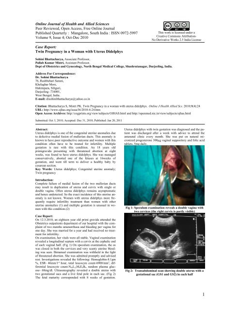
On exam<strong>in</strong>ation, her vitals were all stable. Vag<strong>in</strong>al exam<strong>in</strong>ation<br />
revealed a longitud<strong>in</strong>al septum <strong>with</strong> a cervix at the cephalic end<br />
of each vag<strong>in</strong>al half. (Fig 1) On speculum exam<strong>in</strong>ation, the os<br />
was closed <strong>in</strong> both the cervices and very scanty uter<strong>in</strong>e bleed<strong>in</strong>g<br />
was seen. Bimanual exam<strong>in</strong>ation was <strong>with</strong>held <strong>in</strong> the light<br />
of threatened abortion. She was admitted promptly and advised<br />
rest. Investigations revealed the follow<strong>in</strong>g: Hemoglob<strong>in</strong>-8.1gm<br />
%, ESR- 46mm/1 st hour, total leucocyte count-6900/mm 3 , differential<br />
leucocyte count-N 68L 22M 4E 6B 0, random plasma glucose-<br />
68mg/dl. Ultrasonography revealed a double uterus <strong>with</strong><br />
two gestational sacs and a live fetal pole <strong>in</strong> each sac. (Fig 2)<br />
The fetal maturity corresponded <strong>with</strong> 8 weeks of gestation.<br />
<strong>Uterus</strong> didelphys <strong>with</strong> tw<strong>in</strong> gestation was diagnosed and the patient<br />
was discharged after a week <strong>with</strong> advice to attend the<br />
antenatal cl<strong>in</strong>ic every month. She was put on natural micronized<br />
progesterone 300µg vag<strong>in</strong>al suppository and folic acid<br />
tablets, 5mg daily.<br />
Fig 1: Speculum exam<strong>in</strong>ation reveals a double vag<strong>in</strong>a <strong>with</strong><br />
two cervices (the right cervix is partly visible)<br />
Fig 2: Transabdom<strong>in</strong>al scan show<strong>in</strong>g double uterus <strong>with</strong> a<br />
gestational sac (GS1 and GS2) <strong>in</strong> each half<br />
1
On 20.5.2010, she was admitted aga<strong>in</strong> <strong>in</strong> gynaecological emergency<br />
ward <strong>with</strong> severe bleed<strong>in</strong>g per vag<strong>in</strong>a and expulsion of a<br />
fetus at home. Internal exam<strong>in</strong>ation confirmed the diagnosis of<br />
<strong>in</strong>complete abortion <strong>in</strong> the left hemiuterus. An exploration under<br />
anaesthesia was done on the same day to arrest bleed<strong>in</strong>g.<br />
Two units of blood were transfused. She was discharged after<br />
two days and was followed up closely <strong>in</strong> the antenatal cl<strong>in</strong>ic.<br />
On 20.9.2010, she developed rupture of membranes and gave<br />
birth to a 2.45 kg term baby by cesarean section<br />
Discussion:<br />
Although the true prevalence of congenital uter<strong>in</strong>e anomalies <strong>in</strong><br />
the population is unknown, it is known to vary from 0.1% to<br />
10%. In one study, the 24.2% of the women <strong>with</strong> uter<strong>in</strong>e anomalies<br />
had uterus didelphys.(1) Congenital uter<strong>in</strong>e anomalies are<br />
associated <strong>with</strong> the highest <strong>in</strong>cidence of reproductive failure<br />
and obstetric complications. Authors are not unanimous <strong>in</strong> predict<strong>in</strong>g<br />
the obstetric outcome of uterus didelphys. Accord<strong>in</strong>g to<br />
some, women <strong>with</strong> this form of congenital anomaly required <strong>in</strong>fertility<br />
treatment more frequently than women <strong>with</strong> other uter<strong>in</strong>e<br />
anomalies.(1) and the overall reproductive performance of<br />
uterus didelphys is poor.(3)<br />
Others feel that didelphic uterus offers the best chance for a<br />
successful pregnancy(57%) (4) <strong>with</strong> a fetal survival rate as high<br />
as 64%.(5)<br />
Multifetal gestation is rare <strong>in</strong> women <strong>with</strong> uterus didelphys.(2)<br />
It has been reported <strong>in</strong> the literature. But the preferred route of<br />
term<strong>in</strong>ation of pregnancy <strong>in</strong> these patients is not clear. Spontaneous<br />
vag<strong>in</strong>al delivery as well as cesarean section at term has<br />
been reported.(6-10) There has also been a triplet pregnancy<br />
<strong>with</strong> uterus didelphys <strong>with</strong> 72 days lapse between the delivery<br />
of the first two fetuses and the third.(6)<br />
References:<br />
1. Zhang Yan, Zhao Yang-yu, Qiao Jie. Obstetric outcome<br />
of women <strong>with</strong> uter<strong>in</strong>e anomalies <strong>in</strong> Ch<strong>in</strong>a.<br />
Ch<strong>in</strong>ese Medical Journal. 2010;123(4):418-422.<br />
2. Olah KS. Uter<strong>in</strong>e torsion and ischemia of one horn<br />
of a bicornuate uterus: A rare cause of failed second<br />
trimester term<strong>in</strong>ation of pregnancy. Br J Obstet<br />
Gynecol 2002;109:585.<br />
3. Raga F, Bauset C, Remohi J et al. Reproductive impact<br />
of congenital mullerian anomalies. Hum Reprod<br />
1997;12(10):2277-2281.<br />
4. Musich JR, Behrman SJ. Obsteric outcome before<br />
and after metroplasty <strong>in</strong> women <strong>with</strong> uter<strong>in</strong>e anomalies.<br />
Obstet Gynecol.1978;52:63.<br />
5. He<strong>in</strong>ohen PK, Saarikoski S, Pystynen P. Reproductive<br />
performance of women <strong>with</strong> uter<strong>in</strong>e anomalies.<br />
Acta Obstet gynecol Scand 1982;61:157.<br />
6. Mashiach S, Ben-Rafael Z, Dor J, Serr DM. Triplet<br />
pregnancy <strong>in</strong> uterus didelphys <strong>with</strong> delivery <strong>in</strong>terval<br />
of 72 days. Obstet Gynecol. 1981 Oct;58(4):519-<br />
521.<br />
7. Ahmad FK, Sherman SJ, Hagglund KH. <strong>Tw<strong>in</strong></strong> gestation<br />
<strong>in</strong> a woman <strong>with</strong> a uterus didelphys. A case report.<br />
J Reprod Med. 2000 Apr;45(4):357-359.<br />
8. Kekkonen R, Nuutila M, Laatika<strong>in</strong>en T. <strong>Tw<strong>in</strong></strong> pregnancy<br />
<strong>with</strong> a fetus <strong>in</strong> each half of a uterus didelphys.<br />
Acta Obstet Gynecol Scand. 1991;70(4-5):373-374.<br />
9. Nhân VQ, Huisjes HJ. Double uterus <strong>with</strong> a pregnancy<br />
<strong>in</strong> each half. Obstet Gynecol. 1983<br />
Jan;61(1):115-117.<br />
10. Lewenthal H, Biale Y, Ben-Adereth N. <strong>Uterus</strong><br />
didelphys <strong>with</strong> a pregnancy <strong>in</strong> each horn. Case report.<br />
Br J Obstet Gynaecol. 1977 Feb;84(2):155-<br />
158.<br />
2