The rib cage and abdominal components of respiratory system ...
The rib cage and abdominal components of respiratory system ...
The rib cage and abdominal components of respiratory system ...

Create successful ePaper yourself
Turn your PDF publications into a flip-book with our unique Google optimized e-Paper software.
Eur Respir J<br />
1988, 1, 242-247<br />
<strong>The</strong> <strong>rib</strong> <strong>cage</strong> <strong>and</strong> <strong>abdominal</strong> <strong>components</strong> <strong>of</strong><br />
<strong>respiratory</strong> <strong>system</strong> compliance in tetraplegic patients<br />
J.M. Goldman*, S.J. Williams**, D.M. Denison*<br />
<strong>The</strong> <strong>rib</strong> <strong>cage</strong> <strong>and</strong> <strong>abdominal</strong> <strong>components</strong> <strong>of</strong> <strong>respiratory</strong> <strong>system</strong> compliance in<br />
tetraplegic patients. J.M. Goldman, S.J. Williams. D.M. Denison.<br />
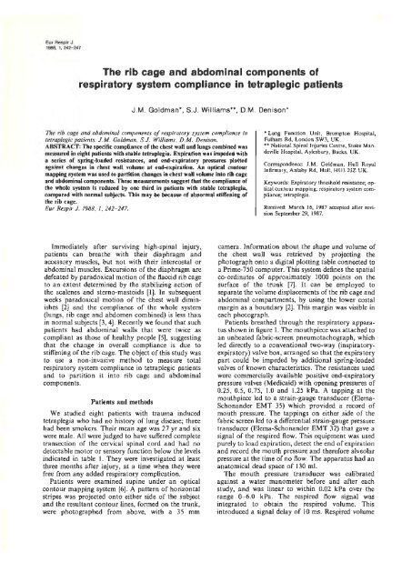
ABSTRACT: <strong>The</strong> specific compliance <strong>of</strong> the chest wall <strong>and</strong> lungs combined was<br />
measured in eight patients with stable tetraplegia. Expiration was impeded with<br />
a series <strong>of</strong> spring-loaded resistances, <strong>and</strong> end-expiratory pressures plotted<br />
against changes in chest wall volume at end-expiration. An optical contour<br />
mapping <strong>system</strong> was used to partition changes in chest wall volume into <strong>rib</strong> <strong>cage</strong><br />
<strong>and</strong> <strong>abdominal</strong> <strong>components</strong>. <strong>The</strong>se measuremen~ suggest tbat the compliance <strong>of</strong><br />
the whole <strong>system</strong> is reduced by one third in pati.ents with stable tetraplegia,<br />
compared with normal subjects. This may be because <strong>of</strong> abnormal stiffening <strong>of</strong><br />
the <strong>rib</strong> <strong>cage</strong>.<br />
Eur Respir J. 1988, I, 242- 247.<br />
• Lung Function Unit, Brompton Hospital,<br />
Fulham Rd, London SW3, UK.<br />
•• National SpinaJ Injuries Centre, Stoke M<strong>and</strong>eville<br />
Hospital, Aylesbury, Bucks, UK.<br />
Correspondence: J.M. Goldman, Hull Royal<br />
Infirmary, Anlaby Rd, Hull, HU3 2JZ UK.<br />
Keywords: Expiratory threshold resistance; optical<br />
contour mapping; <strong>respiratory</strong> <strong>system</strong> compliance;<br />
tetraplegia.<br />
Received: March 16, 1987 accepted after revi·<br />
sian September 29, 1987.<br />
Immediately after survlVlng high-spinal injury,<br />
patients can breathe with their diaphragm <strong>and</strong><br />
accessory muscles, but not with their intercostal or<br />
<strong>abdominal</strong> muscles. Excursions <strong>of</strong> the diaphragm are<br />
defeated by paradoxical motion <strong>of</strong> the flaccid <strong>rib</strong> <strong>cage</strong><br />
to an extent determined by the stabilizing action <strong>of</strong><br />
the scalenes <strong>and</strong> sterno-mastoids [1]. In subsequent<br />
weeks paradoxical motion <strong>of</strong> the chest wall diminishes<br />
[2] <strong>and</strong> the compliance <strong>of</strong> the whole <strong>system</strong><br />
(lungs, <strong>rib</strong> <strong>cage</strong> <strong>and</strong> abdomen combined) is less than<br />
in normal subjects (3, 4). Recently we found that such<br />
patients had <strong>abdominal</strong> walls that were twice as<br />
compliant as those <strong>of</strong> healthy people [5], suggesting<br />
that the change in overall compliance is due to<br />
stiffening <strong>of</strong> the <strong>rib</strong> <strong>cage</strong>. <strong>The</strong> object <strong>of</strong> this study was<br />
to use a non-invasive method to measure total<br />
<strong>respiratory</strong> <strong>system</strong> compliance in tetraplegic patients<br />
<strong>and</strong> to partition it into <strong>rib</strong> <strong>cage</strong> <strong>and</strong> <strong>abdominal</strong><br />
<strong>components</strong>.<br />
Patients <strong>and</strong> methods<br />
We studied eight patients with trauma induced<br />
tetraplegia who had no history <strong>of</strong> lung disease; three<br />
had been smokers. <strong>The</strong>ir mean age was 27 yr <strong>and</strong> six<br />
were male. All were judged to have suffered complete<br />
transection <strong>of</strong> the cervical spinal cord <strong>and</strong> had no<br />
detectable motor or sensory function below the levels<br />
indicated in table 1. <strong>The</strong>y were investigated at least<br />
three months after injury, at a time when they were<br />
free from any added <strong>respiratory</strong> complication.<br />
Patients were examined supine under an optical<br />
contour mapping <strong>system</strong> [6]. A pattern <strong>of</strong> horizontal<br />
stripes was projected onto either side <strong>of</strong> the subject<br />
<strong>and</strong> the resultant contour lines, formed on the trunk,<br />
were photographed from above, with a 35 mm<br />
camera. Information about the shape <strong>and</strong> volume <strong>of</strong><br />
the chest wall was retrieved by projecting the<br />
photograph onto a digital plotting table connected to<br />
a Prime-750 computer. This <strong>system</strong> defines the spatial<br />
co-ordinates <strong>of</strong> approximately 1000 points on the<br />
surface <strong>of</strong> the trunk (7]. It can be employed to<br />
separate the volume displacements <strong>of</strong> the <strong>rib</strong> <strong>cage</strong> <strong>and</strong><br />
<strong>abdominal</strong> compartments, by using the lower costal<br />
margin as a boundary [2]. This margin was visible in<br />
each photograph.<br />
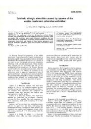
Patients breathed through the <strong>respiratory</strong> apparatus<br />
shown in figure I. <strong>The</strong> mouthpiece was attached to<br />
an unheated fabric-screen pneumotachograph, which<br />
led directly to a conventional two-way (inspiratoryexpiratory)<br />
valve box, arranged so that the expiratory<br />
part could be impeded by additional spring-loaded<br />
valves <strong>of</strong> known characteristics. <strong>The</strong> resistances used<br />
were commercially available positive end-expiratory<br />
pressure valves (Medicaid) with opening pressures <strong>of</strong><br />
0.25, 0.5, 0.75, 1.0 <strong>and</strong> 1.25 kPa. A tapping at the<br />
mouthpiece Jed to a strain-gauge transducer (Elema<br />
Schon<strong>and</strong>er EMT 35) which provided a record <strong>of</strong><br />
mouth pressure. <strong>The</strong> tappings on either side <strong>of</strong> the<br />
fabric screen led to a differential strain-gauge pressure<br />
transducer (Elema-Schon<strong>and</strong>er EMT 32) that gave a<br />
signal <strong>of</strong> the respired flow. This equipment was used<br />
purely to load expiration, detect the end <strong>of</strong> expiration<br />
<strong>and</strong> record the mouth pressure <strong>and</strong> therefore alveolar<br />
pressure at the time <strong>of</strong> no flow. <strong>The</strong> apparatus had an<br />
anatomical dead space <strong>of</strong> 130 mi.<br />
<strong>The</strong> mouth pressure transducer was calibrated<br />
against a water manometer before <strong>and</strong> after each<br />
study, <strong>and</strong> was linear to within 0.02 kPa over the<br />
range 0- 6.0 kPa. <strong>The</strong> respired flow signal was<br />
integrated to obtain the respired volume. This<br />
introduced a signal delay <strong>of</strong> 10 ms. Respired volume
CHEST WALL COMPLIANCE IN TETRAPLEGJCS 243<br />
Table 1.- Tetraplegic patients studied<br />
Patient Sex Age Level Time Predicted<br />
<strong>of</strong> after TLC<br />
yr lesion injury<br />
1 M 24 C6 4yr 6.6<br />
2* F 25 T1 4mth 5.8<br />
3 M 34 C5,6 7mth 7.0<br />
4* F 35 C5,6 6mth 5.0<br />
5 M 31 C5 6mth 6.75<br />
6 M 27 C5 4mth 7.75<br />
7* M 29 C5 9mth 5.8<br />
8 M 20 C5 19 mth 7.0<br />
M: male; F: female; *: smoker, TLC: total lung capacity, C: cervical spine segment, T: thoracic spine segment.<br />
spring loaded valve<br />
O.SkPa ]<br />
airway pressure<br />
2-way tap<br />
2-way valve<br />
mouth<br />
prea1111e<br />
monltOI'iog<br />
port<br />
Fig. I. Diagrammatic representation <strong>of</strong> the apparatus used to<br />
apply an expiratory threshold resistance to tetraplegic patients, <strong>and</strong><br />
record airway pressure <strong>and</strong> phase <strong>of</strong> respiration.<br />
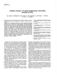
<strong>and</strong> mouth pressure, together with a shutter-opening<br />
signal from the camera, were displayed on a multichannel<br />
thermal pen recorder (Gould 4400), as shown<br />
in figure 2.<br />
Protocol<br />
Patients at rest were supine beneath the optical<br />
frame <strong>and</strong> breathed spontaneollsly throughout. <strong>The</strong><br />
camera was triggered manually to obtain photographs<br />
(<strong>of</strong> <strong>abdominal</strong> <strong>and</strong> <strong>rib</strong> <strong>cage</strong> volume) at the end<br />
<strong>of</strong> unimpeded expiration <strong>and</strong> then at the end <strong>of</strong> the<br />
last five <strong>of</strong> ten breaths opposed by resistances that<br />
were increased progressively from 0.25 to I .25 kPa.<br />
<strong>The</strong> photograph which was closest to true endexpiration<br />
in each series <strong>of</strong> five was selected by<br />
inspection <strong>of</strong> the pen recording <strong>and</strong> used to relate<br />
<strong>abdominal</strong>, <strong>rib</strong> <strong>cage</strong> <strong>and</strong> trunk volumes to the mouth<br />
pressure measured at the time. Patients were allowed<br />
a few minutes unimpeded breathing between each<br />
obstruction. <strong>The</strong> body volumes were calculated from<br />
the photographs as desc<strong>rib</strong>ed in [6].<br />
11 1<br />
ventilation<br />
camera trigger<br />
Ssec<br />
Fig. 2. A typical trace <strong>of</strong> airway pressure, ventilation, <strong>and</strong> the<br />
camera trigger, recorded from a tetraplegic patient breathing with<br />
an expiratory threshold resistance.<br />
Results<br />
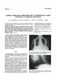
<strong>The</strong> open circles in figure 3 plot the increases in<br />
trunk volume, relative to functional residual capacity<br />
(FRC), seen in each patient as end-expiratory<br />
pressures increased. Data are normalized to predicted<br />
total lung capicity (TLC) [8] to eliminate the effects <strong>of</strong><br />
differences in body size. <strong>The</strong> broken lines show<br />
regressions constrained to pass through the origin <strong>of</strong><br />
the graph in each case. <strong>The</strong> slope <strong>of</strong> these lines have<br />
been taken as a measure <strong>of</strong> specific <strong>respiratory</strong> <strong>system</strong><br />
compliance for each individual. Values for these<br />
slopes are listed in table II. <strong>The</strong> relationship is linear<br />
in seven <strong>of</strong> the eight patients (r > 0.96), but not in the<br />
eighth (r = 0.64 in patient 7). <strong>The</strong> mean slope for all<br />
eight patients is 0.81 ml· kPa ·/- 1 TLC (so= 0.41<br />
ml·kPa·r 1 TLC).<br />
<strong>The</strong> solid circles in figure 3 plot the volume
244 J.M . GOLDMAN, S.J. WILLIAMS, D.M. DENISON<br />
Table 2.- Respiratory <strong>system</strong> compliance in tetraplegic patients<br />
Patient Total Resp. System Abdominal Compartment<br />
Specific Corr. Specific Corr. sCabd/sCrs<br />
compliance coeff. compliance coeff.<br />
mJ.IcPa·r 1 TLC r ml-kPa·I ' 1 TLC r %<br />
1 0.912 0.98 0.956 0.99 105<br />
2 1.102 0.99 1.034 0.99 94<br />
3 1.485 0.99 0.027 0.97 69<br />
4 0.570 0.98 0.306 0.86 54<br />
5 0.787 0.97 0.471 0.89 60<br />
6 0.229 0.93 0.186 0.95 81<br />
7 0.359 0.64 0.402 0.77 110<br />
8 1.102 0.99 0.340 0.95 31<br />
Specific compliance <strong>of</strong> the total <strong>respiratory</strong> <strong>system</strong> <strong>and</strong> its <strong>abdominal</strong> compartment in tetraplegic patients, with the<br />
correlation coefficients <strong>of</strong> each plot. sCabd/sCrs: specific compliance <strong>abdominal</strong>/specific compliance <strong>respiratory</strong> <strong>system</strong> ~<br />
%.<br />
p<br />
I<br />
200 Patient 1 Patient 2<br />
Patient 31/<br />
I<br />
?I<br />
'<br />
I<br />
,<br />
•<br />
•<br />
Patient 4<br />
I<br />
()' '<br />
,<br />
Patient 6<br />
Patient 7<br />
Patient 8<br />
ISO<br />
100<br />
50<br />
0<br />
,'<br />
, ,<br />
,'<br />
,0 P kPa<br />
1.0 1.5 ° 0<br />
1.5<br />
Fig. 3. Specific compliance plots <strong>of</strong> the total <strong>respiratory</strong> <strong>system</strong> <strong>and</strong> its <strong>abdominal</strong> compartments in tetraplegic patients. flY: change in<br />
volume above resting FRC expressed in mH predicted TLC; P: airway pressure in kPa. Black data points <strong>and</strong> solid lines represent data<br />
pertaining to the <strong>abdominal</strong> compartment. White data points <strong>and</strong> broken lines represent data pertaining to the total <strong>respiratory</strong> <strong>system</strong>.
CHEST WALL COMPLIANCE TN TETRAPLEGICS 245<br />
displacements <strong>of</strong> the <strong>abdominal</strong> wall, relative to FRC,<br />
seen in each patient as end-expiratory pressures<br />
increased. Data are again normalized to predicted<br />
TLC. <strong>The</strong> solid lines show regressions constrained to<br />
pass through the origin <strong>of</strong> the graph in each case. <strong>The</strong><br />
slope <strong>of</strong> this line has been taken as a measure <strong>of</strong> the<br />
specific compliance <strong>of</strong> the <strong>abdominal</strong> pathway. It<br />
accounts for all, or almost all, <strong>of</strong> the specific<br />
compliance <strong>of</strong> the whole <strong>system</strong> in patients I, 2, 3, 5,<br />
6, <strong>and</strong> 7, <strong>and</strong> half or slightly less than half <strong>of</strong> the<br />
whole <strong>system</strong> compliance in patients 4 <strong>and</strong> 8 (see<br />
table 2). <strong>The</strong> mean slope for the <strong>abdominal</strong> pathway<br />
represents 75.5 ± 27.2% <strong>of</strong> <strong>system</strong> compliance, in the<br />
group as a whole.<br />
Discussion<br />
At the end <strong>of</strong> an expiration there is no movement <strong>of</strong><br />
air into or out <strong>of</strong> the lungs even though the glottis is<br />
open. Since there is no flow, alveolar pressure <strong>and</strong><br />
mouth pressure are equal. <strong>The</strong> <strong>system</strong> is stationary<br />
<strong>and</strong> the various forces within it must be at equilibrium.<br />
When positive end-expiratory pressure is added<br />
to the <strong>system</strong> <strong>and</strong> quiet breathing continues, we have<br />
supposed that the slope <strong>of</strong> the pressure-volume curve<br />
<strong>of</strong> the trunk measures the compliance <strong>of</strong> the relaxed<br />
<strong>respiratory</strong> <strong>system</strong> at FRC <strong>and</strong> above. We have<br />
assumed that the <strong>respiratory</strong> muscles <strong>of</strong> tetraplegic<br />
patients are relaxed at end-expiration. It is possible<br />
that there is reflex activity in the <strong>abdominal</strong> or<br />
intercostal muscles, but, by definition, these patients<br />
have no control over such activity <strong>and</strong> we feel that<br />
measurement <strong>of</strong> the 'effective' compliance <strong>of</strong> the<br />
<strong>respiratory</strong> <strong>system</strong> is valid.<br />
Our method is analogous to the weighted spirometer<br />
technique <strong>of</strong> HEAF <strong>and</strong> PRIME (9). Instead <strong>of</strong><br />
weights, expiratory threshold resistances were used to<br />
load expiration. Instead <strong>of</strong> a spirometer to measure<br />
respired volume, the optical contour mapping <strong>system</strong><br />
was used to measure changes in chest wall volume.<br />
This <strong>system</strong> is accurate in partitioning chest wall<br />
volume into its <strong>rib</strong> <strong>cage</strong> <strong>and</strong> <strong>abdominal</strong> <strong>components</strong>,<br />
with 95% confidence limits <strong>of</strong> 1% <strong>of</strong> the measured<br />
volume <strong>of</strong> the <strong>abdominal</strong> component [10). Both the<br />
weighted spirometer technique <strong>and</strong> the method used<br />
in this study measure total <strong>respiratory</strong> compliance.<br />
Since airway pressure is plotted against change in<br />
chest wall volume, it is the compliance <strong>of</strong> the whole<br />
<strong>system</strong>, including the lungs, which is represented by<br />
the gradient <strong>of</strong> the plot. To normalize the data for<br />
patients with different body size, results have been<br />
divided by predicted TLC, <strong>and</strong> expressed as specific<br />
compliance.<br />
VELLODY et at. [II) refined the weighted spirometer<br />
method by using magnetometers to measure <strong>rib</strong> <strong>cage</strong><br />
<strong>and</strong> <strong>abdominal</strong> pathway compliance separately in<br />
normal subjects. EsTENNE et al. (4] applied a similar<br />
method to patients with <strong>respiratory</strong> muscle weakness,<br />
including ten patients with complete neurological<br />
lesions <strong>of</strong> the cervical spinal cord (eight due to trauma<br />
<strong>and</strong> two due to transverse myelitis). <strong>The</strong>y measured<br />
lung compliance with an oesophageal balloon to<br />
derive true chest wall compliance, but could not<br />
separate it into <strong>abdominal</strong> <strong>and</strong> <strong>rib</strong> <strong>cage</strong> <strong>components</strong><br />
with any certainty. More recently, the same group (3]<br />
measured chest wall compliance in twenty seated<br />
tetraplegic patients using a weighted spirometer<br />
technique <strong>and</strong>, by using pairs <strong>of</strong> linear magnetometers,<br />
derived <strong>rib</strong> <strong>cage</strong> <strong>and</strong> diaphragm-abdomen compliance.<br />
We have applied the optical contour mapping<br />
<strong>system</strong> in this situation <strong>and</strong> believe it has advantages,<br />
as it measures change in chest wall volume rather than<br />
extrapolating from change in diameter. It will<br />
therefore take into account any asymmetric or<br />
paradoxical movement.<br />
Expiratory threshold resistances were used to load<br />
breathing. CAMPBELL eta/. [12] exposed conscious <strong>and</strong><br />
anaesthetized normal subjects to such resistances, <strong>and</strong><br />
found that FRC increased with addition <strong>of</strong> the load,<br />
<strong>and</strong> decreased to normal after it was removed. In<br />
conscious subjects, this pattern was seen even if the<br />
load was present for only 4-I 0 breaths. An increase in<br />
end-expiratory volume occurred from the first loaded<br />
breath <strong>and</strong> was complete after three or more breaths.<br />
In the present study, loads were applied for ten<br />
breaths <strong>and</strong> end-expiratory volume measured after at<br />
least five breaths. D'ANGELO <strong>and</strong> AGOSTONl [ 13] noted<br />
that this response to expiratory threshold resistances<br />
was abolished by vagotomy in dogs, <strong>and</strong> that it was<br />
not altered by Tl cordotomy. This evidence suggests<br />
that response to expiratory threshold resistanee is<br />
mediated via the vagus <strong>and</strong> should therefore be intact<br />
in patients with cervical cord injury. AxEN [14] has<br />
made detailed studies <strong>of</strong> loading inspiration in<br />
tetraplegic patients <strong>and</strong> found their response similar<br />
to normal subjects.<br />
In the present study FRC rose linearly with the<br />
addition <strong>of</strong> expiratory threshold resistance in seven<br />
out <strong>of</strong> eight patients. VELLODY et al. [II] found a<br />
linear increase in end-expiratory volume as airway<br />
pressure increased in normal subjects whether they<br />
were relaxed (0.0077 I· kPa - 1 ) or anaesthetized <strong>and</strong><br />
paralysed (0.0078 l· kPa - 1 ). Using the data from<br />
VELLODY et al. [11], specific compliance <strong>of</strong> the<br />
<strong>respiratory</strong> <strong>system</strong> has been calculated for each <strong>of</strong> the<br />
paralysed <strong>and</strong> anaesthetized normal subjects that they<br />
studied. This data is displayed in table 3, <strong>and</strong> may be<br />
compared to the data for tetraplegic patients in table<br />
I. <strong>The</strong> mean specific compliance <strong>of</strong> the <strong>respiratory</strong><br />
<strong>system</strong> for paralysed normal subjects was 1.237<br />
ml· kPa- 1 ·I TLC (so 0.336 ml· kPa- 1 ·I TLC), whilst<br />
for the tetraplegic patients <strong>of</strong> the present study, it was<br />
0.813 ml · kPa- 1 ·I TLC (so 0.41 ml· kPa- 1 ·I TLC),<br />
which is 65.7% <strong>of</strong> normal. <strong>The</strong> data for normal<br />
subjects <strong>and</strong> tetraplegic patients were compared using<br />
an unpaired Student's t-test. <strong>The</strong>re was a significant<br />
difference between the two groups (p < 0.05).<br />
A lo·w effective <strong>respiratory</strong> <strong>system</strong> compliance<br />
amongst supine tetraplegic patients supports the<br />
findings <strong>of</strong> ESTENNE et a/. [4] in a similar seated group.<br />
<strong>The</strong>ir published data <strong>of</strong> lung <strong>and</strong> chest wall compliance<br />
were used to derive <strong>respiratory</strong> <strong>system</strong> compli-
246 J.M. GOLDMAN, S.J. WILLIAMS, D.M. DENISON<br />
Table 3.- Respiratory <strong>system</strong> compliance in normal subjects<br />
Subject Height TLC sCrs % partitioned to <strong>abdominal</strong><br />
em ml·kPa·1·1·1 TLC compartment<br />
1 175 6.35 1.024 46.2<br />
2 183 6.97 1.019 37.2<br />
3 162 5.34 0.749 60.6<br />
4 188 7.36 1.421 58.0<br />
5 170 5.96 1.628 46.4<br />
6 183 6.97 1.365 52.6<br />
7 170 5.96 1.040 53.2<br />
8 168 5.80 1.310 42.1<br />
9 173 6.19 1.309 46.9<br />
10 175 6.35 1.874 47.1<br />
11 178 6.58 0.866 62.2<br />
Specific compliance <strong>of</strong> the total <strong>respiratory</strong> <strong>system</strong>, <strong>and</strong> the percentage partitioned to the <strong>abdominal</strong> compartment in<br />
normal paralysed anaesthetized subjects from the data <strong>of</strong> YEUODY et al. [11).<br />
ance for their ten patients with tetraplegia. <strong>The</strong> mean<br />
compliance <strong>of</strong> the <strong>respiratory</strong> <strong>system</strong> was 0.0082<br />
l·kPa- 1 , while in normal subjects it was 0.0135<br />
I· kPa - 1 • <strong>The</strong>y suggested that this decrease was due<br />
predominantly to a decrease in <strong>rib</strong> <strong>cage</strong> compliance,<br />
<strong>and</strong> subsequently confirmed this by direct measurement<br />
using magnetometers [3]. <strong>The</strong> tetraplegic patients<br />
in the present study partitioned 24.5% <strong>of</strong> the specific<br />
compliance <strong>of</strong> the <strong>respiratory</strong> <strong>system</strong> to the <strong>rib</strong> <strong>cage</strong><br />
pathway, whilst the normal subjects in the study <strong>of</strong><br />
VELLODY et al. [II] partitioned a mean <strong>of</strong> 50% to the<br />
<strong>rib</strong> <strong>cage</strong>. We did not measure lung compliance in our<br />
patients, as we were aiming to develop a non-invasive<br />
technique for clinical application. A decrease in lung<br />
compliance in tetraplegia <strong>of</strong> up to 30% is well<br />
desc<strong>rib</strong>ed [4], but this will not have a preferential effect<br />
on <strong>rib</strong> <strong>cage</strong> or <strong>abdominal</strong> pathway compliance. <strong>The</strong><br />
ratio between absolute <strong>rib</strong> <strong>cage</strong> <strong>and</strong> <strong>abdominal</strong><br />
compliance will be the same as that between the <strong>rib</strong><br />
<strong>cage</strong> <strong>and</strong> <strong>abdominal</strong> pathway~ <strong>of</strong> total <strong>respiratory</strong><br />
<strong>system</strong> compliance. This ratio in our study was 25% to<br />
75%, i.e. 3 to 1. In normal subjects it is I to 1. Since we<br />
know that <strong>abdominal</strong> wall compliance is doubled in<br />
tetraplegic patients [5] <strong>and</strong> we have found <strong>respiratory</strong><br />
<strong>system</strong> compliance to be reduced, we would suggest<br />
that <strong>rib</strong> <strong>cage</strong> compliance is less than normal.<br />
Our findings agree those <strong>of</strong> EsTENNE <strong>and</strong> DE<br />
TROYER [3] who measured chest wall compliance in<br />
twenty seated tetraplegic patients, <strong>and</strong> found it<br />
reduced to 72% <strong>of</strong> normal. This was due to a decrease<br />
in <strong>rib</strong> <strong>cage</strong> compliance to 55% <strong>of</strong> normal, whilst<br />
<strong>abdominal</strong> compliance was increased to 170% <strong>of</strong><br />
normal. We have observed that, in our patients,<br />
<strong>respiratory</strong> <strong>system</strong> compliance is made up almost<br />
entirely by the <strong>abdominal</strong> pathway. In normal supine<br />
subjects, the <strong>rib</strong> <strong>cage</strong> is stiffer <strong>and</strong> the abdomen more<br />
compliant than it is in the erect posture [15, 16}. This<br />
is compatible with our findings, <strong>and</strong> additionally we<br />
know that the <strong>abdominal</strong> wall compliance is twice the<br />
normal in tetraplegic patients [5] whilst the <strong>rib</strong> <strong>cage</strong> is<br />
known to be less compliant than normal [3]. In this<br />
situation, if the <strong>respiratory</strong> <strong>system</strong> is passively<br />
inflated, the increase in volume will be partitioned to<br />
its most compliant pathway until the compliances <strong>of</strong><br />
the two pathways equilibrate.<br />
Residual <strong>respiratory</strong> muscle activity might also<br />
effect the relative compliances <strong>of</strong> the <strong>rib</strong> <strong>cage</strong> <strong>and</strong><br />
abdomen. DE TROYER et a/. [17] have shown that<br />
seated tetraplegic patients may use the clavicular<br />
portion <strong>of</strong> the pectoralis major during active expiration.<br />
This muscle, if activated by end-expiratory<br />
pressure, could decrease <strong>rib</strong> <strong>cage</strong> compliance. However,<br />
since our patients were studied supine during<br />
relaxed breathing, we think this unlikely. Another<br />
possible explanation for our findings would be the<br />
<strong>system</strong>atic overestimation <strong>of</strong> the increment in chest<br />
wall volume partitioned to the <strong>abdominal</strong> compartment.<br />
This seems unlikely, given the 95% confidence<br />
limits we have quoted for our method. However, it<br />
must be remembered that each point plotted repre-
CHEST WALL COMPLIANCE IN TETRAPLEGICS 247<br />
sents a value derived from photographs taken at FRC<br />
<strong>and</strong> at a given end-expiratory pressure. A combination<br />
<strong>of</strong> two sets <strong>of</strong> errors might explain the results in the two<br />
patients who partitioned more than 100% <strong>of</strong> <strong>respiratory</strong><br />
<strong>system</strong> compliance to the <strong>abdominal</strong> pathway, but<br />
will not alter the general trend <strong>of</strong> our results.<br />
Conclusion<br />
In normal subjects who have been anaesthetized <strong>and</strong><br />
paralysed, the volume <strong>of</strong> the <strong>rib</strong> <strong>cage</strong> <strong>and</strong> abdomen<br />
increases in parallel when expiration is loaded [16]. In<br />
six out <strong>of</strong> the eight tetraplegic patients studied, the<br />
<strong>abdominal</strong> compartment cont<strong>rib</strong>uted almost all the<br />
increase in chest wall volume. Since the intercostal <strong>and</strong><br />
<strong>abdominal</strong> muscles are paralysed in both these groups,<br />
the decrease in <strong>respiratory</strong> <strong>system</strong> compliance in<br />
tetraplegic patients may be due to the <strong>rib</strong> <strong>cage</strong> itself<br />
becoming stiffer (for example, due to honey ankylosis <strong>of</strong><br />
joints), or to spasticity <strong>of</strong> the intercostals. We conclude<br />
that the specific compliance <strong>of</strong> the total <strong>respiratory</strong><br />
<strong>system</strong> <strong>of</strong> tetraplegic patients is less than that <strong>of</strong> normal<br />
subjects, probably because <strong>of</strong> a decrease in <strong>rib</strong> <strong>cage</strong><br />
compliance. This stiffness will limit paradoxical motion<br />
<strong>of</strong> the <strong>rib</strong> <strong>cage</strong> <strong>and</strong> may be an advantage.<br />
Ackll()wkdgements: We wish to thank J.R. Silver, H.<br />
Frankel <strong>and</strong> I. Nusiebeh for allowing us to study their<br />
patients, L: Rose for help in carrying out the studies, R.<br />
Robinson <strong>and</strong> the medical electronics department for<br />
technical assistance, <strong>and</strong> the staff <strong>and</strong> patients <strong>of</strong> the<br />
spinal injuries unit for their co-operation. J.M.<br />
Goldman was supported by a Brompton Hospital<br />
Clinical Research Committee grant.<br />
References<br />
I. Estenne M, De Troyer A. - Relationship between <strong>respiratory</strong><br />
muscle electromyogram <strong>and</strong> <strong>rib</strong> <strong>cage</strong> motion in tetraplegia. Am Rev<br />
Respir Dis, 1985, 132, 53- 59.<br />
2. Morgan MDL, Gourlay AR, Silver JR, Williams SJ, Denison<br />
DM. - Cont<strong>rib</strong>ution <strong>of</strong> the <strong>rib</strong> <strong>cage</strong> to breathing in tetraplegia.<br />
Thorax, 1985, 40, 613- 617.<br />
3. Estenne M, De Troyer A. - <strong>The</strong> effect <strong>of</strong> tetraplegia on chest<br />
wall statistics. Am Rev Respir Dis, 1986, 134, 121- 124.<br />
4. Estenne M, Yernault JC, De Troyer A. - Rib <strong>cage</strong> <strong>and</strong><br />
diaphragm-abdomen compliance in humans: effect <strong>of</strong> age <strong>and</strong><br />
posture. J Appl Physiol, 1985, 59, 1842- 1848.<br />
5. Goldman JM, Morgan MDL, Denison DM. - <strong>The</strong> measurement<br />
<strong>of</strong> <strong>abdominal</strong> wall compliance in normal subjects <strong>and</strong><br />
tetraplegic patients. Thorax, 1986, 41, 513- 518.<br />
6. Morgan MDL, Gourlay AR, Denison DM. - An optical<br />
method <strong>of</strong> studying the shape <strong>and</strong> movement <strong>of</strong> the chest wall in<br />
recumbent patients. Thorax, 1984, 39, 101 - 106.<br />
7. Gourlay AR, Kaye G, Morgan MDL, Denison DM, Peacock<br />
AJ. - Analysis <strong>of</strong> an optical mapping technique for lung function<br />
studies. Comput Bioi Med, 1984, 14, 47- 58.<br />
8. Cotes JE. - Lung function assessment <strong>and</strong> application in<br />
medicine. 4th edition. Blackwell, Oxford, 1979, 32~- 387.<br />
9. HeafPJD, Prime FJ. - <strong>The</strong> compliance <strong>of</strong> the thorax in normal<br />
human subjects. C/in Sci, 1956, 15, 319- 327.<br />
10. Goldman JM. - <strong>The</strong> role <strong>of</strong> the abdomen in breathing in<br />
normal subjects <strong>and</strong> tetraplegic patients. MD <strong>The</strong>sis, London<br />
University 1986.<br />
II. Vellody VPS, Nassery M, Bialasaraswathi K, Goldberg NB,<br />
Sharp JT. - Compliance <strong>of</strong> human <strong>rib</strong> <strong>cage</strong> ani! diaphragm<br />
abdomen pathways relaxed ver.rus paralysed states. Am Rev Respir<br />
Dis, 1978, I 18, 479-491.<br />
12. Campbell EJM, Dickinson CJ, Dinnick OP, Howell JBL. -<br />
<strong>The</strong> immediate effects <strong>of</strong> threshold loads on the breathing <strong>of</strong> men<br />
<strong>and</strong> dogs. Clin Sci, 1961, 21, 309-320.<br />
13. D'Angelo E, Agostoni E. - Immediate response to expiratory<br />
threshold load. Respir Physiol, 1975, 25, 269-284.<br />
14. Axen K. - Adaptation <strong>of</strong> quadriplegic men to consecutively<br />
loaded breaths. J Appl Physiol: Respirat Environ Exercise Physiol,<br />
1984, 56, 1099- 1103.<br />
15. Estenne M, Heilpom A, Delhez L, Yernault JC, De Troyer A.<br />
- Chest wall stiffness in patients with chronic <strong>respiratory</strong> muscle<br />
weakness. Am Rev Respir Dis, 1983, 120, 1002-1100.<br />
16. Konno K, Mead J. - Static volume pressure characteristics<br />
<strong>of</strong> the <strong>rib</strong> <strong>cage</strong> <strong>and</strong> abdomen. J Appl Physio/, 1968, 24, 544-<br />
548.<br />
17. De Troyer A, Estenne M, Heilpom A. - Mechanism <strong>of</strong> active<br />
expiration in tetraplegic patients. N Eng/ J Med, 1986, 314,<br />
740-744.