Nervous System I
Nervous System I
Nervous System I

You also want an ePaper? Increase the reach of your titles
YUMPU automatically turns print PDFs into web optimized ePapers that Google loves.
10<br />
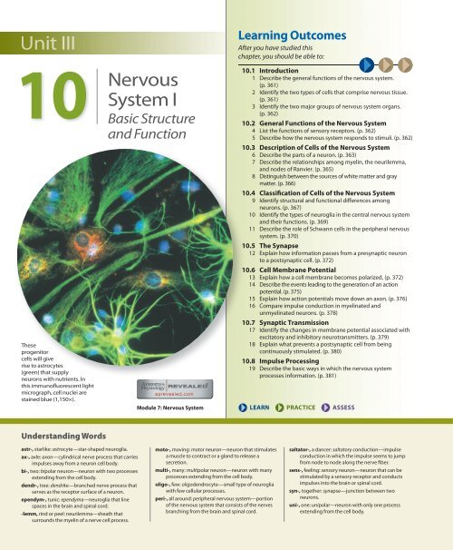
These<br />
progenitor<br />
cells will give<br />
rise to astrocytes<br />
(green) that supply<br />
neurons with nutrients. In<br />
this immunofluorescent light<br />
micrograph, cell nuclei are<br />
stained blue (1,150×).<br />
<strong>Nervous</strong><br />
<strong>System</strong> I<br />
Basic Structure<br />
and Function<br />
Module 7: <strong>Nervous</strong> <strong>System</strong><br />
Learning Outcomes<br />
After you have studied this<br />
chapter, you should be able to:<br />
10.1 Introduction<br />
1 Describe the general functions of the nervous system.<br />
(p. 361)<br />
2 Identify the two types of cells that comprise nervous tissue.<br />
(p. 361)<br />
3 Identify the two major groups of nervous system organs.<br />
(p. 362)<br />
10.2 General Functions of the <strong>Nervous</strong> <strong>System</strong><br />
4 List the functions of sensory receptors. (p. 362)<br />
5 Describe how the nervous system responds to stimuli. (p. 362)<br />
10.3 Description of Cells of the <strong>Nervous</strong> <strong>System</strong><br />
6 Describe the parts of a neuron. (p. 363)<br />
7 Describe the relationships among myelin, the neurilemma,<br />
and nodes of Ranvier. (p. 365)<br />
8 Distinguish between the sources of white matter and gray<br />
matter. (p. 366)<br />
10.4 Classification of Cells of the <strong>Nervous</strong> <strong>System</strong><br />
9 Identify structural and functional differences among<br />
neurons. (p. 367)<br />
10 Identify the types of neuroglia in the central nervous system<br />
and their functions. (p. 369)<br />
11 Describe the role of Schwann cells in the peripheral nervous<br />
system. (p. 370)<br />
10.5 The Synapse<br />
12 Explain how information passes from a presynaptic neuron<br />
to a postsynaptic cell. (p. 372)<br />
10.6 Cell Membrane Potential<br />
13 Explain how a cell membrane becomes polarized. (p. 372)<br />
14 Describe the events leading to the generation of an action<br />
potential. (p. 375)<br />
15 Explain how action potentials move down an axon. (p. 376)<br />
16 Compare impulse conduction in myelinated and<br />
unmyelinated neurons. (p. 378)<br />
10.7 Synaptic Transmission<br />
17 Identify the changes in membrane potential associated with<br />
excitatory and inhibitory neurotransmitters. (p. 379)<br />
18 Explain what prevents a postsynaptic cell from being<br />
continuously stimulated. (p. 380)<br />
10.8 Impulse Processing<br />
19 Describe the basic ways in which the nervous system<br />
processes information. (p. 381)<br />
LEARN PRACTICE ASSESS<br />
Understanding Words<br />
astr-, starlike: astrocyte—star-shaped neuroglia.<br />
ax-, axle: axon—cylindrical nerve process that carries<br />
impulses away from a neuron cell body.<br />
bi-, two: bipolar neuron—neuron with two processes<br />
extending from the cell body.<br />
dendr-, tree: dendrite—branched nerve process that<br />
serves as the receptor surface of a neuron.<br />
ependym-, tunic: ependyma—neuroglia that line<br />
spaces in the brain and spinal cord.<br />
-lemm, rind or peel: neurilemma—sheath that<br />
surrounds the myelin of a nerve cell process.<br />
moto-, moving: motor neuron—neuron that stimulates<br />
a muscle to contract or a gland to release a<br />
secretion.<br />
multi-, many: multipolar neuron—neuron with many<br />
processes extending from the cell body.<br />
oligo-, few: oligodendrocyte—small type of neuroglia<br />
with few cellular processes.<br />
peri-, all around: peripheral nervous system—portion<br />
of the nervous system that consists of the nerves<br />
branching from the brain and spinal cord.<br />
saltator-, a dancer: saltatory conduction—impulse<br />
conduction in which the impulse seems to jump<br />
from node to node along the nerve fiber.<br />
sens-, feeling: sensory neuron—neuron that can be<br />
stimulated by a sensory receptor and conducts<br />
impulses into the brain or spinal cord.<br />
syn-, together: synapse—junction between two<br />
neurons.<br />
uni-, one: unipolar—neuron with only one process<br />
extending from the cell body.
Brain Banks<br />
In a large room at the Croatian Institute for Brain Research, rows of shelves<br />
hold a variety of fluid-filled jars, a human brain suspended in each. Their<br />
sizes differ, reflecting their origins from embryos up to the elderly.<br />
Researchers can use the more than 1,000 brains and more than 130,000<br />
histological slides at the bank to investigate brain-based diseases and injuries<br />
that affect many millions of people worldwide and also to better understand<br />
the functioning of the normal human brain.<br />
In the United States, several brain banks offer tissue sections from thousands<br />
of people who willed their brains to science. Unlike donated hearts, lungs,<br />
or corneas, which directly help other people, donated brains go to research labs.<br />
Many brain banks are specialized. The bank at Harvard University is devoted<br />
to neurodegenerative diseases, such as Alzheimer and Parkinson diseases, while<br />
the resource at the University of Maryland in Baltimore focuses on developmental<br />
disorders, including Down syndrome and autism. The brain bank at the<br />
University of Miami has brains from people who had schizophrenia, depression,<br />
amyotrophic lateral sclerosis, and several other disorders, as well as undiseased<br />
brains for comparison.<br />
Brains must be removed from the skull within twelve hours of death. Then<br />
they are halved and cut into one-centimeter thick sections and frozen in plastic<br />
bags. The specimens are provided free to researchers.<br />
Study of brain function and malfunction is also possible at the cellular<br />
level. The National Human Neural Stem Cell Resource provides neural stem<br />
cells, which function after death longer than neurons because their energy<br />
and oxygen requirements are not as high as those of the more specialized cells.<br />
Hospitals collect brain material upon autopsy and send it to the facility, where a<br />
special protocol is used to obtain and preserve the cells from several brain areas.<br />
Investigators use the human neural stem cells to study neurodegenerative disorders,<br />
stroke, traumatic brain injury, rare inborn errors of metabolism, as well as<br />
the development of the incredibly complex human brain from initial stem and<br />
Neurospheres cultured in the laboratory consist of neural stem cells. These<br />
cells can divide and differentiate to give rise to neural progenitor cells, which<br />
in turn divide and differentiate, yielding neurons and neuroglia. In the brain,<br />
neural stem cells occupy certain areas but are exceedingly rare. Researchers are<br />
attempting to harness the natural ability of neural stem and progenitor cells to<br />
divide and replace damaged or diseased neural tissue.<br />
progenitor cells. The material in brain and stem cell banks is also being used in<br />
drug discovery and in developing new treatments based on cell implants. The<br />
chapter opening image shows neural progenitor cells and the photo accompanying<br />
this vignette shows neural stem cells. <br />
10.1 INTRODUCTION<br />
The nervous system oversees all that we do and largely determines<br />
who we are. Through a vast communicating network<br />
of cells and the information that they send and receive, the<br />
nervous system can detect changes in the body, make decisions,<br />
and stimulate muscles or glands to respond. Typically,<br />
these responses counteract the effects of the changes, and<br />
in this way, the nervous system helps maintain homeostasis.<br />
Clinical Application 10.1 discusses how environmental<br />
changes may trigger migraine headaches, a common medical<br />
problem attributed to the nervous system that may involve<br />
its blood supply as well as neurons.<br />
The nervous system is composed predominantly of neural<br />
tissue, but also includes blood vessels and connective<br />
tissue. Neural tissue consists of two cell types: nerve cells,<br />
or neurons (nu′ronz), and neuroglia (nu-ro′gle-ah) (or neuroglial<br />
cells).<br />
Neurons are specialized to react to physical and chemical<br />
changes in their surroundings. Small cellular processes<br />
called dendrites (den′drītz) receive the input. A longer<br />
process called an axon (ak′son), or nerve fiber, carries the<br />
information away from the cell in the form of bioelectric<br />
signals, often called impulses, which allow the neuron to<br />
communicate with other neurons and with cells outside the<br />
nervous system (fig. 10.1). Nerves are bundles of axons.<br />
Neuroglia are found throughout the nervous system,<br />
and in the brain they greatly outnumber neurons. It was<br />
once thought that neuroglia only fill spaces and surround<br />
or support neurons. Today we know that they have many<br />
other functions, including nourishing neurons and sending<br />
and receiving messages.<br />
An important part of the nervous system at the cellular<br />
level is not a cell at all, but the small space between a neuron<br />
and the cell(s) with which it communicates, called a<br />
synapse (sin′aps). Much of the work of the nervous system<br />
is to send and receive electrochemical messages across synapses.<br />
Biological messenger molecules called neurotransmitters<br />
(nu″ro-trans-mit′erz) convey this neural information.<br />
The organs of the nervous system can be divided into<br />
two groups. One group, consisting of the brain and spinal<br />
cord, forms the central nervous system (CNS). The other,<br />
composed of the nerves (cranial and spinal nerves) that<br />
CHAPTER TEN<br />
<strong>Nervous</strong> <strong>System</strong> I<br />
361
10.1 CLINICAL APPLICATION<br />
Migraine<br />
The signs of a migraine are unmistakable—<br />
a pounding head, waves of nausea, shimmering<br />
images in the peripheral visual field,<br />
and extreme sensitivity to light or sound. Inherited<br />
susceptibilities and environmental factors probably<br />
cause migraines. Environmental triggers include<br />
sudden exposure to bright light, eating a particular<br />
food (chocolate, red wine, nuts, and processed<br />
meats top the list), lack of sleep, stress, high altitude,<br />
stormy weather, and excessive caffeine or<br />
alcohol intake. Hormonal influences may also be<br />
involved, because two-thirds of the 300 million<br />
people who suffer from migraines worldwide are<br />
women between the ages of 15 and 55.<br />
A migraine attack may last only a few hours,<br />
or days. It is due to a phenomenon called “cortical<br />
spreading depression,” in which an intense wave of<br />
excitation followed by a brief period of unresponsiveness<br />
in certain neurons stimulates the trigeminal<br />
nuclei at the base of the brain to produce pain sensations.<br />
The excitation and dampening of the activity<br />
level of these neurons also triggers changes in blood<br />
flow in the brain that were once thought to be the<br />
direct cause of migraine.<br />
Drugs called triptans can very effectively halt a<br />
migraine attack, but must be taken as soon as symptoms<br />
begin. Triptans block the release of neurotransmitter<br />
from the neurons in the trigeminal nerves.<br />
Because triptans constrict blood vessels throughout<br />
the body, making them dangerous for some people,<br />
newer migraine drugs have been developed that<br />
block the specific neurotransmitter that the trigeminal<br />
nerves release (calcitonin gene-related peptide),<br />
better targeting the therapeutic effect.<br />
Several drugs developed to treat other conditions<br />
are used on a long-term, daily basis to lessen<br />
the frequency of migraines. These drugs include<br />
certain antidepressants, anticonvulsants, and drugs<br />
used to treat high blood pressure (calcium channel<br />
blockers and beta blockers). A physician must consider<br />
an individual’s family and health history before<br />
prescribing these drugs to prevent migraine.<br />
Nuclei of<br />
neuroglia<br />
Dendrites<br />
Cell body<br />
Axon<br />
FIGURE 10.1<br />
Neurons are the structural and functional units<br />
of the nervous system (600×). Neuroglia are cells that surround and support<br />
a neuron, appearing as dark dots. Note the locations of the neuron processes<br />
(dendrites and a single axon).<br />
Q: What structure forms the outer portion of the axon and dendrites<br />
of a neuron?<br />
Answer can be found in Appendix G on page 938.<br />
connect the central nervous system to other body parts, is<br />
the peripheral nervous system (PNS) (fig. 10.2).<br />
PRACTICE<br />
1. Name two cell types in neural tissue.<br />
2. Name two groups of nervous system organs.<br />
10.2 GENERAL FUNCTIONS<br />
OF THE NERVOUS SYSTEM<br />
The three general functions of the nervous system—receiving<br />
information, deciding what to do, and acting on those<br />
decisions—are termed sensory, integrative, and motor.<br />
Structures called sensory receptors at the ends of neurons<br />
in the peripheral nervous system (peripheral neurons) provide<br />
the sensory function of the nervous system (see chapter<br />
11, p. 396). These receptors gather information by detecting<br />
changes inside and outside the body. They monitor external<br />
environmental factors such as light and sound intensities as<br />
well as the temperature, oxygen concentration, and other<br />
conditions of the body’s internal environment.<br />
Sensory receptors convert (or transduce) their information<br />
into impulses, which are then conducted along peripheral<br />
nerves to the CNS. There the signals are integrated. That<br />
is, they are brought together, creating sensations, adding to<br />
memory, or helping produce thoughts. Following integration,<br />
conscious or subconscious decisions are made and then<br />
acted upon by means of motor functions.<br />
Neurons that conduct impulses from the CNS to responsive<br />
structures called effectors carry out the motor functions<br />
of the nervous system. These effectors are outside the nervous<br />
system and include muscles and glands whose actions<br />
are either controlled or modified by nerve activity. The motor<br />
portion of the PNS can be subdivided into the somatic and the<br />
autonomic nervous systems. The somatic nervous system<br />
communicates voluntary (conscious) instructions originating<br />
in the CNS to skeletal muscles, causing contraction. The<br />
autonomic nervous system communicates instructions from<br />
the CNS that control viscera, such as the heart and various<br />
glands, and thus causes involuntary subconscious actions.<br />
PRACTICE<br />
3. List the general functions of the nervous system.<br />
362 UNIT THREE
Brain<br />
Cranial<br />
nerves<br />
Central <strong>Nervous</strong> <strong>System</strong><br />
(Brain and Spinal Cord)<br />
Peripheral <strong>Nervous</strong> <strong>System</strong><br />
(Cranial and Spinal Nerves)<br />
Sensory division<br />
Sensory receptors<br />
Spinal<br />
cord<br />
Spinal<br />
nerves<br />
Motor division<br />
Somatic<br />
<strong>Nervous</strong><br />
<strong>System</strong><br />
Skeletal muscle<br />
Autonomic<br />
<strong>Nervous</strong><br />
<strong>System</strong><br />
Smooth muscle<br />
Cardiac muscle<br />
Glands<br />
(a)<br />
(b)<br />
FIGURE 10.2 A diagrammatic representation of the nervous system. (a) The nervous system includes the central nervous system (brain and spinal cord) and the<br />
peripheral nervous system (cranial nerves and spinal nerves). (b) The nervous system receives information from sensory receptors and initiates responses through<br />
effector organs (muscles and glands).<br />
An action potential is often referred to as a "nerve impulse." This may<br />
be misleading, because in anatomy the term nerve refers to a bundle<br />
of axons, not an individual cell. A similar situation arises when referring<br />
to a neuron as a "nerve cell." We may sometimes use the terms<br />
nerve impulse and nerve cell because they are familiar to teachers and<br />
students, but we feel it is important to point out this departure from<br />
strict anatomical terminology.<br />
10.3 DESCRIPTION OF CELLS<br />
OF THE NERVOUS SYSTEM<br />
Neurons vary in size and shape. They may differ in the<br />
lengths and sizes of their axons and dendrites and in the<br />
number of processes. Despite this variability, neurons share<br />
certain features. Every neuron has a cell body, dendrites,<br />
and an axon. Figure 10.3 shows some of the other structures<br />
common to neurons.<br />
A neuron’s cell body (soma or perikaryon) contains<br />
granular cytoplasm, mitochondria, lysosomes, a Golgi apparatus,<br />
and many microtubules. A network of fine threads<br />
called neurofibrils extends into the axon and supports it.<br />
Scattered throughout the cytoplasm are many membranous<br />
packets of chromatophilic substance (Nissl bodies), which<br />
consist mainly of rough endoplasmic reticulum. Cytoplasmic<br />
inclusions in neurons include glycogen, lipids, and pigments<br />
such as melanin. Near the center of the neuron cell body is a<br />
large, spherical nucleus with a conspicuous nucleolus.<br />
Dendrites are typically highly branched, providing receptive<br />
surfaces with which processes from other neurons communicate.<br />
(In some types of neurons, the cell body provides<br />
such a receptive surface.) Some dendrites have tiny, thornlike<br />
spines (dendritic spines) on their surfaces, which are<br />
contact points for other neurons.<br />
A neuron may have many dendrites, but only one axon.<br />
In most neurons the axon arises from the cell body as a coneshaped<br />
thickening called the axon hillock. The cytoplasm of<br />
the axon includes many mitochondria, microtubules, and<br />
neurofibrils (ribosomes are found only in the cell body). The<br />
axon may give off branches, called collaterals. Near its end,<br />
an axon may have many fine extensions, each with a specialized<br />
ending called an axon terminal. The axon terminal ends<br />
as a synaptic knob close to the receptive surface of another<br />
cell, separated only by a space called the synaptic cleft. The<br />
general pattern is that neurons receive input through the<br />
dendrites and the cell body, and send output in the form of<br />
an impulse conducted away from the cell body, down the<br />
axon.<br />
An axon, in addition to conducting impulses, conveys<br />
biochemicals produced in the neuron cell body, which can<br />
be quite a task in these long cells. In this activity, called axonal<br />
transport, vesicles, mitochondria, ions, nutrients, and<br />
CHAPTER TEN<br />
<strong>Nervous</strong> <strong>System</strong> I<br />
363
Chromatophilic<br />
substance<br />
(Nissl bodies)<br />
Dendrites<br />
Cell body<br />
Nucleus<br />
Nucleolus<br />
Axon<br />
hillock<br />
Neurofibrils<br />
Impulse<br />
Axon<br />
Synaptic knob of<br />
axon terminal<br />
Nodes of Ranvier<br />
Myelin (cut)<br />
Axon<br />
Nucleus of<br />
Schwann cell<br />
Schwann<br />
cell<br />
Portion of a<br />
collateral<br />
FIGURE 10.3<br />
A common neuron.<br />
Q: How do the genes in the nucleus of a neuron compare with the genes in the nuclei of its Schwann cells?<br />
Answer can be found in Appendix G on page 938.<br />
364 UNIT THREE
neuro transmitters move from the cell body to the ends of the<br />
axon. It is a highly regulated process.<br />
In the PNS, neuroglia called Schwann cells encase<br />
the large axons of peripheral neurons in lipid-rich sheaths.<br />
These tight coverings form as Schwann cell membranes<br />
wind and wrap around axons. The layers are composed of<br />
myelin (mi′ĕ-lin), which consists of several types of lipids<br />
and proteins. Myelin gives the cell membranes of Schwann<br />
cells a higher proportion of lipid than other cell mem-<br />
branes. This coating is called a myelin sheath. The parts of<br />
the Schwann cells that contain most of the cytoplasm and<br />
the nuclei remain outside the myelin sheath and comprise<br />
a neurilemma (nur″īlem′ah), or neurilemmal sheath, which<br />
surrounds the myelin sheath. Narrow gaps in the myelin<br />
sheath between Schwann cells are called nodes of Ranvier<br />
(fig. 10.4).<br />
Schwann cells also enclose, but do not wind around, the<br />
smallest axons of peripheral neurons. Consequently, these<br />
Dendrite<br />
Unmyelinated<br />
region of axon<br />
Myelinated region of axon<br />
Node of Ranvier<br />
Axon<br />
Schwann cell<br />
(a)<br />
Neuron<br />
cell body<br />
Neuron<br />
nucleus<br />
Schwann cell<br />
nucleus<br />
Myelin sheath<br />
Axon<br />
Neurofibrils<br />
Neurilemma<br />
Myelin<br />
Node of Ranvier<br />
(b)<br />
Axon<br />
FIGURE 10.4<br />
A myelinated axon. (a) The part of a<br />
Schwann cell that winds tightly around an axon forms the myelin<br />
sheath. The cytoplasm and nucleus of the Schwann cell, remaining<br />
on the outside, form the neurilemma. (b) Light micrograph of a<br />
myelinated axon (longitudinal section) (650×). (c) An axon lying in a<br />
longitudinal groove of a Schwann cell lacks a myelin sheath.<br />
Longitudinal<br />
groove<br />
(c)<br />
Enveloping<br />
Schwann cell<br />
Schwann<br />
cell nucleus<br />
Unmyelinated<br />
axon<br />
CHAPTER TEN<br />
<strong>Nervous</strong> <strong>System</strong> I<br />
365
10.2 CLINICAL APPLICATION<br />
Multiple Sclerosis<br />
Multiple sclerosis (MS) is a disorder of the<br />
CNS that affects 2.5 million people worldwide,<br />
and 400,000 in North America.<br />
In addition to overt nervous system symptoms,<br />
affected individuals experience disability, mood<br />
problems such as depression, and great fatigue. Four<br />
subtypes of MS are recognized, based on the pattern<br />
of symptomatic periods over time.<br />
In MS, the myelin coating in various sites<br />
throughout the brain and spinal cord becomes<br />
inflamed due to an immune response and is eventually<br />
destroyed, leaving hard scars, called scleroses,<br />
that block the underlying neurons from transmitting<br />
messages. Muscles that no longer receive input from<br />
motor neurons stop contracting, and eventually,<br />
they atrophy. Symptoms reflect the specific neurons<br />
affected. Short-circuiting in one part of the brain<br />
affects fine coordination in one hand; in another part<br />
of the brain, malfunctioning neurons alter vision.<br />
The first symptoms of MS are often blurred<br />
vision and numb limbs. Because in many cases these<br />
symptoms are intermittent, diagnosis may take a<br />
while. It is based on symptoms and repeated magnetic<br />
resonance (MR) scans that track the development<br />
of lesions. A diagnostic workup for MS might<br />
also include a lumbar puncture to rule out infection<br />
and an evoked potential test to measure electrical<br />
signals sent from the brain. About 70% of affected<br />
individuals first notice symptoms between the ages<br />
of twenty and forty; the earliest known age of onset<br />
is three years, and the latest, sixty-seven years. Some<br />
affected individuals eventually become permanently<br />
paralyzed. Women are twice as likely to develop MS<br />
as men, and Caucasians are more often affected than<br />
people of other races.<br />
MS may develop when particular infections in<br />
certain individuals stimulate T cells (a type of white<br />
blood cell that takes part in immune responses) in the<br />
periphery, which then cross the blood-brain barrier.<br />
Here, the T cells attack myelin-producing cells through<br />
a flood of inflammatory molecules and by stimulating<br />
other cells to produce antibodies against myelin.<br />
A virus may lie behind the misplaced immune<br />
attack that is MS. Evidence includes the observations<br />
that viral infection can cause repeated bouts of symptoms,<br />
as can MS, and that MS is much more common<br />
in some geographical regions (the temperate zones<br />
of Europe, South America, and North America) than<br />
others, suggesting a pattern of infection.<br />
Various drugs are used to manage MS. Drugs to<br />
decrease bladder spasms can temper problems of<br />
urinary urgency and incontinence. Antidepressants<br />
are sometimes prescribed, and short-term steroid<br />
drugs are used to shorten the length of acute disabling<br />
relapses. Muscle relaxants ease stiffness and<br />
spasms.<br />
Several drugs are used for long-term treatment<br />
of MS. Beta interferons are immune system biochemicals<br />
that diminish the intensity of flare-ups, but they<br />
may cause flulike adverse effects. Beta interferons<br />
are self-injected once to several times a week.<br />
Glatiramer is an alternative to beta interferon<br />
that is prescribed if the course of the disease is “relapsing<br />
remitting,” with periodic flare-ups. Glatiramer is<br />
self-injected daily and dampens the immune system’s<br />
attack on myelin. It consists of part of myelin<br />
basic protein, the most abundant protein of myelin.<br />
In response, T cells decrease inflammation. Glatiramer<br />
also stimulates increased production of brain-derived<br />
neurotrophic factor, which protects axons.<br />
Mitoxantrone is another drug that halts the<br />
immune response against CNS myelin, but because<br />
it can damage the heart, it is typically used in severe<br />
cases of MS and only for up to two years. Another<br />
drug, natalizumab, prevents T cells from binding<br />
blood vessels in the brain, also quelling the abnormal<br />
immune response against myelin. It too may<br />
have rare but serious adverse effects.<br />
axons lack myelin sheaths. Instead, the axon or a group of<br />
axons may lie partially or completely in a longitudinal groove<br />
of a Schwann cell.<br />
Axons that have myelin sheaths are called myelinated<br />
(medullated) axons, and those that lack these sheaths are<br />
unmyelinated axons (fig. 10.5). Groups of myelinated axons<br />
appear white. Masses of such axons impart color to the white<br />
matter in the brain and spinal cord. In the CNS, myelin is<br />
produced by a type of neuroglia called an oligodendrocyte<br />
rather than by a Schwann cell. In the brain and spinal cord,<br />
myelinated axons lack neurilemmae.<br />
Unmyelinated nerve tissue appears gray. Thus, the gray<br />
matter in the CNS contains many unmyelinated axons and<br />
neuron cell bodies. Clinical Application 10.2 discusses multiple<br />
sclerosis, in which neurons in the brain and spinal cord<br />
lose their myelin.<br />
Schwann<br />
cell cytoplasm<br />
Myelin<br />
sheath<br />
Myelinated<br />
axon<br />
PRACTICE<br />
4 Describe a neuron.<br />
5 Explain how an axon in the peripheral nervous system becomes<br />
myelinated.<br />
Unmyelinated<br />
axon<br />
FIGURE 10.5<br />
A falsely colored transmission electron micrograph<br />
of myelinated and unmyelinated axons in cross section (30,000×).<br />
366 UNIT THREE
Myelin begins to form on axons during the fourteenth week of prenatal<br />
development. At the time of birth, many axons are not completely<br />
myelinated. All myelinated axons have begun to develop sheaths by the<br />
time a child starts to walk, and myelination continues into adolescence.<br />
Excess myelin seriously impairs nervous system functioning. In<br />
Tay-Sachs disease, an inherited defect in a lysosomal enzyme causes<br />
myelin to accumulate, burying neurons in lipid. The affected child<br />
begins to show symptoms by six months of age, gradually losing sight,<br />
hearing, and muscle function until death occurs by age four. Thanks to<br />
genetic screening among people of eastern European descent who are<br />
most likely to carry this gene, Tay-Sachs disease is extremely rare.<br />
10.4 CLASSIFICATION OF CELLS<br />
OF THE NERVOUS SYSTEM<br />
The cells of nervous tissue, neurons and neuroglia, are intimately<br />
related. They descend from the same neural stem cells<br />
and remain associated throughout their existence.<br />
Classification of Neurons<br />
Neurons can be classified into three major groups based<br />
on structural differences, as figure 10.6 shows. Each type<br />
of neuron is specialized to conduct an impulse in one<br />
direction.<br />
1. A multipolar neuron has many processes arising from<br />
its cell body. Only one is an axon; the rest are dendrites.<br />
Most neurons whose cell bodies lie within the brain or<br />
spinal cord are of this type. The neuron illustrated in<br />
figure 10.3 is multipolar.<br />
2. The cell body of a bipolar neuron has only two<br />
processes, one arising from either end. Although these<br />
processes are similar in structure, one is an axon and<br />
the other is a dendrite. Bipolar neurons are found in<br />
specialized parts of the eyes, nose, and ears.<br />
3. Each unipolar neuron has a single process extending<br />
from its cell body. A short distance from the cell body,<br />
this process divides into two branches, which really<br />
function as a single axon: One branch (peripheral<br />
process) is associated with dendrites near a peripheral<br />
body part. The other branch (central process) enters the<br />
brain or spinal cord. The cell bodies of some unipolar<br />
neurons aggregate in specialized masses of nerve tissue<br />
called ganglia, which are located outside the brain and<br />
spinal cord.<br />
Neurons can also be classified by functional differences<br />
into the following groups, depending on whether they carry<br />
Dendrites<br />
Peripheral<br />
process<br />
Direction<br />
of impulse<br />
Central<br />
process<br />
Axon<br />
Axon<br />
Axon<br />
(a) Multipolar<br />
(b) Bipolar<br />
(c) Unipolar<br />
FIGURE 10.6 Structural types of neurons include (a) the multipolar neuron, (b) the bipolar neuron, and (c) the unipolar neuron.<br />
CHAPTER TEN<br />
<strong>Nervous</strong> <strong>System</strong> I<br />
367
Central <strong>Nervous</strong> <strong>System</strong><br />
Peripheral <strong>Nervous</strong> <strong>System</strong><br />
Cell body<br />
Interneurons<br />
Cell body<br />
Axon<br />
(central process)<br />
Dendrites<br />
Sensory<br />
receptor<br />
Axon<br />
(peripheral process)<br />
Sensory (afferent) neuron<br />
FIGURE 10.7 Neurons are classified by function<br />
as well as structure. Sensory (afferent) neurons carry<br />
information into the central nervous system (CNS),<br />
interneurons are completely within the CNS, and motor<br />
(efferent) neurons carry instructions to effectors.<br />
Axon<br />
Axon<br />
Motor (efferent) neuron<br />
Axon<br />
terminal<br />
Effector<br />
(muscle or gland)<br />
information into the CNS, completely within the CNS, or out<br />
of the CNS (fig. 10.7).<br />
1. Sensory neurons (afferent neurons) conduct impulses<br />
from peripheral body parts into the brain or spinal cord.<br />
At their distal ends, the dendrites of these neurons<br />
or specialized structures associated with them act as<br />
sensory receptors, detecting changes in the outside<br />
world (for example, eyes, ears, or touch receptors in<br />
the skin) or in the body (for example, temperature or<br />
blood pressure receptors). When sufficiently stimulated,<br />
sensory receptors trigger impulses that travel on sensory<br />
neuron axons into the brain or spinal cord. Most sensory<br />
neurons are unipolar, as shown in figure 10.7, although<br />
some are bipolar.<br />
2. Interneurons (also called association or internuncial<br />
neurons) lie within the brain or spinal cord. They<br />
are multipolar and form links with other neurons.<br />
Interneurons relay information from one part of the<br />
brain or spinal cord to another. That is, they may direct<br />
incoming sensory information to appropriate regions<br />
for processing and interpreting. Other instructions are<br />
transferred to motor neurons.<br />
3. Motor neurons (efferent neurons) are multipolar and<br />
conduct impulses out of the brain or spinal cord to<br />
effectors. For example, when motor neurons stimulate<br />
muscle cells, the muscle cells contract; when motor<br />
neurons stimulate glands, the glands release secretions.<br />
Motor neurons of the somatic nervous system<br />
(see fig. 10.2) that control skeletal muscle contraction<br />
are under voluntary (conscious) control. Those that<br />
control cardiac and smooth muscle contraction and the<br />
secretions of glands are part of the autonomic nervous<br />
system and are largely under involuntary control.<br />
Table 10.1 summarizes the classification of neurons.<br />
TABLE 10.1 | Types of Neurons<br />
A. Classified by Structure<br />
Type Structural Characteristics Location<br />
1. Multipolar neuron Cell body with many processes, one of which is an axon, the rest<br />
dendrites<br />
2. Bipolar neuron Cell body with a process, arising from each end, one axon and one<br />
dendrite<br />
3. Unipolar neuron Cell body with a single process that divides into two branches and<br />
functions as an axon<br />
Most common type of neuron in the brain and spinal cord<br />
In specialized parts of the eyes, nose, and ears<br />
Found in ganglia outside the brain or spinal cord<br />
B. Classified by Function<br />
Type Functional Characteristics Structural Characteristics<br />
1. Sensory neuron Conducts impulses from receptors in peripheral body parts into the<br />
brain or spinal cord<br />
Most unipolar; some bipolar<br />
2. Interneuron Relays information between neurons in the brain and spinal cord Multipolar<br />
3. Motor neuron Conducts impulses from the brain or spinal cord out to effectors—<br />
muscles or glands<br />
Multipolar<br />
368 UNIT THREE
Fluid-filled cavity<br />
of the brain or<br />
spinal cord<br />
Neuron<br />
Ependymal<br />
cell<br />
Oligodendrocyte<br />
Astrocyte<br />
Microglial cell<br />
Capillary<br />
Axon<br />
Myelin<br />
sheath (cut)<br />
Node of<br />
Ranvier<br />
FIGURE 10.8 Types of<br />
neuroglia in the central nervous<br />
system include the astrocyte,<br />
oligodendrocyte, microglial cell,<br />
and ependymal cell. Cilia are on<br />
most ependymal cells during<br />
development and early childhood,<br />
but in the adult are mostly on<br />
ependymal cells in the ventricles of<br />
the brain.<br />
Classification of Neuroglia<br />
Neuroglia were once thought to be mere bystanders to neural<br />
function, providing scaffolding and controlling the sites<br />
at which neurons contact one another (figs. 10.8 and 10.9).<br />
These important cells have additional functions. In the<br />
embryo, neuroglia guide neurons to their positions and may<br />
stimulate them to specialize. Neuroglia also produce the<br />
growth factors that nourish neurons and remove excess ions<br />
and neurotransmitters that accumulate between neurons. In<br />
cell culture experiments, certain types of neuroglia (astrocytes)<br />
signal neurons to form and maintain synapses.<br />
Neuroglia of the CNS<br />
The four types of CNS neuroglia are astrocytes, oligodendrocytes,<br />
microglia, and ependyma:<br />
1. As their name implies, astrocytes are star-shaped<br />
cells. They are commonly found between neurons and<br />
blood vessels, where they provide support and hold<br />
Neuron<br />
cell body<br />
Neuroglia<br />
FIGURE 10.9 A scanning electron micrograph of a neuron cell body and<br />
some of the neuroglia associated with it (10,000×). (Tissues and Organs: A Text-<br />
Atlas of Scanning Electron Microscopy, by R. G. Kessel and R. H. Kardon, © 1979<br />
W. H. Freeman and Company.)<br />
CHAPTER TEN<br />
<strong>Nervous</strong> <strong>System</strong> I<br />
369
structures together with abundant cellular processes.<br />
Astrocytes aid metabolism of certain substances, such as<br />
glucose, and they may help regulate the concentrations<br />
of important ions, such as potassium ions, in the<br />
interstitial space of nervous tissue. Astrocytes also<br />
respond to injury of brain tissue and form a special type<br />
of scar tissue, which fills spaces and closes gaps in the<br />
CNS. These multifunctional cells also have a nutritive<br />
function, regulating movement of substances from<br />
blood vessels to neurons and bathing nearby neurons<br />
in growth factors. Astrocytes play an important role<br />
in the blood-brain barrier, which restricts movement<br />
of substances between the blood and the CNS (see<br />
From Science to Technology 5.1, p. 153). Gap junctions<br />
link astrocytes to one another, forming protein-lined<br />
channels through which calcium ions travel, possibly<br />
stimulating neurons.<br />
2. Oligodendrocytes resemble astrocytes but are smaller<br />
and have fewer processes. They form in rows along<br />
myelinated axons, and produce myelin in the brain and<br />
spinal cord.<br />
Unlike the Schwann cells of the PNS, oligodendrocytes<br />
can send out a number of processes, each of<br />
which forms a myelin sheath around a nearby axon. In<br />
this way, a single oligodendrocyte may myelinate many<br />
axons. However, these cells do not form neurilemmae.<br />
3. Microglia are small cells and have fewer processes<br />
than other types of neuroglia. These cells are scattered<br />
throughout the CNS, where they help support neurons<br />
and phagocytize bacterial cells and cellular debris. They<br />
usually proliferate whenever the brain or spinal cord is<br />
inflamed because of injury or disease.<br />
4. Ependyma are cuboidal or columnar cells in shape and<br />
may have cilia. They form the inner lining of the central<br />
canal that extends downward through the spinal cord.<br />
Ependymal cells also form a one-cell-thick epitheliallike<br />
membrane that covers the inside of spaces in the<br />
brain called ventricles (see chapter 11, p. 392). Here,<br />
gap junctions join ependymal cells, allowing free<br />
exchange between cells. The ependymal layer itself is<br />
porous, allowing substances to diffuse freely between<br />
the interstitial fluid of the brain tissues and the fluid<br />
(cerebrospinal fluid) in the ventricles.<br />
Ependymal cells also cover the specialized<br />
capillaries called choroid plexuses associated with the<br />
ventricles of the brain. Here they help regulate the<br />
composition of the cerebrospinal fluid.<br />
Neuroglia, which comprise more than half of the volume of<br />
the brain and outnumber neurons 10 to 1, are critical to<br />
neuron function. Abnormal neuroglia are associated with<br />
certain disorders. Most brain tumors, for example, consist<br />
of neuroglia that divide too often. Neuroglia that produce<br />
toxins may lie behind some neurodegenerative disorders.<br />
In one familial form of amyotrophic lateral sclerosis (ALS,<br />
or Lou Gehrig’s disease), astrocytes release a toxin that<br />
destroys motor neurons, causing progressive weakness.<br />
In Huntington disease (HD), which causes uncontrollable<br />
movements and cognitive impairment, microglia in the brain<br />
release a toxin that damages neurons. In both ALS and HD,<br />
only specific sets of neurons are affected. Identifying the<br />
roles of neuroglia in nervous system disorders suggests new<br />
targets for treatments.<br />
Neuroglia of the PNS<br />
The two types of neuroglia in the peripheral nervous system<br />
are Schwann cells and satellite cells:<br />
1. Schwann cells produce the myelin on peripheral<br />
myelinated neurons, as described earlier.<br />
2. Satellite cells support clusters of neuron cell bodies<br />
called ganglia, in the PNS.<br />
Table 10.2 summarizes the characteristics and functions of<br />
neuroglia.<br />
TABLE 10.2 | Types of Neuroglia<br />
Type Characteristics Functions<br />
CNS<br />
Astrocytes Star-shaped cells between neurons and blood vessels Structural support, formation of scar tissue, transport of substances<br />
between blood vessels and neurons, communicate with one another<br />
and with neurons, mop up excess ions and neurotransmitters, induce<br />
synapse formation<br />
Oligodendrocytes Shaped like astrocytes, but with fewer cellular processes, in rows<br />
along axons<br />
Form myelin sheaths in the brain and spinal cord, produce nerve<br />
growth factors<br />
Microglia Small cells with few cellular processes and found throughout the CNS Structural support and phagocytosis (immune protection)<br />
Ependyma<br />
PNS<br />
Schwann cells<br />
Cuboidal and columnar cells in the lining of the ventricles of the<br />
brain and the central canal of the spinal cord<br />
Cells with abundant, lipid-rich membranes that wrap tightly around<br />
the axons of peripheral neurons<br />
Form a porous layer through which substances diffuse between the<br />
interstitial fluid of the brain and spinal cord and the cerebrospinal<br />
fluid<br />
Speed neurotransmission<br />
Satellite cells Small, cuboidal cells that surround cell bodies of neurons in ganglia Support ganglia in the PNS<br />
370 UNIT THREE
Neuroglia and Axonal Regeneration<br />
Injury to the cell body usually kills the neuron, and because<br />
mature neurons do not divide, the destroyed cell is not<br />
replaced unless neural stem cells are stimulated to proliferate.<br />
However, a damaged peripheral axon may regenerate.<br />
For example, if injury or disease separates an axon in<br />
a peripheral nerve from its cell body, the distal portion of<br />
the axon and its myelin sheath deteriorate within a few<br />
weeks, although the Schwann cells and neurilemma remain.<br />
Macrophages remove the fragments of myelin and other cellular<br />
debris. The proximal end of the injured axon develops<br />
sprouts shortly after the injury. Influenced by nerve growth<br />
factors that nearby neuroglia secrete, one of these sprouts<br />
may grow into a tube formed by remaining Schwann cells. At<br />
the same time, Schwann cells along the length of the regenerating<br />
portion form new myelin around the growing axon.<br />
Growth of a regenerating axon is slow (up to 4 millimeters<br />
per day), but eventually the new axon may reestablish<br />
the former connection (fig. 10.10). Nerve growth factors,<br />
secreted by neuroglia, may help direct the growing axon.<br />
However, the regenerating axon may still end up in the<br />
wrong place, so full function often does not return.<br />
If an axon of a neuron within the CNS is separated from<br />
its cell body, the distal portion of the axon will degenerate,<br />
but more slowly than a separated axon in the PNS. However,<br />
axons in the CNS lack neurilemmae, and the myelinproducing<br />
oligodendrocytes do not proliferate following<br />
injury. Consequently, the proximal end of a damaged axon<br />
that begins to grow has no tube of sheath cells to guide it.<br />
Therefore, regeneration is unlikely.<br />
If a peripheral nerve is severed, it is important that the two cut ends<br />
be connected as soon as possible so that the regenerating sprouts of<br />
the axons can more easily reach the tubes formed by the Schwann<br />
cells on the distal side of the gap. When the gap exceeds 3 millimeters,<br />
the regenerating axons may not line up correctly with the distal<br />
side of the gap and may form a tangled mass called a neuroma.<br />
A neuroma is composed of sensory axons and is painfully sensitive to<br />
pressure.<br />
PRACTICE<br />
6 What are neuroglia?<br />
7 Name and describe four types of neuroglia.<br />
8 What are some functions of neuroglia?<br />
9 Explain how an injured peripheral axon might regenerate.<br />
10 Explain why functionally significant regeneration is unlikely in<br />
the CNS.<br />
Motor neuron<br />
cell body<br />
Skeletal<br />
muscle fiber<br />
Changes<br />
over time<br />
Axon<br />
Site of injury<br />
Schwann cells<br />
(a)<br />
Distal portion of<br />
axon degenerates<br />
(b)<br />
Proximal end of injured axon<br />
regenerates into tube of sheath cells<br />
(c)<br />
Schwann cells<br />
degenerate<br />
(d)<br />
Schwann cells<br />
regenerate<br />
(e)<br />
Former connection<br />
reestablished<br />
FIGURE 10.10 If a myelinated axon is injured, the following events may occur over several weeks to months: (a) The proximal portion of the axon may survive, but<br />
(b) the portion distal to the injury degenerates. (c and d) In time, the proximal portion may develop extensions that grow into the tube formed by remaining Schwann<br />
cells and (e) possibly reestablish the former connection. Nerve growth factors that neuroglia secrete assist in the regeneration process.<br />
CHAPTER TEN<br />
<strong>Nervous</strong> <strong>System</strong> I<br />
371
Neurons do not divide. New neural tissue arises from neural stem<br />
cells, which give rise to neural progenitor cells that can give rise to<br />
neurons or neuroglia. In the adult brain, the rare neural stem cells<br />
are in a region called the dentate gyrus and near fluid-filled cavities<br />
called ventricles.<br />
Neural stem cells were discovered in the 1980s, in songbirds—<br />
the cells were inferred to exist because the numbers of neurons<br />
waxed and waned with the seasons, peaking when the birds learned<br />
songs. Moving songbirds far from food, forcing them to sing longer,<br />
resulted in more brain neurons, thanks to the stem cells. In the 1990s,<br />
researchers identified the cells in brain slices from marmosets and<br />
tree shrews given a drug that marks dividing cells. Then they were<br />
discovered in humans when a researcher learned of patients taking<br />
a drug to mark their tongue and larynx cancer cells. The drug also<br />
marked neural stem cells. When five of the patients donated their<br />
brains, researchers were able to see human neural stem cells for the<br />
first time. Today, human neural stem and progenitor cells are being<br />
used to screen drugs and are being delivered as implants to experimentally<br />
treat a variety of brain disorders. One day, a person’s neural<br />
stem cells may be coaxed to help heal from within.<br />
Axon of<br />
presynaptic<br />
neuron<br />
Synaptic<br />
cleft<br />
Dendrites<br />
Impulse<br />
Axon of<br />
presynaptic<br />
neuron<br />
Axon of<br />
postsynaptic<br />
neuron<br />
10.5 THE SYNAPSE<br />
Neurons communicate with one another (or with other cells)<br />
at synapses (fig. 10.11). When you receive a text message,<br />
the person texting is the sender and you are the receiver.<br />
Similarly, the neuron carrying an impulse to the synapse is<br />
the sender, or presynaptic neuron. The neuron receiving<br />
input at the synapse is the postsynaptic neuron. A synaptic<br />
cleft, or gap, separates the two cells, which are connected<br />
functionally, but not physically. The process by which the<br />
impulse in the presynaptic neuron signals the postsynaptic<br />
cell is called synaptic transmission. As a result of synaptic<br />
transmission, the presynaptic neuron stimulates or inhibits a<br />
postsynaptic cell (fig. 10.12).<br />
Synaptic transmission is a one-way process carried out<br />
by neurotransmitters. An impulse travels along the axon of<br />
the presynaptic neuron to the axon terminal. Most axons<br />
have several rounded synaptic knobs at their terminals,<br />
which dendrites lack. These knobs have arrays of membranous<br />
sacs, called synaptic vesicles, that contain neurotransmitter<br />
molecules. When an impulse reaches a synaptic knob,<br />
voltage-sensitive calcium channels open and calcium diffuses<br />
inward from the extracellular fluid. The increased calcium<br />
concentration inside the cell initiates a series of events<br />
that fuses the synaptic vesicles with the cell membrane,<br />
where they release their neurotransmitter by exocytosis.<br />
Once the neurotransmitter binds to receptors on a postsynaptic<br />
cell, the action is either excitatory (turning a process<br />
on) or inhibitory (turning a process off). The net effect<br />
on the postsynaptic cell depends on the combined effect<br />
of the excitatory and inhibitory inputs from as few as 1 to<br />
100,000 or more presynaptic neurons.<br />
Impulse<br />
Cell body of<br />
postsynaptic<br />
neuron<br />
Impulse<br />
FIGURE 10.11<br />
For an impulse to continue from one neuron to<br />
another, it must cross the synaptic cleft. A synapse usually separates an axon<br />
and a dendrite or an axon and a cell body.<br />
10.6 CELL MEMBRANE POTENTIAL<br />
A cell membrane is usually electrically charged, or polarized,<br />
so that the inside is negatively charged with respect to<br />
the outside. This polarization is due to an unequal distribution<br />
of positive and negative ions across the membrane. It<br />
is important in the conduction of impulses in both muscle<br />
fibers and neurons.<br />
Distribution of Ions<br />
Potassium ions (K + ) are the major intracellular positive ion<br />
(cation), and sodium ions (Na + ) are the major extracellular<br />
cation. The distribution is created largely by the sodium–<br />
potassium pump (Na + /K + pump), which actively transports<br />
sodium ions out of the cell and potassium ions into the cell.<br />
It is also in part due to channels in the cell membrane that<br />
determine membrane permeability to these ions. These<br />
channels, formed by membrane proteins, can be selective;<br />
that is, a particular channel may allow only one type of ion<br />
to pass through and exclude all other ions of different size<br />
and charge. Thus, even though concentration gradients are<br />
372 UNIT THREE
Direction of<br />
nerve impulse<br />
Gatelike mechanism<br />
Protein<br />
Axon<br />
Ca + 2<br />
Synaptic<br />
vesicles<br />
Ca + 2<br />
Presynaptic neuron<br />
Synaptic knob<br />
Cell body or dendrite<br />
of postsynaptic neuron<br />
Mitochondrion<br />
(a) Channel closed<br />
Cell<br />
membrane<br />
Fatty acid<br />
tail<br />
Phosphate<br />
head<br />
Ca + 2<br />
Synaptic<br />
vesicle<br />
Vesicle releasing<br />
neurotransmitter<br />
Axon<br />
membrane<br />
Neurotransmitter<br />
(a)<br />
+ + + + +<br />
+ + + + +<br />
– –<br />
–<br />
– –<br />
+ + + + +<br />
–<br />
– – – –<br />
Synaptic cleft<br />
Polarized<br />
membrane<br />
Depolarized<br />
membrane<br />
(b) Channel open<br />
FIGURE 10.13 A gatelike mechanism can (a) close or (b) open some of the<br />
channels in cell membranes through which ions pass.<br />
present for sodium and potassium, the ability of these ions<br />
to diffuse across the cell membrane depends on the presence<br />
of channels.<br />
RECONNECT<br />
To Chapter 3, Cell Membrane, page 88.<br />
Mitochondrion<br />
Some channels are always open, whereas others may be<br />
either open or closed, somewhat like a gate. Both chemical<br />
and electrical factors can affect the opening and closing of<br />
these gated channels (fig. 10.13).<br />
(b)<br />
Synaptic<br />
vesicle<br />
Synaptic<br />
cleft<br />
Postsynaptic<br />
membrane<br />
FIGURE 10.12<br />
The synapse. (a) When a nerve impulse<br />
reaches the synaptic knob at the end of an axon, synaptic vesicles release<br />
a neurotransmitter that diffuses across the synaptic cleft. In this case the<br />
neurotransmitter is excitatory. (b) A transmission electron micrograph of a<br />
synaptic knob filled with synaptic vesicles (37,500×).<br />
Resting Potential<br />
A resting nerve cell is not being stimulated to send an<br />
impulse. Under resting conditions, non-gated (always open)<br />
channels determine the membrane permeability to sodium<br />
and potassium ions.<br />
Sodium and potassium ions follow the laws of diffusion<br />
described in chapter 3 (pp. 100 and 101) and show a net movement<br />
from areas of high concentration to areas of low concentration<br />
across a membrane as their permeabilities permit. The<br />
resting cell membrane is only slightly permeable to these ions,<br />
but the membrane is more permeable to potassium ions than to<br />
sodium ions (fig. 10.14a). Also, the cytoplasm of these cells has<br />
many negatively charged ions (anions), which include phosphate<br />
(PO 4<br />
–2<br />
), sulfate (SO 4<br />
–2<br />
), and proteins, that are synthesized<br />
inside the cell and cannot diffuse through cell membranes.<br />
If we consider a hypothetical neuron, before a membrane<br />
potential has been established, we would expect potassium<br />
CHAPTER TEN<br />
<strong>Nervous</strong> <strong>System</strong> I<br />
373
to diffuse out of the cell more rapidly than sodium could<br />
diffuse in. This means that every millisecond (as the membrane<br />
potential is being established in our hypothetical<br />
cell), a few more positive ions leave the cell than enter it<br />
(fig. 10.14a). As a result, the outside of the membrane gains<br />
High Na +<br />
a slight surplus of positive charges, and the inside reflects<br />
a surplus of the impermeant negatively charged ions. This<br />
creates a separation of positive and negative electrical<br />
charges between the inside and outside surfaces of the cell<br />
membrane (fig. 10.14b). All this time, the cell continues<br />
to expend metabolic energy in the form of ATP to actively<br />
transport sodium and potassium ions in opposite directions,<br />
thus maintaining the concentration gradients for those ions<br />
responsible for their diffusion in the first place.<br />
The difference in electrical charge between two points is<br />
measured in units called volts. It is called a potential difference<br />
because it represents stored electrical energy that can be<br />
Low Na +<br />
Impermeant<br />
negative ions<br />
Low K + High K +<br />
Cell body<br />
Axon<br />
Axon terminal<br />
(a) If we imagine a cell before the membrane potential<br />
is established, concentration gradients are such that<br />
diffusion of potassium ions out of the cell exceeds<br />
diffusion of sodium ions into the cell, causing a net<br />
loss of positive charge from the cell.<br />
+<br />
–<br />
+ –<br />
–<br />
+<br />
+<br />
–<br />
+ –<br />
+<br />
–<br />
+ –<br />
+ –<br />
+<br />
–<br />
+ –<br />
+<br />
–<br />
+<br />
–<br />
+ – –<br />
+<br />
+<br />
–<br />
–<br />
+<br />
+<br />
–<br />
–<br />
+<br />
–70 mV<br />
+<br />
–<br />
+<br />
–<br />
+<br />
–<br />
+<br />
–<br />
(b) The net loss of positive charges from the inside<br />
of the cell has left the inside of the cell membrane<br />
slightly negative compared to the outside of the<br />
membrane, which is left slightly positive. This<br />
difference (an electrical “potential difference”) is<br />
measured as –70 millivolts (mV) in a typical neuron,<br />
and is called the resting membrane potential.<br />
High Na + Low Na +<br />
Na + Pump<br />
K +<br />
Low K + High K+<br />
+ –<br />
+ –<br />
+ –<br />
+ –<br />
+<br />
–<br />
+<br />
–<br />
+ – –<br />
+<br />
+<br />
–<br />
–<br />
+<br />
+<br />
–<br />
–<br />
+<br />
–70 mV<br />
(c) With the membrane potential established, sodium diffusion into the cell<br />
is now aided, and potassium diffusion opposed, by the negative charge on<br />
the inside of the membrane. As a result, slightly more sodium ions enter the<br />
cell than potassium ions leave, but the action of the sodium/potassium pump<br />
balances these movements, and as a result the concentrations of these ions,<br />
and the resting membrane potential, are maintained.<br />
FIGURE 10.14 The resting potential. (a) Conditions that<br />
lead to the resting potential. (b) In the resting neuron, the inside<br />
of the membrane is negative relative to the outside. (c) The Na + /K +<br />
pump maintains the concentration gradients for Na + and K + ions.<br />
Q: Constant activity of the Na + /K + pump requires a<br />
constant supply of which substance?<br />
Answer can be found in Appendix G on page 938.<br />
374 UNIT THREE
used to do work at some future time. The potential difference<br />
across the cell membrane is called the membrane potential<br />
(transmembrane potential) and is measured in millivolts.<br />
In the case of a resting neuron, one that is not sending<br />
impulses or responding to other neurons, the membrane<br />
potential is termed the resting potential (resting<br />
membrane potential) and has a value of –70 millivolts (fig.<br />
10.14b). The negative sign is relative to the inside of the<br />
cell and is due to the excess negative charges on the inside<br />
of the cell membrane. To understand how the resting potential<br />
provides the energy for sending a nerve impulse down<br />
the axon, we must first understand how neurons respond to<br />
signals called stimuli.<br />
With the resting membrane potential established, a few<br />
sodium ions and potassium ions continue to diffuse across<br />
the cell membrane. The negative membrane potential helps<br />
sodium ions enter the cell despite sodium’s low permeability,<br />
but it hinders potassium ions from leaving the cell despite<br />
potassium’s higher permeability. The net effect is that three<br />
sodium ions “leak” into the cell for every two potassium ions<br />
that “leak” out. The Na + /K + pump exactly balances these<br />
leaks by pumping three sodium ions out for every two potassium<br />
ions it pumps in (fig. 10.14c).<br />
Local Potential Changes<br />
Neurons are excitable; that is, they can respond to changes in<br />
their surroundings. Some neurons, for example, detect changes<br />
in temperature, light, or pressure outside the body, whereas<br />
others respond to signals from inside the body, often from<br />
other neurons. In either case, such changes or stimuli usually<br />
affect the membrane potential in the region of the membrane<br />
exposed to the stimulus, causing a local potential change.<br />
Typically, the environmental change affects the membrane<br />
potential by opening a gated ion channel. The effect<br />
will depend on the ion that can pass through the channel. If,<br />
as a result, the membrane potential becomes more negative<br />
than the resting potential, the membrane is hyperpolarized.<br />
If the membrane becomes less negative (more positive) than<br />
the resting potential, the membrane is depolarized.<br />
Local potential changes are graded. This means that the<br />
degree of change in the resting potential is directly proportional<br />
to the intensity of the stimulation. For example, if the<br />
membrane is being depolarized, the greater the stimulus, the<br />
greater the depolarization. If neurons are sufficiently depolarized,<br />
the membrane potential reaches a level called the<br />
threshold potential, which is approximately –55 millivolts in<br />
a neuron. If threshold is reached, an action potential results.<br />
In many cases, a single depolarizing stimulus is not sufficient<br />
to bring the postsynaptic cell to threshold. For example,<br />
if presynaptic neurons release enough neurotransmitter to<br />
open some chemically gated sodium channels for a moment,<br />
the depolarization that results might be insufficient to reach<br />
threshold (fig. 10.15a). Such a subthreshold depolarization<br />
will not result in an action potential.<br />
Na +<br />
Na +<br />
Neurotransmitter<br />
Chemically-gated<br />
Na + channel<br />
–62 mV<br />
Presynaptic<br />
neuron<br />
(a) If sodium or potassium channels open, more of that<br />
particular ion will cross the cell membrane, and the<br />
resting membrane potential will be altered. Here we<br />
see the effect of sodium channels opening in response<br />
to neurotransmitter. As sodium ions enter the cell, the<br />
membrane potential becomes more positive (or less<br />
negative), changing from –70 millivolts to –62 millivolts<br />
in this example. This change in a positive direction is<br />
called depolarization.<br />
Na +<br />
Voltage-gated<br />
Na + channel<br />
Na + Na +<br />
Na + Na +<br />
–55 mV<br />
Trigger zone<br />
FIGURE 10.15 Action potentials. (a) A subthreshold<br />
depolarization will not result in an action potential.<br />
(b) Multiple stimulation by presynaptic neurons may<br />
reach threshold, opening voltage-gated channels at the<br />
trigger zone.<br />
(b) If enough sodium ions enter the cell, the membrane potential depolarizes to a<br />
value called threshold, here shown to be –55 millivolts. A threshold depolarization<br />
causes another type of sodium channel to open. These channels are found along<br />
the axon of the neuron, especially near the beginning in an area known as the<br />
“trigger zone,” and their opening is what triggers the action potential.<br />
CHAPTER TEN<br />
<strong>Nervous</strong> <strong>System</strong> I<br />
375
If the presynaptic neurons release more neurotransmitter,<br />
or if other neurons that synapse with the same cell join<br />
in the effort to depolarize, threshold may be reached, and an<br />
action potential results. The mechanism uses another type<br />
of ion channel, a voltage-gated sodium channel that opens<br />
when threshold is reached (fig. 10.15b).<br />
Action Potentials<br />
In a multipolar neuron, the first part of the axon, the initial<br />
segment, is often referred to as the trigger zone because it<br />
contains many voltage-gated sodium channels. At the resting<br />
membrane potential, these sodium channels remain<br />
closed, but when threshold is reached, they open for an<br />
instant, briefly increasing sodium permeability. Sodium<br />
ions diffuse inward across that part of the cell membrane,<br />
down their concentration gradient, aided by the attraction<br />
of the sodium ions to the negative electrical condition on<br />
the inside of the membrane.<br />
As the sodium ions diffuse inward, the membrane<br />
potential changes from its resting value (fig. 10.16a) and<br />
momentarily becomes positive on the inside (still considered<br />
depolarization). At the peak of the action potential, the<br />
membrane potential may reach +30mV (fig. 10.16b).<br />
The voltage-gated sodium channels quickly close, but at<br />
almost the same time, slower voltage-gated potassium channels<br />
open and briefly increase potassium permeability. As<br />
potassium ions diffuse outward across that part of the membrane,<br />
the inside of the membrane becomes negatively charged<br />
once more. The membrane is thus repolarized (note in fig.<br />
10.16c that it hyperpolarizes for an instant). The voltage-gated<br />
potassium channels then close as well. In this way, the resting<br />
potential is quickly reestablished, and it remains in the resting<br />
state until it is stimulated again (fig. 10.17). The active transport<br />
mechanism in the membrane works to restore and maintain<br />
the original concentrations of sodium and potassium ions.<br />
Axons are capable of action potentials, but the cell body<br />
and dendrites are not. An action potential at the trigger zone<br />
Na + Na + Na + Na + Na + Na + Na + Na + Na + Na + Na +<br />
K + K + K + K + K + K + K + K +<br />
–0<br />
(a)<br />
K + K + K + K + K + K + K + K +<br />
Na + Na + Na + Na + Na + Na + Na + Na + Na + Na + Na +<br />
–70<br />
Na + Na + Na + Na + Na + Na + Na + Na +<br />
K + Na + Na + Na + K + K + K + K + K +<br />
K + K +<br />
Threshold<br />
stimulus K + K +<br />
K + Na + Na + Na + K + K + K + K + K +<br />
Na + Na + Na + Na + Na + Na + Na + Na +<br />
–0<br />
–70<br />
Na + channels open<br />
K + channels closed<br />
Region of depolarization<br />
(b)<br />
K + K + K + Na + Na + Na + Na + Na + Na + Na + Na +<br />
K + Na + Na + Na + K + K + K + K + K +<br />
–0<br />
K + Na + Na + Na + K + K + K + K + K +<br />
–70<br />
K + K + K + Na + Na + Na + Na + Na + Na + Na + Na +<br />
K + channels open<br />
Na + channels closed<br />
Region of repolarization<br />
(c)<br />
FIGURE 10.16<br />
At rest (a), the membrane potential is about –70 millivolts. When the membrane reaches threshold (b), voltage-sensitive sodium<br />
channels open, some Na + diffuses inward, and the membrane is depolarized. Soon afterward (c), voltage-sensitive potassium channels open, K + diffuses out, and the<br />
membrane is repolarized. (Negative ions not shown.)<br />
376 UNIT THREE
Membrane potential (millivolts)<br />
+40<br />
+20<br />
0<br />
−20<br />
−40<br />
−60<br />
−80<br />
0<br />
Resting<br />
potential<br />
1<br />
Action potential<br />
Resting potential<br />
reestablished<br />
Hyperpolarization<br />
2 3 4 5 6 7 8<br />
Milliseconds<br />
FIGURE 10.17 An oscilloscope records an action potential.<br />
causes an electric current to flow a short distance down the<br />
axon, which stimulates the adjacent membrane to reach its<br />
threshold level, triggering another action potential. The second<br />
action potential causes another electric current to flow<br />
farther down the axon. This sequence of events results in a<br />
series of action potentials occurring sequentially all the way<br />
to the end of the axon without decreasing in amplitude, even<br />
if the axon branches. The propagation of action potentials (or<br />
impulse conduction) continues along the axon (fig. 10.18).<br />
Conduction of an impulse along an axon is similar to<br />
the muscle impulse mentioned in chapter 9, page 298. In the<br />
muscle fiber, stimulation at the motor end plate triggers an<br />
impulse to travel over the surface of the fiber and down into<br />
its transverse tubules. See table 10.3 for a summary of the<br />
events leading to the conduction of a series of action potentials<br />
down an axon.<br />
All-or-None Response<br />
An action potential is an all-or-none response. In other<br />
words, if a neuron responds at all, it responds completely.<br />
Thus, an impulse is conducted whenever a stimulus of<br />
threshold intensity or above is applied to an axon and<br />
all impulses carried on that axon are the same strength. A<br />
greater intensity of stimulation produces more impulses per<br />
second, not a stronger impulse.<br />
Refractory Period<br />
For a short time during the passage of an impulse, an axon<br />
becomes unresponsive to a normal threshold stimulus. This<br />
brief period, called the refractory period, has two parts.<br />
During the absolute refractory period, which lasts about<br />
1/2,500 of a second, the axon’s voltage-gated sodium channels<br />
are not responsive at all, and the axon cannot be stimulated.<br />
This is followed by a relative refractory period, during the time<br />
when the membrane reestablishes its resting potential. While<br />
the membrane is in the relative refractory period, even though<br />
(a)<br />
(b)<br />
Region of<br />
action potential<br />
+ +<br />
+ +<br />
Direction of nerve impulse<br />
+ + + + + + + + +<br />
– – – – – – – – –<br />
– – – – – – – – –<br />
+ + + + + + + + +<br />
+ + + + + + + + +<br />
– – – + + – – – – – –<br />
– – – + + – – – – – –<br />
+ + + + + + + + +<br />
+ + + + + + + + +<br />
– – – – – – – + + – –<br />
– – – – – – – + + – –<br />
+ + + + + + + + +<br />
(c)<br />
FIGURE 10.18<br />
Impulse conduction. (a) An action potential<br />
in one region stimulates the adjacent region, and (b and c) a wave of action<br />
potentials moves along the axon.<br />
TABLE 10.3 | Events Leading to<br />
Impulse Conduction<br />
1. Nerve cell membrane maintains resting potential<br />
by diffusion of Na + and K + down their concentration<br />
gradients as the cell pumps them up the gradients.<br />
2. Neurons receive stimulation, causing local potentials,<br />
which may sum to reach threshold.<br />
3. Sodium channels in the trigger zone of the axon<br />
open.<br />
4. Sodium ions diffuse inward, depolarizing the<br />
membrane.<br />
5. Potassium channels in the membrane open.<br />
6. Potassium ions diffuse outward, repolarizing the<br />
membrane.<br />
7. The resulting action potential causes an electric<br />
current that stimulates adjacent portions of the<br />
membrane.<br />
8. Action potentials occur sequentially along the length<br />
of the axon.<br />
CHAPTER TEN<br />
<strong>Nervous</strong> <strong>System</strong> I<br />
377
Released neurotransmitter molecules diffuse across the synaptic<br />
cleft and react with specific molecules called receptors<br />
in the postsynaptic neuron membrane. Effects of neurotransrepolarization<br />
is incomplete, a stimulus of higher than usual<br />
intensity may trigger an impulse.<br />
Rapidly, the intensity of stimulation required to trigger<br />
an impulse decreases until the axon’s original excitability is<br />
restored. This return to the resting state usually takes from 1<br />
to 3 milliseconds.<br />
The refractory period limits how many action potentials<br />
may be generated in a neuron in a given period. Remembering<br />
that the action potential takes about a millisecond, and adding<br />
the time of the absolute refractory period, the maximum<br />
theoretical frequency of impulses in a neuron is about 700 per<br />
second. In the body, this limit is rarely achieved—frequencies<br />
of about 100 impulses per second are common.<br />
Impulse Conduction<br />
An unmyelinated axon conducts an impulse over its entire<br />
surface. A myelinated axon functions differently. Myelin<br />
contains a high proportion of lipid that excludes water and<br />
water-soluble substances. Thus, myelin serves as an electrical<br />
insulator and prevents almost all flow of ions through the<br />
membrane that it encloses.<br />
It might seem that the myelin sheath would prevent conduction<br />
of an impulse, and this would be true if the sheath<br />
were continuous. However, nodes of Ranvier between<br />
Schwann cells or oligodendrocytes interrupt the sheath (see<br />
fig. 10.3). At these nodes, the axon membrane has channels<br />
for sodium and potassium ions that open during a threshold<br />
depolarization.<br />
When a myelinated axon is stimulated to threshold, an<br />
action potential occurs at the trigger zone. This causes a bioelectric<br />
current to flow away from the trigger zone through<br />
the cytoplasm of the axon. As this local current reaches the<br />
first node, it stimulates the membrane to its threshold level,<br />
and an action potential occurs there, sending a bioelectric<br />
current to the next node. Consequently, as an impulse is conducted<br />
along a myelinated axon, action potentials occur only<br />
at the nodes. Because the action potentials appear to jump<br />
from node to node, this type of impulse conduction is called<br />
saltatory conduction. Conduction on myelinated axons is<br />
many times faster than conduction on unmyelinated axons<br />
(fig. 10.19).<br />
The diameter of the axon also affects the speed of nerve<br />
impulse conduction—the greater the diameter, the faster the<br />
impulse. An impulse on a thick, myelinated axon, such as<br />
that of a motor neuron associated with a skeletal muscle,<br />
might travel 120 meters per second, whereas an impulse on<br />
a thin, unmyelinated axon, such as that of a sensory neuron<br />
associated with the skin, might move only 0.5 meter per<br />
second. Clinical Application 10.3 discusses factors that influence<br />
nerve impulse conduction.<br />
PRACTICE<br />
11 Summarize how a resting potential is achieved.<br />
12 Explain how a polarized axon responds to stimulation.<br />
13 List the major events of an action potential.<br />
14 Define refractory period.<br />
15 Explain how impulse conduction differs in myelinated and<br />
unmyelinated axons.<br />
10.7 SYNAPTIC TRANSMISSION<br />
Action<br />
potential<br />
Bioelectric current Nodes Axon<br />
++<br />
++<br />
+ + +<br />
––<br />
––<br />
––<br />
––<br />
++<br />
++<br />
(a)<br />
Action<br />
potential<br />
Schwann cells<br />
++<br />
––<br />
––<br />
++<br />
++<br />
––<br />
––<br />
++<br />
++<br />
––<br />
––<br />
++<br />
+ + +<br />
++<br />
––<br />
––<br />
++<br />
++<br />
––<br />
––<br />
++<br />
++<br />
––<br />
––<br />
++<br />
(b)<br />
Action<br />
potential<br />
+ +<br />
––<br />
––<br />
++<br />
+ +<br />
––<br />
––<br />
++<br />
+ + +<br />
++<br />
––<br />
––<br />
++<br />
++<br />
––<br />
––<br />
++<br />
(c)<br />
FIGURE 10.19 On a myelinated axon, an impulse appears to jump from node to node.<br />
378 UNIT THREE
10.3 CLINICAL APPLICATION<br />
Factors Affecting Impulse<br />
Conduction<br />
Convulsions, paralysis, and anesthesia can<br />
each result from changes in the permeability<br />
of axons to particular ions. A number of substances<br />
alter axon membrane permeability to ions.<br />
A small increase in the concentration of extracellular<br />
potassium ions causes the resting potential<br />
of nerve fibers to be less negative (partially depolarized).<br />
As a result, the threshold potential is reached<br />
with a less intense stimulus than usual. The affected<br />
fibers are excitable, and the person may experience<br />
convulsions.<br />
If the extracellular potassium ion concentration<br />
is greatly decreased, the resting potentials<br />
of the nerve fibers may become so negative that<br />
action potentials are not generated. In this case,<br />
impulses are not triggered, and muscles become<br />
paralyzed.<br />
Certain anesthetic drugs, such as procaine,<br />
decrease membrane permeability to sodium ions.<br />
In the tissue fluids surrounding an axon, these drugs<br />
prevent impulses from passing through the affected<br />
region. Consequently, the drugs keep impulses from<br />
reaching the brain, preventing perception of touch<br />
and pain. <br />
mitters vary. Some open ion channels, and others close them.<br />
Ion channels that respond to neurotransmitter molecules are<br />
called chemically gated, in contrast to the voltage-gated ion<br />
channels that participate in action potentials. Changes in<br />
chemically gated ion channels create local potentials, called<br />
synaptic potentials, which enable one neuron to affect<br />
another.<br />
Synaptic Potentials<br />
Synaptic potentials can depolarize or hyperpolarize the<br />
receiving cell membrane. For example, if a neurotransmitter<br />
binds to a postsynaptic receptor and opens sodium ion channels,<br />
the ions diffuse inward, depolarizing the membrane,<br />
possibly triggering an action potential. This type of membrane<br />
change is called an excitatory postsynaptic potential<br />
(EPSP), and it lasts for about 15 milliseconds.<br />
If a different neurotransmitter binds other receptors<br />
and increases membrane permeability to potassium ions,<br />
these ions diffuse outward, hyperpolarizing the membrane.<br />
Because an action potential is now less likely to occur,<br />
this change is called an inhibitory postsynaptic potential<br />
(IPSP). Some inhibitory neurotransmitters open chloride ion<br />
channels. In this case, if sodium ions enter the cell, negative<br />
chloride ions are free to follow, opposing the depolarization.<br />
In the brain and spinal cord, each neuron may receive<br />
the synaptic knobs of a thousand or more axons on its<br />
dendrites and cell body (fig. 10.20). Furthermore, at any<br />
moment, some of the postsynaptic potentials are excitatory<br />
on a particular neuron, while others are inhibitory.<br />
The integrated sum of the EPSPs and IPSPs determines<br />
whether an action potential results. If the net effect is more<br />
excitatory than inhibitory, threshold may be reached and<br />
an action potential triggered. Conversely, if the net effect is<br />
inhibitory, an action potential does not occur.<br />
Summation of the excitatory and inhibitory effects of<br />
the postsynaptic potentials commonly takes place at the<br />
trigger zone. This is usually in a proximal region of the<br />
Neuron<br />
cell body<br />
Nucleus<br />
Presynaptic<br />
knob<br />
Presynaptic<br />
axon<br />
FIGURE 10.20 The synaptic knobs of many axons may communicate with<br />
the cell body of a neuron.<br />
axon, but in some sensory neurons it may be in the distal<br />
peripheral process. This region has an especially low threshold<br />
for triggering an action potential. In this way, it serves<br />
as a decision-making part of the neuron.<br />
PRACTICE<br />
16 Describe a synapse.<br />
17 Explain the function of a neurotransmitter.<br />
18 Distinguish between an EPSP and an IPSP.<br />
19 Describe the net effects of EPSPs and IPSPs.<br />
CHAPTER TEN<br />
<strong>Nervous</strong> <strong>System</strong> I<br />
379
TABLE 10.4 | Some Neurotransmitters and<br />
Representative Actions<br />
Neurotransmitter Location Major Actions<br />
Acetylcholine CNS Controls skeletal muscle actions<br />
PNS Stimulates skeletal muscle<br />
contraction at neuromuscular<br />
junctions. May excite or inhibit<br />
at autonomic nervous system<br />
synapses<br />
Biogenic amines<br />
Norepinephrine CNS Creates a sense of well-being; low<br />
levels may lead to depression<br />
PNS May excite or inhibit autonomic<br />
nervous system actions,<br />
depending on receptors<br />
Dopamine CNS Creates a sense of well-being;<br />
deficiency in some brain areas<br />
associated with Parkinson disease<br />
PNS Limited actions in autonomic<br />
nervous system; may excite or<br />
inhibit, depending on receptors<br />
Serotonin CNS Primarily inhibitory; leads to<br />
sleepiness; action is blocked<br />
by LSD, enhanced by selective<br />
serotonin reuptake inhibitor<br />
antidepressant drugs<br />
Histamine CNS Release in hypothalamus<br />
promotes alertness<br />
Amino acids<br />
GABA CNS Generally inhibitory<br />
Glutamate CNS Generally excitatory<br />
Neuropeptides<br />
Enkephalins, endorphins CNS Generally inhibitory; reduce pain<br />
by inhibiting substance P release<br />
Substance P PNS Excitatory; pain perception<br />
Gases<br />
Nitric oxide CNS May play a role in memory<br />
PNS Vasodilation<br />
Neurotransmitters<br />
The nervous system produces at least 100 different types<br />
of neurotransmitters in the brain alone. Some neurons<br />
release only one type, whereas others produce two or three.<br />
Neurotransmitters include acetylcholine, which stimulates<br />
skeletal muscle contractions (see chapter 9, p. 298); a group<br />
of compounds called monoamines (such as epinephrine, norepinephrine,<br />
dopamine, and serotonin), which are modified<br />
amino acids; a group of unmodified amino acids (such as glycine,<br />
glutamic acid, aspartic acid, and gamma-aminobutyric<br />
acid—GABA); and a large group of peptides (such as enkephalins<br />
and substance P), which are short chains of amino acids.<br />
Peptide neurotransmitters are synthesized in the rough<br />
endoplasmic reticulum of a neuron cell body and transported<br />
in vesicles down the axon to the nerve terminal. Other neurotransmitters<br />
are synthesized in the cytoplasm of the nerve<br />
terminals and stored in vesicles. When an action potential<br />
passes along the membrane of a synaptic knob, it increases<br />
the membrane’s permeability to calcium ions by opening<br />
calcium ion channels. Calcium ions diffuse inward, and in<br />
response, some of the synaptic vesicles fuse with the presynaptic<br />
membrane and release their contents by exocytosis<br />
into the synaptic cleft. If multiple action potentials reach the<br />
synaptic knob, more calcium will enter. The more calcium<br />
that enters the synaptic knob, the more vesicles release neurotransmitter.<br />
Table 10.4 lists the major neurotransmitters<br />
and their actions. Tables 10.5 and 10.6, respectively, list disorders<br />
and drugs that alter neurotransmitter levels.<br />
RECONNECT<br />
To Chapter 3, Exocytosis, page 106.<br />
A vesicle becomes part of the cell membrane after it<br />
releases its neurotransmitter. Endocytosis eventually returns<br />
the membrane material to the cytoplasm, where it can provide<br />
material to form new secretory vesicles. Table 10.7 summarizes<br />
this process, called vesicle trafficking.<br />
To keep signal duration short, enzymes in synaptic<br />
clefts and on postsynaptic membranes rapidly decompose<br />
TABLE 10.5 | Disorders Associated with Neurotransmitter Imbalances<br />
Condition Symptoms Imbalance of Neurotransmitter in Brain<br />
Clinical depression Debilitating, inexplicable sadness Deficient norepinephrine and/or serotonin<br />
Epilepsy Seizures, loss of consciousness Excess GABA leads to excess norepinephrine and dopamine<br />
Huntington disease Cognitive and behavioral changes, loss of coordination, uncontrollable Deficient GABA<br />
dancelike movements, death<br />
Hypersomnia Excessive sleeping Excess serotonin<br />
Insomnia Inability to sleep Deficient serotonin<br />
Mania Elation, irritability, overtalkativeness, increased movements Excess norepinephrine<br />
Parkinson disease Tremors of hands, slowed movements, muscle rigidity Deficient dopamine<br />
Schizophrenia Inappropriate emotional responses, hallucinations Deficient GABA leads to excess dopamine<br />
Tardive dyskinesia Uncontrollable movements of facial muscles Deficient dopamine<br />
380 UNIT THREE
TABLE 10.6 | Drugs That Alter Neurotransmitter Levels<br />
Drug Neurotransmitter Affected* Mechanism of Action Effect<br />
Tryptophan Serotonin Stimulates neurotransmitter synthesis Sleepiness<br />
Reserpine Norepinephrine Decreases packaging of neurotransmitter into vesicles Decreases blood pressure<br />
Curare Acetylcholine Blocks receptor binding Muscle paralysis<br />
Valium GABA Enhances receptor binding Decreases anxiety<br />
Nicotine Acetylcholine Activates receptors Increases alertness<br />
Dopamine Elevates levels Sense of pleasure<br />
Cocaine Dopamine Blocks reuptake Euphoria<br />
Tricyclic antidepressants Norepinephrine Blocks reuptake Antidepressant<br />
Serotonin Blocks reuptake Antidepressant<br />
Monoamine<br />
oxidase inhibitors<br />
Norepinephrine Blocks enzymatic degradation of neurotransmitter in presynaptic cell Antidepressant<br />
Selective serotonin<br />
reuptake inhibitors<br />
Serotonin Blocks reuptake Antidepressant,<br />
Anti-anxiety agent<br />
Dual reuptake inhibitors Serotonin, norepinephrine Blocks reuptake Mood elevation<br />
*Others may be affected as well.<br />
TABLE 10.7 | Events Leading to<br />
Neurotransmitter Release<br />
1. Action potential passes along an axon and over the surface of its<br />
synaptic knob.<br />
2. Synaptic knob membrane becomes more permeable to calcium<br />
ions, and they diffuse inward.<br />
3. In the presence of calcium ions, synaptic vesicles fuse to synaptic<br />
knob membrane.<br />
4. Synaptic vesicles release their neurotransmitter by exocytosis into<br />
the synaptic cleft.<br />
5. Synaptic vesicles become part of the membrane.<br />
6. The added membrane provides material for endocytotic vesicles.<br />
some neurotransmitters. The enzyme acetylcholinesterase,<br />
for example, decomposes acetylcholine on postsynaptic<br />
membranes. Other neurotransmitters are transported back<br />
into the synaptic knob of the presynaptic neuron or into<br />
nearby neurons or neuroglia, a process called reuptake. The<br />
enzyme monoamine oxidase inactivates the mono amine<br />
neurotransmitters epinephrine and norepinephrine after<br />
reuptake. This enzyme is found in mitochondria in the synaptic<br />
knob. Destruction or removal of neurotransmitter prevents<br />
continuous stimulation of the postsynaptic neuron.<br />
Neuropeptides<br />
Neurons in the brain or spinal cord synthesize neuropeptides.<br />
These peptides act as neurotransmitters or as neuromodulators,<br />
which are substances that alter a neuron’s<br />
response to a neurotransmitter or block the release of a<br />
neurotransmitter.<br />
Among the neuropeptides are the enkephalins, which are<br />
present throughout the brain and spinal cord. Each enkephalin<br />
molecule is a chain of five amino acids. Synthesis of enkephalins<br />
increases during periods of painful stress, and they bind<br />
to the same receptors in the brain (opiate receptors) as the<br />
narcotic morphine. Enkephalins relieve pain sensations and<br />
probably have other functions. Another morphinelike peptide,<br />
beta endorphin, is found in the brain and cerebrospinal<br />
fluid. It acts longer than enkephalins and is a much more<br />
potent pain reliever (Clinical Application 10.4).<br />
Substance P is a neuropeptide that consists of eleven<br />
amino acids and is widely distributed. It functions as a<br />
neuro transmitter (or perhaps as a neuromodulator) in the<br />
neurons that transmit pain impulses into the spinal cord and<br />
on to the brain. Enkephalins and endorphins may relieve<br />
pain by inhibiting the release of substance P from paintransmitting<br />
neurons.<br />
10.8 IMPULSE PROCESSING<br />
The way the nervous system collects, processes, and<br />
responds to information reflects, in part, the organization of<br />
neurons and axons in the brain and spinal cord.<br />
Neuronal Pools<br />
Interneurons, which are the neurons completely within the<br />
CNS, are organized into neuronal pools. These are groups<br />
of neurons that synapse with each other and perform a common<br />
function, even though their cell bodies may be in different<br />
parts of the CNS. Each neuronal pool receives input<br />
from neurons (which may be part of other pools), and each<br />
pool generates output. Neuronal pools may have excitatory<br />
or inhibitory effects on other pools or on peripheral effectors.<br />
As a result of incoming impulses and neurotransmitter<br />
release, a particular neuron of a neuronal pool may be<br />
CHAPTER TEN<br />
<strong>Nervous</strong> <strong>System</strong> I<br />
381
10.4 CLINICAL APPLICATION<br />
Opiates in the Human Body<br />
Opiate drugs, such as morphine, heroin,<br />
codeine, and opium, are potent painkillers<br />
derived from the poppy plant. These drugs<br />
alter pain perception, making it easier to tolerate,<br />
and they elevate mood.<br />
The human body produces opiates, called<br />
endorphins (for “endogenous morphine”), that are<br />
peptides. Like the poppy-derived opiates that they<br />
structurally resemble, endorphins influence mood<br />
and perception of pain.<br />
The discovery of endorphins began in 1971 at<br />
Stanford University and the Johns Hopkins School of<br />
Medicine, where researchers exposed pieces of brain<br />
tissue from experimental mammals to morphine.<br />
The morphine was radioactively labeled (some of<br />
the atoms were radioactive isotopes) so researchers<br />
could follow its destination in the brain.<br />
The morphine bound receptors on neurons<br />
that transmit pain. Why, the investigators wondered,<br />
would an animal’s brain cells have receptors for a<br />
plant chemical? One explanation was that a mammal’s<br />
body could manufacture opiates. The opiate<br />
receptors, then, would normally bind the body’s opiates<br />
(the endorphins) but would also bind the chemically<br />
similar compounds from poppies. Researchers<br />
have since identified several types of endorphins in<br />
the human brain and associated their release with<br />
situations involving pain relief, such as acupuncture,<br />
analgesia to mother and child during childbirth, and<br />
the feeling of well-being that accompanies exercise.<br />
PET scans reveal endorphins binding opiate receptors<br />
after conditioned athletes run for two hours.<br />
Endorphins explain why some people addicted<br />
to opiate drugs, such as heroin, experience withdrawal<br />
pain when they stop taking the drug. Initially<br />
the body interprets the frequent binding of heroin to<br />
its endorphin receptors as an excess of endorphins.<br />
To lower the level, the body slows its own production<br />
of endorphins. Then, when the person stops taking<br />
the heroin, the body becomes short of opiates<br />
(heroin and endorphins). The result is pain.<br />
Opiate drugs can be powerfully addicting when<br />
they are taken repeatedly by a person who is not<br />
in pain. These same drugs, however, are extremely<br />
useful in dulling severe pain, particularly in terminal<br />
illnesses.<br />
excited by some presynaptic neurons and inhibited by others.<br />
If the net effect is excitatory, threshold may be reached,<br />
and an impulse triggered. If the net effect is excitatory, but<br />
subthreshold, an impulse will not be triggered, but another<br />
immediate release of neurotransmitter will be more likely to<br />
bring the postsynaptic cell to threshold. This is called facilitation<br />
(fah-sil″ĭ-tă′shun).<br />
1 2<br />
4<br />
Convergence<br />
Any single neuron in a neuronal pool may receive input from<br />
two or more other neurons. Axons originating from different<br />
parts of the nervous system leading to the same neuron<br />
exhibit convergence (kon-ver′jens).<br />
Incoming impulses often represent information from<br />
various sensory receptors that detect changes. Convergence<br />
allows the nervous system to collect, process, and respond to<br />
information.<br />
Convergence makes it possible for a neuron to sum<br />
impulses from different sources. For example, if a neuron<br />
receives subthreshold stimulation from one input neuron, it<br />
may reach threshold if it receives additional stimulation from<br />
a second input neuron. Thus, output from this neuron may<br />
reflect summation of input from two sources (fig. 10.21a).<br />
Such output may reach a particular effector and evoke a<br />
response.<br />
Divergence<br />
A neuron has a single axon, but axons may branch at several<br />
points. Therefore, impulses conducted by a neuron of a<br />
neuronal pool may exhibit divergence (di-ver′jens) by reach-<br />
3<br />
(a)<br />
(b)<br />
FIGURE 10.21 Impulse processing in neuronal pools. (a) Axons of<br />
neurons 1 and 2 converge to the cell body of neuron 3. (b) The axon of neuron 4<br />
diverges to the cell bodies of neurons 5 and 6.<br />
ing several other neurons. One neuron stimulates two others,<br />
each of which stimulates several others, and so forth. Such<br />
a pattern of diverging axons amplifies an impulse—that is,<br />
spreads it to increasing numbers of neurons within the pool<br />
(fig. 10.21b).<br />
As a result of divergence, an impulse originating from a<br />
single neuron in the CNS may cause several motor units in<br />
a skeletal muscle to contract. Similarly, an impulse originating<br />
from a sensory receptor may diverge and reach several<br />
5<br />
6<br />
382 UNIT THREE
10.5 CLINICAL APPLICATION<br />
Drug Addiction<br />
Drug abuse and addiction have a long history.<br />
A 3,500-year-old Egyptian document decries<br />
reliance on opium. In the 1600s, a smokable<br />
form of opium enslaved many Chinese, and the<br />
Japanese and Europeans discovered the addictive<br />
nature of nicotine. During the American Civil War,<br />
morphine was a widely used painkiller, and soon<br />
cocaine was introduced to relieve veterans addicted<br />
to morphine. Today, abuse of drugs intended for<br />
medical use continues. LSD, originally used in psychotherapy,<br />
was abused in the 1960s as a hallucinogen.<br />
PCP was an anesthetic before being abused in<br />
the 1980s.<br />
Why do certain drugs compel a person to<br />
repeatedly use them, even when knowing their dangers?<br />
Eating hot fudge sundaes is highly enjoyable,<br />
but we usually don’t feel driven to consume them<br />
repeatedly. The biology of neurotransmission helps<br />
to explain drug addiction.<br />
When a drug alters the activity of a neurotransmitter<br />
on a postsynaptic neuron, it either halts<br />
or enhances synaptic transmission. A drug that binds<br />
to a receptor, blocking a neurotransmitter from binding,<br />
is called an antagonist. A drug that activates the<br />
receptor, triggering an action potential, or that helps<br />
a neurotransmitter to bind, is called an agonist.<br />
The effect of a drug depends upon whether it is an<br />
antagonist or an agonist; on the particular behaviors<br />
the affected neurotransmitter normally regulates;<br />
and in which parts of the brain the drug affects neurotransmitters<br />
and their binding to receptors. Many<br />
addictive substances bind to receptors for the neurotransmitter<br />
dopamine, in a brain region called the<br />
nucleus accumbens.<br />
With repeated use of an addictive substance,<br />
the number of receptors it targets can decline. When<br />
this happens, the person must use more of the drug<br />
to feel the same effect. For example, neural pathways<br />
that use the neurotransmitter norepinephrine<br />
control arousal, dreaming, and mood. Amphetamine<br />
enhances norepinephrine activity, thereby heightening<br />
alertness and mood. Amphetamine’s structure<br />
is so similar to that of norepinephrine that it binds<br />
to norepinephrine receptors and triggers the same<br />
changes in the postsynaptic membrane.<br />
Cocaine has a complex mechanism of action,<br />
both blocking reuptake of norepinephrine and binding<br />
to molecules that transport dopamine to postsynaptic<br />
cells. The drug valium causes relaxation<br />
and inhibits seizures and anxiety by helping GABA,<br />
an inhibitory neurotransmitter used in a third of the<br />
brain’s synapses, bind to receptors on postsynaptic<br />
neurons. Valium is therefore a GABA agonist.<br />
Nicotine is highly addictive, and the chemicals<br />
in cigarette smoke can destroy health. An<br />
activated form of nicotine binds postsynaptic<br />
nicotinic receptors that normally receive acetylcholine.<br />
When sufficient nicotine binds, a receptor<br />
channel opens, allowing positive ions into<br />
the cell (fig. 10A). When a certain number of<br />
Outside<br />
nerve cell<br />
Nicotine<br />
Membrane<br />
lipid bilayer<br />
Inside<br />
nerve cell<br />
Cigarette<br />
α protein<br />
subunit<br />
positive ions enter, the neuron releases dopamine<br />
from its other end, which provides the<br />
pleasurable feelings associated with smoking.<br />
A drug that helps some people stop smoking (varenicline,<br />
or Chantix) works by partially activating the<br />
nicotinic receptors, which decreases both the enjoyment<br />
of smoking and the craving.<br />
When a smoker increases the number of cigarettes<br />
smoked, the number of nicotinic receptors<br />
increases. This happens because nicotine binding<br />
impairs the recycling of receptor proteins, so that<br />
receptors are produced faster than they are taken<br />
apart. After a period of steady nicotine exposure,<br />
many of the receptors malfunction and no longer<br />
admit the positive ions that trigger dopamine<br />
release. This may be why as time goes on it takes<br />
more nicotine to produce the same effects—a hallmark<br />
of addiction. <br />
Ion channel<br />
+<br />
+<br />
Receptor<br />
β protein<br />
subunit<br />
FIGURE 10A Nicotine binds and transiently alters postsynaptic receptors that normally bind the<br />
neurotransmitter acetylcholine. As a result, positive ions enter the cell, triggering dopamine release.<br />
With frequent smoking, receptors accumulate and soon become nonfunctional. Nicotine’s effects on<br />
the nervous system are complex.<br />
different regions of the CNS, where the information can be<br />
processed and evoke a response.<br />
The nervous system enables us to experience the world<br />
and to think and feel emotion. This organ system is also<br />
sensitive to outside influences. Clinical Application 10.5 discusses<br />
one way that an outside influence can affect the nervous<br />
system—drug addiction.<br />
PRACTICE<br />
20 Define neuropeptide.<br />
21 What is a neuronal pool?<br />
22 Define facilitation.<br />
23 What is convergence?<br />
24 What is the relationship between divergence and amplification?<br />
CHAPTER TEN<br />
<strong>Nervous</strong> <strong>System</strong> I<br />
383
INNERCONNECTIONS<br />
<strong>Nervous</strong> <strong>System</strong><br />
Integumentary <strong>System</strong><br />
Sensory receptors<br />
provide the nervous<br />
system with<br />
information<br />
about the outside<br />
world.<br />
Lymphatic <strong>System</strong><br />
Stress may<br />
impair the immune<br />
response.<br />
Skeletal <strong>System</strong><br />
Bones protect the<br />
brain and spinal<br />
cord and help<br />
maintain plasma<br />
calcium, important<br />
to neuron function.<br />
Digestive <strong>System</strong><br />
The nervous system<br />
can influence<br />
digestive function.<br />
Muscular <strong>System</strong><br />
The nervous<br />
system controls<br />
movement<br />
and processes<br />
information about<br />
the position of<br />
body parts.<br />
Respiratory <strong>System</strong><br />
The nervous system<br />
alters respiratory<br />
activity to control<br />
oxygen levels and<br />
blood pH.<br />
Endocrine <strong>System</strong><br />
The hypothalamus<br />
controls secretion<br />
of many hormones.<br />
Urinary <strong>System</strong><br />
The nervous system<br />
affects urine production<br />
and elimination.<br />
<strong>Nervous</strong> <strong>System</strong><br />
Nerves conduct impulses that allow body<br />
systems to communicate.<br />
Cardiovascular <strong>System</strong><br />
The nervous system<br />
helps control blood<br />
flow and blood<br />
pressure.<br />
Reproductive <strong>System</strong><br />
The nervous system<br />
plays a role in<br />
egg and sperm<br />
formation, sexual<br />
pleasure, childbirth,<br />
and nursing.
CHAPTER SUMMARY<br />
10.1 INTRODUCTION (PAGE 361)<br />
1. The nervous system is a network of cells that<br />
sense and respond to stimuli in ways that maintain<br />
homeostasis.<br />
2. The nervous system is composed of neural tissue<br />
(neurons and neuroglia), blood vessels, and<br />
connective tissue.<br />
3. Neurons have processes that receive (dendrites) and<br />
send (axons) bioelectric (impulses) and chemical<br />
(neurotransmitters) signals. The chemical signals<br />
cross spaces (synapses) between neurons and the<br />
cells they affect.<br />
4. Organs of the nervous system are divided into the<br />
central and peripheral nervous systems.<br />
10.2 GENERAL FUNCTIONS OF THE NERVOUS<br />
SYSTEM (PAGE 362)<br />
1. Sensory receptors detect changes in internal and<br />
external body conditions.<br />
2. Integrative functions gather sensory information and<br />
make decisions that affect motor functions.<br />
3. Motor impulses stimulate effectors to respond.<br />
a. The motor portion of the PNS that carries out<br />
voluntary activities is the somatic nervous<br />
system.<br />
b. The motor portion of the PNS that carries out<br />
involuntary activities is the autonomic nervous<br />
system.<br />
10.3 DESCRIPTION OF CELLS OF THE NERVOUS<br />
SYSTEM (PAGE 363)<br />
1. Neurons vary in size, shape, sizes and lengths of<br />
axons and dendrites, and number of dendrites.<br />
2. A neuron includes a cell body, cell processes, and the<br />
organelles usually found in cells.<br />
3. Neurofibrils support axons.<br />
4. Chromatophilic substance is mostly rough ER and is<br />
scattered throughout the cytoplasm of neurons.<br />
5. Dendrites and the cell body provide receptive<br />
surfaces.<br />
6. A single axon arises from the cell body and may be<br />
enclosed in a myelin sheath and a neurilemma.<br />
7. White matter consists of myelinated axons, and<br />
gray matter consists of unmyelinated axons and cell<br />
bodies.<br />
10.4 CLASSIFICATION OF CELLS OF THE<br />
NERVOUS SYSTEM (PAGE 367)<br />
1. Classification of neurons<br />
a. Neurons are structurally classified as multipolar,<br />
bipolar, or unipolar.<br />
b. Neurons are functionally classified as sensory<br />
neurons, interneurons, or motor neurons.<br />
2. Classification of neuroglia<br />
a. Neuroglia are abundant and have several<br />
functions.<br />
b. They fill spaces, support neurons, hold nervous<br />
tissue together, help metabolize glucose, help<br />
regulate potassium ion concentration, produce<br />
myelin, carry on phagocytosis, rid synapses<br />
of excess ions and neurotransmitters, nourish<br />
neurons, and stimulate synapse formation.<br />
c. They include astrocytes, oligodendrocytes,<br />
microglia, and ependymal cells in the CNS and<br />
Schwann cells and satellite cells in the PNS.<br />
d. Malfunctioning neuroglia can cause disease.<br />
e. Neuroglia are involved in axonal regeneration.<br />
(1) If a neuron cell body is injured, the neuron is<br />
likely to die; neural stem cells may proliferate<br />
and produce replacements.<br />
(2) If a peripheral axon is severed, its distal<br />
portion will die, but under the influence of<br />
nerve growth factors, the proximal portion<br />
may regenerate and reestablish connections, if<br />
a tube of remaining Schwann cells guides it.<br />
(3) Significant regeneration is not likely in the<br />
CNS.<br />
10.5 THE SYNAPSE (PAGE 372)<br />
A synapse is a functional connection between two cells.<br />
A synaptic cleft is the gap between parts of two cells<br />
at a synapse. Synaptic transmission is the process by<br />
which the impulse in the presynaptic neuron signals the<br />
postsynaptic cell.<br />
1. An impulse is conducted along the axon to a synapse.<br />
2. Axons have synaptic knobs at their distal ends that<br />
release neurotransmitters.<br />
3. The neurotransmitter is released when an impulse<br />
reaches the end of an axon, and the neurotransmitter<br />
diffuses across the synaptic cleft.<br />
4. A neurotransmitter reaching a postsynaptic neuron or<br />
other cell may be excitatory or inhibitory.<br />
10.6 CELL MEMBRANE POTENTIAL<br />
(PAGE 372)<br />
A cell membrane is usually polarized as a result of an<br />
unequal distribution of ions on either side. Channels<br />
in membranes that allow passage of some ions but not<br />
others control ion distribution.<br />
1. Distribution of ions<br />
a. Membrane ion channels, formed by proteins, may<br />
be always open or sometimes open and sometimes<br />
closed.<br />
b. Potassium ions pass more readily through resting<br />
neuron cell membranes than do sodium and<br />
calcium ions.<br />
c. A high concentration of sodium ions is on the<br />
outside of the membrane, and a high concentration<br />
of potassium ions is on the inside.<br />
CHAPTER TEN<br />
<strong>Nervous</strong> <strong>System</strong> I<br />
385
2. Resting potential<br />
a. Large numbers of negatively charged ions, which<br />
cannot diffuse through the cell membrane, are<br />
inside the cell.<br />
b. In a resting cell, more positive ions leave the<br />
cell than enter it, causing the inside of the cell<br />
membrane to develop a negative charge with<br />
respect to the outside.<br />
3. Local potential changes<br />
a. Stimulation of a membrane affects its resting<br />
potential in a local region.<br />
b. The membrane is depolarized if it becomes less<br />
negative; it is hyperpolarized if it becomes more<br />
negative.<br />
c. Local potential changes are graded and subject to<br />
summation.<br />
d. Reaching threshold potential triggers an action<br />
potential.<br />
4. Action potentials<br />
a. At threshold, sodium channels open and sodium<br />
ions diffuse inward, depolarizing the membrane.<br />
b. At almost the same time, potassium channels open<br />
and potassium ions diffuse outward, repolarizing<br />
the membrane.<br />
c. This rapid change in potential is an action<br />
potential.<br />
d. Active transport reestablishes and maintains the<br />
original concentrations of ions across the cell<br />
membrane.<br />
e. The propagation of action potentials along a nerve<br />
fiber is also called impulse conduction.<br />
5. All-or-none response<br />
a. An action potential is an all-or-none response.<br />
An action potential is generated if a stimulus of<br />
threshold intensity is applied to an axon.<br />
b. All the impulses conducted on an axon are the<br />
same strength.<br />
6. Refractory period<br />
a. The refractory period is a brief time during the<br />
passage of an impulse when the membrane is<br />
unresponsive to an ordinary stimulus.<br />
b. During the absolute refractory period, the<br />
membrane cannot respond to a stimulus; during<br />
the relative refractory period, the membrane can<br />
respond to a high-intensity stimulus.<br />
7. Impulse conduction<br />
a. An unmyelinated axon conducts impulses that<br />
travel over its entire surface.<br />
b. A myelinated axon conducts impulses that travel<br />
from node to node.<br />
c. Impulse conduction is more rapid on myelinated<br />
axons with large diameters.<br />
10.7 SYNAPTIC TRANSMISSION (PAGE 378)<br />
Neurotransmitter molecules diffuse across the synaptic<br />
cleft and react with receptors in the postsynaptic neuron<br />
membrane.<br />
1. Synaptic potentials<br />
a. Some neurotransmitters can depolarize the<br />
postsynaptic membrane, possibly triggering an<br />
action potential. This is an excitatory postsynaptic<br />
potential (EPSP).<br />
b. Others hyperpolarize the membrane, inhibiting an<br />
action potential. This is an inhibitory postsynaptic<br />
potential (IPSP).<br />
c. EPSPs and IPSPs are summed in a trigger zone of<br />
the neuron.<br />
2. Neurotransmitters<br />
a. The nervous system produces at least thirty types<br />
of neurotransmitters.<br />
b. Calcium ions diffuse into synaptic knobs<br />
in response to action potentials, releasing<br />
neurotransmitters.<br />
c. Neurotransmitters are quickly decomposed or<br />
removed from synaptic clefts.<br />
3. Neuropeptides<br />
a. Neuropeptides are chains of amino acids.<br />
b. Some neuropeptides are neurotransmitters or<br />
neuromodulators.<br />
c. They include enkephalins, endorphins, and<br />
substance P.<br />
10.8 IMPULSE PROCESSING (PAGE 381)<br />
The way impulses are processed reflects the organization<br />
of neurons in the brain and spinal cord.<br />
1. Neuronal pools<br />
a. Neurons are organized into pools in the CNS.<br />
b. Each pool receives input, processes the<br />
information, and may generate output.<br />
c. Each neuron in a pool may receive excitatory and<br />
inhibitory stimuli.<br />
d. A neuron is facilitated when it receives<br />
subthreshold stimuli and becomes more excitable.<br />
2. Convergence<br />
a. Impulses from two or more axons may converge<br />
on a single postsynaptic neuron.<br />
b. Convergence enables a neuron to sum input from<br />
different sources.<br />
3. Divergence<br />
a. Impulses from a presynaptic neuron may reach<br />
several postsynaptic neurons.<br />
b. Divergence allows a single neuron reaching<br />
threshold to have potentially widespread effects.<br />
386 UNIT THREE
CHAPTER ASSESSMENTS<br />
10.1 Introduction<br />
1 Describe how the nervous system detects change associated<br />
with the body and reacts to that change to maintain<br />
homeostasis. (p. 361)<br />
2 Distinguish between neurons and neuroglia. (p. 361)<br />
3 Which of the following descriptions is accurate? (p. 361)<br />
a. A neuron has a single dendrite, which sends information.<br />
b. A neuron has a single axon, which sends information.<br />
c. A neuron has many axons, which receive information.<br />
d. A neuron has many dendrites, which send information.<br />
e. A neuron has only axons, no dendrites.<br />
4 Explain the difference between the central nervous system<br />
(CNS) and the peripheral nervous system (PNS). (p. 362)<br />
10.2 General Functions of the <strong>Nervous</strong> <strong>System</strong><br />
5 List three general functions of the nervous system. (p. 362)<br />
6 Distinguish a sensory receptor from an effector. (p. 362)<br />
7 Distinguish between the types of activities that the somatic<br />
and autonomic nervous systems control. (p. 362)<br />
10.3 Description of Cells of the <strong>Nervous</strong> <strong>System</strong><br />
8 Match the part of a neuron on the left with the description on<br />
the right (p. 363):<br />
(1) dendrites<br />
(2) chromatophilic<br />
substance<br />
(3) axon<br />
(4) cell body<br />
(5) neurofibrils<br />
A. fine threads in an axon<br />
B. part of neuron from which<br />
axon and dendrites extend<br />
C. highly branched, multiple processes<br />
that may have spines<br />
D. conducts impulses<br />
E. rough endoplasmic reticulum<br />
9 Explain how Schwann cells encase large axons, including<br />
the formation of myelin, the neurilemma, and the nodes of<br />
Ranvier. (p. 365)<br />
10 What do Schwann cells and oligodendrocytes have in<br />
common, and how do they differ? (p. 366)<br />
11 Distinguish between myelinated and unmyelinated axons.<br />
(p. 366)<br />
10.4 Classification of Cells of the <strong>Nervous</strong> <strong>System</strong><br />
12 Describe the three types of neurons classified on the basis of<br />
structure. (p. 367)<br />
13 Describe the three types of neurons classified on the basis of<br />
function (p. 368)<br />
14 List six functions of neuroglia. (p. 368)<br />
15 Describe the neuroglia of the CNS. (p. 369)<br />
16 Explain how malfunctioning neuroglia can harm health.<br />
(p. 369)<br />
17 Describe the neuroglia of the PNS. (p. 370)<br />
18 Explain how an injured neuron may regenerate. (p. 371)<br />
10.5 The Synapse<br />
19 The _________ _________ brings the impulse to the synapse,<br />
whereas the __________ _________ on the other side of the<br />
synapse is stimulated or inhibited as a result of the synaptic<br />
transmission. (p. 372)<br />
20 Explain how information is passed from a presynaptic neuron<br />
to a postsynaptic cell. (p. 372)<br />
21 Diffusion of which of the following ions into the synaptic knob<br />
triggers the release of neurotransmitter? (p. 372)<br />
a. Na +<br />
b. Ca +2<br />
c. Cl –<br />
d. K +<br />
e. H +<br />
10.6 Cell Membrane Potential<br />
22 Define resting potential. (p. 373)<br />
23 Distinguish among polarized, hyperpolarized, and<br />
depolarized. (p. 375)<br />
24 Explain why the “trigger zone” of a neuron is named as such.<br />
(p. 376)<br />
25 List in correct order the changes that occur during an action<br />
potential. (p. 376)<br />
26 Explain the relationship between an action potential and<br />
impulse conduction down an axon. (p. 377)<br />
27 Define refractory period. (p. 377)<br />
28 Explain the importance of the nodes of Ranvier and<br />
conduction in myelinated fibers as opposed to conduction in<br />
unmyelinated fibers. (p. 378)<br />
10.7 Synaptic Transmission<br />
29 Distinguish between excitatory and inhibitory postsynaptic<br />
potentials. (p. 379)<br />
30 Explain how enzymes within synaptic clefts and reuptake<br />
of neurotransmitter prevents continuous stimulation of the<br />
postsynaptic cell. (p. 380)<br />
10.8 Impulse Processing<br />
31 Explain what determines the output of a neuronal pool in<br />
terms of input neurons, excitation, and inhibition.<br />
(p. 381)<br />
32 Define facilitation. (p. 382)<br />
33 Distinguish between convergence and divergence. (p. 382)<br />
CHAPTER TEN<br />
<strong>Nervous</strong> <strong>System</strong> I<br />
387
INTEGRATIVE ASSESSMENTS/CRITICAL THINKING<br />
OUTCOMES 10.3, 10.4<br />
1. Why are rapidly growing cancers that originate in nervous<br />
tissue more likely to be composed of neuroglia than of<br />
neurons?<br />
OUTCOMES 10.3, 10.4, 10.6<br />
2. In Tay-Sachs disease, an infant rapidly loses nervous system<br />
functions as neurons in the brain become covered in too much<br />
myelin. In multiple sclerosis, cells in the CNS have too little<br />
myelin. Identify the type of neuroglia implicated in each of<br />
these conditions.<br />
OUTCOMES 10.4, 10.5, 10.7<br />
3. How would you explain the following observations?<br />
a. When motor nerve fibers in the leg are severed, the muscles<br />
they innervate become paralyzed; however, in time, control<br />
over the muscles often returns.<br />
b. When motor nerve fibers in the spinal cord are severed, the<br />
muscles they control become permanently paralyzed.<br />
OUTCOMES 10.5, 10.6, 10.7<br />
4. Drugs that improve early symptoms of Alzheimer disease do<br />
so by slowing the breakdown of acetylcholine in synaptic clefts<br />
in certain parts of the brain. From this information, suggest<br />
a neurotransmitter imbalance that lies behind Alzheimer<br />
disease.<br />
OUTCOME 10.6<br />
5. People who inherit familial periodic paralysis often develop<br />
very low blood potassium concentrations. How would you<br />
explain that the paralysis may disappear quickly when<br />
potassium ions are administered intravenously?<br />
Visit this book’s website at www.mhhe.com/shier13 for chapter quizzes, interactive<br />
learning exercises, and other study tools.<br />
388 UNIT THREE