Dopaminergic Agonists and Muscarinic Antagonists Improve ...
Dopaminergic Agonists and Muscarinic Antagonists Improve ...
Dopaminergic Agonists and Muscarinic Antagonists Improve ...

You also want an ePaper? Increase the reach of your titles
YUMPU automatically turns print PDFs into web optimized ePapers that Google loves.
738 Nakagawa et al.<br />
drastically enhance turning. Direct dopamine agonists<br />
such as apomorphine act on up-regulated dopamine receptors<br />
<strong>and</strong> elicit contralateral turning, whereas indirect<br />
dopaminergic stimulants such as methamphetamine increase<br />
ipsilateral turning (Ungerstedt <strong>and</strong> Arbuthnott,<br />
1970; Gerlach <strong>and</strong> Riederer, 1996; Schwarting <strong>and</strong> Huston,<br />
1996). The potency of dopaminergic agents can be easily<br />
quantified in terms of this turning behavior of unilaterally<br />
lesioned rats. Because drug-induced turning occurs as a<br />
result of dopaminergic receptor supersensitivity, it is difficult<br />
to observe behavioral/motor deficits due to dopamine<br />
depletion within the striatum in the absence of challenge<br />
with dopaminergic agents, <strong>and</strong> therefore this method cannot<br />
be used for the pharmacological evaluation of nondopaminergic<br />
agents. Moreover, the relation of the druginduced<br />
turning to the clinical symptoms of PD is unclear.<br />
Animals with a unilateral dopaminergic lesion must have<br />
some impairment of contralateral motor function, which<br />
might correspond to clinical PD symptoms, such as akinesia<br />
<strong>and</strong> bradykinesia. If we can evaluate the degree of the motor<br />
dysfunction <strong>and</strong> assess the degree to which a drug can improve<br />
it, we may be able to underst<strong>and</strong> better how drugs<br />
work clinically to improve akinesia <strong>and</strong> bradykinesia. Recently,<br />
a new evaluation method, the so-called stepping test<br />
(Olsson et al., 1995; Kirik et al., 1998; Mukhida et al., 2001),<br />
has been proposed. Although this method can assess akinesia<br />
or rigidity of PD model rats very sensitively, the successful<br />
use of such a model depends on the investigator’s technique.<br />
In this study, we have developed a novel method named the<br />
exploratory Y-maze, with which the lateralization of<br />
hemiparkinsonian rats can be easily assessed in terms of<br />
biased selection of arms in the maze. We have used this<br />
model to examine various antiparkinsonian drugs <strong>and</strong> compounds,<br />
to see whether they improve the lateralization in<br />
unilaterally 6-OHDA-lesioned rats. We found, interestingly,<br />
that not only dopaminergic agonists but also muscarinic antagonists<br />
alleviated the bias of these rats.<br />
Materials <strong>and</strong> Methods<br />
Animals. Male Wistar rats (5 weeks old, weighing 130–150 g;<br />
Charles River Japan, Inc., Kanagawa, Japan) were used. Before the<br />
study, rats were acclimatized to quarters maintained at a room<br />
temperature of 23 1°C with 55 5% relative humidity under a<br />
12-h light/dark cycle for 1 week. During periods of acclimatization<br />
<strong>and</strong> for 4 weeks after operation, solid diet (FM; Oriental Yeast<br />
Industry, Tokyo, Japan) <strong>and</strong> water were provided ad libitum. During<br />
experiments, the solid diet was limited to 20 g/animal/day for body<br />
weight control (300–400 g).<br />
Surgical Procedure for Medial Forebrain Bundle Lesion.<br />
Animals were anesthetized with pentobarbital (Nembtal; Abbott<br />
Laboratories, Chicago, IL) (50 mg/kg i.p.) <strong>and</strong> placed in rat stereotaxic<br />
apparatus with the incisor bar fixed at 2.0 mm. Each animal<br />
received a unilateral injection of 4 l of 6-OHDA HCl (Sigma-Aldrich,<br />
St. Louis, MO) (4 mg/ml in 0.1% ascorbic acid) into the right<br />
medial forebrain bundle (MFB) through a stainless steel cannula at<br />
a rate of 0.4 l/min using a microliter syringe pump. The stereotaxic<br />
coordinates, based on Paxinos <strong>and</strong> Watson (1996), were 2.5 mm<br />
posterior from the bregma, 1.8 mm lateral to the midline, <strong>and</strong> 7.8<br />
mm from the dura. The cannula was left for 5 min after the end of the<br />
injection to prevent backward flow <strong>and</strong> to allow for toxin diffusion.<br />
Thirty minutes before <strong>and</strong> immediately after operation, rats were<br />
injected i.p. with 25 mg/kg desipramine to prevent concurrent damage<br />
to noradrenergic pathways by 6-OHDA infusion (Roberts et al.,<br />
1975).<br />
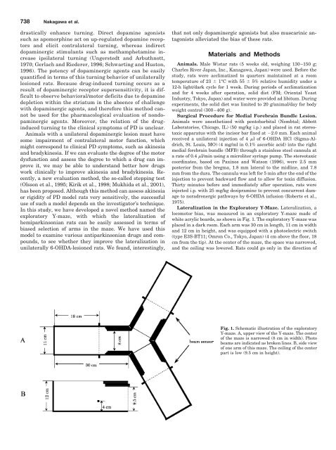
Lateralization in the Exploratory Y-Maze. Lateralization, a<br />
locomotor bias, was measured in an exploratory Y-maze made of<br />
white acrylic boards, as shown in Fig. 1. The exploratory Y-maze was<br />
placed in a dark room. Each arm was 30 cm in length, 11 cm in width<br />
<strong>and</strong> 12 cm in height, <strong>and</strong> was equipped with a photoelectric switch<br />
(type E3S-BT11; Omron Co., Tokyo, Japan) (4 cm above the floor, 18<br />
cm from the tip). At the center of the maze, the space was narrowed,<br />
<strong>and</strong> the ceiling was lowered. Rats could go only in the direction of<br />
Fig. 1. Schematic illustration of the exploratory<br />
Y-maze. A, upper view of the Y-maze. The center<br />
of the maze is narrowed (8 cm in width). Photo<br />
beams are indicated as broken lines. B, side view<br />
of one arm of this maze. The ceiling of the center<br />
part is low (9.5 cm in height).