Chapter 26 - McGraw-Hill Professional
Chapter 26 - McGraw-Hill Professional
Chapter 26 - McGraw-Hill Professional

You also want an ePaper? Increase the reach of your titles
YUMPU automatically turns print PDFs into web optimized ePapers that Google loves.
492 Management of Specific Injuries<br />
SECTION 3 X<br />
■<br />
Follow-Up<br />
As discussed above, secondary sequelae in survivors of cardiac<br />
trauma include valvular abnormalities and intracardiac<br />
fistulas. 4 , 19 , 21 Early postoperative clinical examination and ECG<br />
findings are unreliable. 4 , 21 Thus, echocardiography is recommended<br />
during the initial hospitalization in all patients to<br />
identify occult injury and establish a baseline study. Because the<br />
incidence of late sequelae can be as high as 56%, follow-up<br />
echocardiography 3–4 weeks after injury has been recommended<br />
by<br />
19 , 21<br />
some.<br />
THORACIC GREAT VESSEL INJURY<br />
Injuries to the thoracic great vessels—the aorta and its brachiocephalic<br />
branches, the pulmonary arteries and veins, the superior<br />
and intrathoracic inferior vena cava, and the innominate<br />
and azygos veins—occur following both blunt and penetrating<br />
trauma. Exsanguinating hemorrhage, the primary acute manifestation,<br />
also occurs in the chronic setting when the injured<br />
great vessel forms a fistula involving an adjacent structure or<br />
when a post-traumatic pseudoaneurysm ruptures.<br />
Current knowledge regarding the treatment of injured thoracic<br />
great vessels has been derived primarily from experience<br />
with civilian injuries. Great vessel injuries have been repaired<br />
with increasing frequency, a phenomenon that has paralleled<br />
the development of techniques for elective surgery of the thoracic<br />
aorta and its major branches.<br />
A detailed understanding of normal and variant anatomy<br />
and structural relationships is important for the surgeon and<br />
any one who is a consultant to the surgeon in the evaluation of<br />
imaging studies. Venous anomalies are infrequent with the most<br />
common being absence of the left innominate vein and persistent<br />
left superior vena cava. Thoracic aortic arch anomalies are<br />
relatively common ( Table <strong>26</strong>-3 ). Knowledge of such anomalies<br />
is essential for both open and catheter-based therapies.<br />
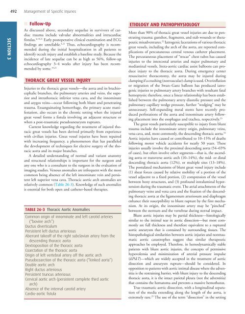
TABLE <strong>26</strong>-3 Thoracic Aortic Anomalies<br />
Common origin of innominate and left carotid arteries<br />
(“bovine arch”)<br />
Ductus diverticulum<br />
Persistent left ductus arteriosus<br />
Aberrant takeoff of the right subclavian artery from the<br />
descending thoracic aorta<br />
Dextroposition of the thoracic aorta<br />
Coarctation of the thoracic aorta<br />
Origin of left vertebral artery off the aortic arch<br />
Pseudocoarction of the thoracic aorta (“kinked aorta”)<br />
Double aortic arch<br />
Right ductus arteriosus<br />
Persistent truncus arteriosus<br />
Cervical aortic arch (persistent complete third aortic<br />
arch)<br />
Absence of the internal carotid artery<br />
Cardio-aortic fistula<br />
ETIOLOGY AND PATHOPHYSIOLOGY<br />
More than 90% of thoracic great vessel injuries are due to penetrating<br />
trauma: gunshot, fragments, and stab wounds or therapeutic<br />
misadventures. 22 Iatrogenic lacerations of various thoracic<br />
great vessels, including the arch of the aorta, are reported complications<br />
of percutaneous central venous catheter placement.<br />
The percutaneous placement of “trocar” chest tubes has caused<br />
injuries to the intercostal arteries and major pulmonary and<br />
mediastinal vessels. Intra-aortic cardiac assist balloons can produce<br />
injury to the thoracic aorta. During emergency center<br />
resuscitative thoracotomy, the aorta may be injured during<br />
clamping if a crushing (nonvascular) clamp is used. Overinflation<br />
or migration of the Swan–Ganz balloon has produced iatrogenic<br />
injuries to pulmonary artery branches with resultant fatal<br />
hemoptysis; therefore, once a linear relationship has been established<br />
between the pulmonary artery diastolic pressure and the<br />
pulmonary capillary wedge pressure, further “wedging” may be<br />
unnecessary. Self-expanding metal stents have recently produced<br />
perforations of the aorta and innominate artery following<br />
placement into the esophagus and trachea, respectively. 23<br />
The great vessels particularly susceptible to injury from blunt<br />
trauma include the innominate artery origin, pulmonary veins,<br />
vena cava, and, most commonly, the descending thoracic aorta. 24<br />
Aortic injuries have caused or contributed to 10–15% of deaths<br />
following motor vehicle accidents for nearly 50 years. These<br />
injuries usually involve the proximal descending aorta (54–65%<br />
of cases), but often involve other segments—that is, the ascending<br />
aorta or transverse aortic arch (10–14%), the mid- or distal<br />
descending thoracic aorta (12%), or multiple sites (13–18%).<br />
The postulated mechanisms of blunt great vessel injury include<br />
(1) shear forces caused by relative mobility of a portion of the<br />
vessel adjacent to a fixed portion, (2) compression of the vessel<br />
between bony structures, and (3) profound intraluminal hypertension<br />
during the traumatic event. The atrial attachments of the<br />
pulmonary veins and vena cava and the fixation of the descending<br />
thoracic aorta at the ligamentum arteriosum and diaphragm<br />
enhance their susceptibility to blunt rupture by the first mechanism.<br />
At its origin, the innominate artery may be “pinched”<br />
between the sternum and the vertebrae during sternal impact.<br />
Blunt aortic injuries may be partial thickness—histologically<br />
similar to the intimal tear in aortic dissection—but most commonly<br />
are full thickness and therefore equivalent to a ruptured<br />
aortic aneurysm that is contained by surrounding tissues. The<br />
histopathological similarities between aortic injuries and nontraumatic<br />
aortic catastrophes suggest that similar therapeutic<br />
approaches be employed. Therefore, in hemodynamically stable<br />
patients with blunt aortic injuries, the concepts of permissive<br />
hypovolemia and minimization of arterial pressure impulse<br />
(d P /dT )—which are widely accepted in the treatment of aortic<br />
dissection and aneurysm rupture—should be considered. In<br />
opposition to patients with aortic intimal disease where the adventitia<br />
is the restraining barrier, with blunt injury to the descending<br />
thoracic aorta, it is the intact parietal pleura (not the adventitia)<br />
that contains the hematoma and prevents a massive hemothorax.<br />
True traumatic aortic dissection, with a longitudinal separation<br />
of the media extending along the length of the aorta, is<br />
extremely rare. 25 The use of the term “dissection” in the setting