THE CELLULAR BASIS OF HYDROID MORPHOGENESIS
THE CELLULAR BASIS OF HYDROID MORPHOGENESIS
THE CELLULAR BASIS OF HYDROID MORPHOGENESIS

Create successful ePaper yourself
Turn your PDF publications into a flip-book with our unique Google optimized e-Paper software.
Cellular Basis of Hydroid Morphogenesis 231<br />
E. Special Considerations. The passive movement of gastroderm cells through<br />
the hydroplasm was observed by GOLDIZEN (ibid.) in time lapse films of growing stolons.<br />
He describes intact cells floating along in the hydroplasm. These cells adhered to the<br />
gastroderm wall of the stolon, displaced adjacent cells and adhered to the mesoglea.<br />
HALE (ibid.) makes virtually the same observation of cells floating in Clytia's<br />
hydroplasm.<br />
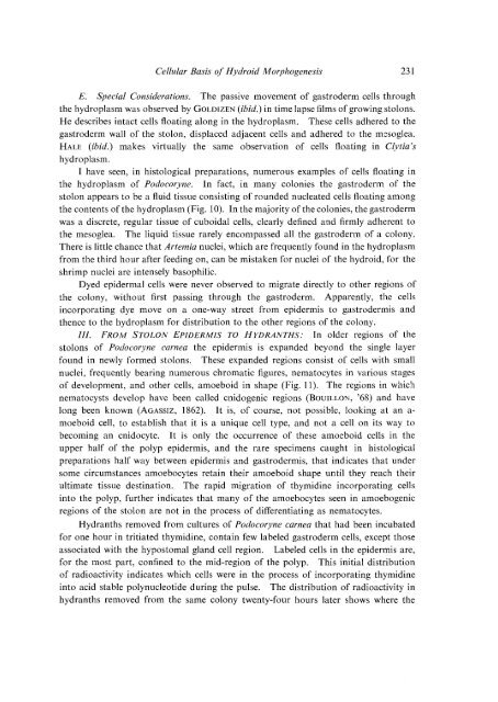
I have seen, in histological preparations, numerous examples of cells floating in<br />
the hydroplasm of Podocoryne. In fact, in many colonies the gastroderm of the<br />
stolon appears to be a fluid tissue consisting of rounded nucleated cells floating among<br />
the contents of the hydroplasm (Fig. 10). In the majority of the colonies, the gastroderm<br />
was a discrete, regular tissue of cuboidal cells, clearly defined and firmly adherent to<br />
the mesoglea. The liquid tissue rarely encompassed all the gastroderm of a colony.<br />
There is little chance that Artemia nuclei, which are frequently found in the hydroplasm<br />
from the third hour after feeding on, can be mistaken for nuclei of the hydroid, for the<br />
shrimp nuclei are intensely basophilic.<br />
Dyed epidermal cells were never observed to migrate directly to other regions of<br />
the colony, without first passing through the gastroderm. Apparently, the cells<br />
incorporating dye move on a one-way street from epidermis to gastrodermis and<br />
thence to the hydroplasm for distribution to the other regions of the colony.<br />
Ill. FROM STOLON EPIDERMIS TO HYDRANTHS: In older regions of the<br />
stolons of Podocoryne carnea the epidermis is expanded beyond the single layer<br />
found in newly formed stolons. These expanded regions consist of cells with small<br />
nuclei, frequently bearing numerous chromatic figures, nematocytes in various stages<br />
of development, and other cells, amoeboid in shape (Fig. 11 ). The regions in which<br />
nematocysts develop have been called cnidogenic regions (BoUILLON, '68) and have<br />
long been known (AGASSIZ, 1862). It is, of course, not possible, looking at an a<br />
moeboid cell, to establish that it is a unique cell type, and not a cell on its way to<br />
becoming an cnidocyte. It is only the occurrence of these amoeboid cells in the<br />
upper half of the polyp epidermis, and the rare specimens caught in histological<br />
preparations half way between epidermis and gastrodermis, that indicates that under<br />
some circumstances amoebocytes retain their amoeboid shape until they reach their<br />
ultimate tissue destination. The rapid migration of thymidine incorporating cells<br />
into the polyp, further indicates that many of the amoebocytes seen in amoebogenic<br />
regions of the stolon are not in the process of differentiating as nematocytes.<br />
Hydranths removed from cultures of Podocoryne carnea that had been incubated<br />
for one hour in tritiated thymidine, contain few labeled gastroderm cells, except those<br />
associated with the hypostomal gland cell region. Labeled cells in the epidermis are,<br />
for the most part, confined to the mid-region of the polyp. This initial distribution<br />
of radioactivity indicates which cells were in the process of incorporating thymidine<br />
into acid stable polynucleotide during the pulse. The distribution of radioactivity in<br />
hydranths removed from the same colony twenty-four hours later shows where the