THE CELLULAR BASIS OF HYDROID MORPHOGENESIS
THE CELLULAR BASIS OF HYDROID MORPHOGENESIS
THE CELLULAR BASIS OF HYDROID MORPHOGENESIS

You also want an ePaper? Increase the reach of your titles
YUMPU automatically turns print PDFs into web optimized ePapers that Google loves.
Cellular Basis of Hydroid Morphogenesis 241<br />
the stolon density is considerably less (Fig. 17).<br />
Although the new branches/new stolon length were reduced in the starved colonies<br />
to about 1/3 the number of those in the fed, the number of new hydranths/new stolon<br />
length was the same in starved and in fed colonies. From this data a morphogenetic<br />
priority can be constructed. Stolon extension will continue despite starvation and new<br />
hydranth formation will occur on stolons of the appropriate age, in proportion to the<br />
length of new stolon formed. Branch formation, however, will be curtailed in response<br />
to limited nutrition.<br />
Startlingly similar results regarding growth priorities were reported by Sears<br />
CROWELL ('57) on the basis of experiments similar to, but considerably more extensive<br />
and more elegantly controlled than, these. CROWELL observed, "There is good<br />
correspondence between the total quantity of the stolonic system and the quantity of<br />
food," but he showed that branching to form new uprights is severely retarded under<br />
starvation conditions. This is true, he reports, in both the calyptoblast Campanularia<br />
flexulosa and the Cordylophora sp. CROWELL's analysis of the growth priorities, taking<br />
into account an earlier description by BERRILL ('50) is lucid: "it seems to be a clear<br />
rule that locations where proliferation is occurring have precedence over zones of<br />
prospective growth." This was written before CROWELL's vital dye studies led him to<br />
question that growth regions are proliferation regions (CROWELL, WYTTENBACH and<br />
SUDDITH, '65).<br />
The Nature of Growth Regions<br />
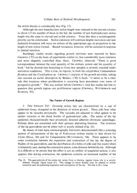
I. <strong>THE</strong> STOLON TIP: Growing stolon tips are characterized by a cap of<br />
epidermal tissue, elongated in the direction of stolon growth. These cells bear what<br />
appear to be vacuoles proximally. The vacuoles are adjacent to what appear to be<br />
similar vacuoles at the distal border of gastrodermal cells. The nuclei of the tip<br />
epidermis characteristically bear proximally directed adherent chromatic appendages.<br />
Perhaps these are associated with their perisarc depositing function. The structure<br />
of the tip gastroderm can be either well or poorly defined (Fig. 18).<br />
By means of time lapse cinematography GoLDIZEN demonstrated ('66) a pulsating<br />
pattern of advancement of the tip of Podocoryne stolons similar to that shown for<br />
Clytia (HALE, '64) and for Campanularia (WYTTENBACH, '64). There appears to be<br />
some correlation between the expansion and contraction of the tip, the degree of<br />
fluidity of the gastroderm, and the distribution of a bolus of cells and free nuclei which<br />
is frequently seen, during the contraction phase, some distance behind the tip. Although<br />
it is difficult to be certain that the effect is not an artifact of tangential section, it also<br />
appears that during contraction, the epidermis contains an increased number of the<br />
Fig. 18. The gastrodermis of the stolon tip varies from a discrete, regular tissue (A) to a poorly<br />
defined, virtually liquid tissue (C). This change in tissue fluidity may be related to stolon<br />
extension and the pulsating expansion and contraction of the tip. The bar is 50 f1 long.