THE CELLULAR BASIS OF HYDROID MORPHOGENESIS
THE CELLULAR BASIS OF HYDROID MORPHOGENESIS
THE CELLULAR BASIS OF HYDROID MORPHOGENESIS

Create successful ePaper yourself
Turn your PDF publications into a flip-book with our unique Google optimized e-Paper software.
Cellular Basis of Hydroid Morphogenesis 233<br />
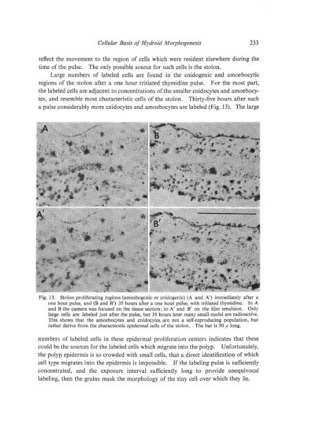
reflect the movement to the region of cells which were resident elsewhere during the<br />
time of the pulse. The only possible source for such cells is the stolon.<br />
Large numbers of labeled cells are found in the cnidogenic and amoebocytic<br />
regions of the stolon after a one hour tritiated thymidine pulse. For the most part,<br />
the labeled cells are adjacent to concentrations of the smaller cnidocytes and amoebocytes,<br />
and resemble most characteristic cells of the stolon. Thirty-five hours after such<br />
a pulse considerably more cnidocytes and amoebocytes are labeled (Fig. 13). The large<br />
Fig. 13. Stolon proliferating regions (amoebogenic or cnidogenic) (A and A') immediately after a<br />
one hour pulse, and (B and B') 35 hours after a one hour pulse, with tritiated thymidine. In A<br />
and B the camera was focused on the tissue section; in A' and B' on the film emulsion. Only<br />
large cells are labeled just after the pulse, but 35 hours later many small nuclei are radioactive.<br />
This shows that the amoebocytes and cnidocytes are not a self-reproducing population, but<br />
rather derive from the characterstic epidermal cells of the stolon. The bar is 50 f1 long.<br />
numbers of labeled cells in these epidermal proliferation centers indicates that these<br />
could be the sources for the labeled cells which migrate into the polyp. Unfortunately,<br />
the polyp epidermis is so crowded with small cells, that a direct identification of which<br />
cell type migrates into the epidermis is impossible. If the labeling pulse is sufficiently<br />
concentrated, and the exposure interval sufficiently long to provide unequivocal<br />
labeling, then the grains mask the morphology of the tiny cell over which they lie.