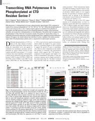
Reconstitution of the Transport of Protein between Successive ...
Reconstitution of the Transport of Protein between Successive ...
Reconstitution of the Transport of Protein between Successive ...

You also want an ePaper? Increase the reach of your titles
YUMPU automatically turns print PDFs into web optimized ePapers that Google loves.
Cell<br />
408<br />
<strong>the</strong>ir donor activity; i.e., for <strong>the</strong>ir ability to provide G protein<br />
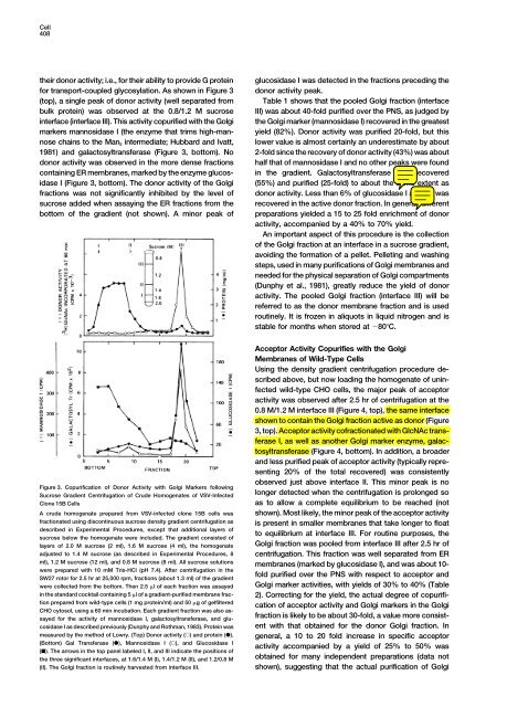
for transport-coupled glycosylation. As shown in Figure 3<br />
(top), a single peak <strong>of</strong> donor activity (well separated from<br />
bulk protein) was observed at <strong>the</strong> 0.8/1.2 M sucrose<br />
interface (interface III). This activity copurified with <strong>the</strong> Golgi<br />
markers mannosidase I (<strong>the</strong> enzyme that trims high-mannose<br />
chains to <strong>the</strong> Man 5 intermediate; Hubbard and Ivatt,<br />
1981) and galactosyltransferase (Figure 3, bottom). No<br />
donor activity was observed in <strong>the</strong> more dense fractions<br />
containing ER membranes, marked by <strong>the</strong> enzyme glucosidase<br />
I (Figure 3, bottom). The donor activity <strong>of</strong> <strong>the</strong> Golgi<br />
fractions was not significantly inhibited by <strong>the</strong> level <strong>of</strong><br />
sucrose added when assaying <strong>the</strong> ER fractions from <strong>the</strong><br />
bottom <strong>of</strong> <strong>the</strong> gradient (not shown). A minor peak <strong>of</strong><br />
glucosidase I was detected in <strong>the</strong> fractions preceding <strong>the</strong><br />
donor activity peak.<br />
Table 1 shows that <strong>the</strong> pooled Golgi fraction (interface<br />
III) was about 40-fold purified over <strong>the</strong> PNS, as judged by<br />
<strong>the</strong> Golgi marker (mannosidase I) recovered in <strong>the</strong> greatest<br />
yield (82%). Donor activity was purified 20-fold, but this<br />
lower value is almost certainly an underestimate by about<br />
2-fold since <strong>the</strong> recovery <strong>of</strong> donor activity (43%) was about<br />
half that <strong>of</strong> mannosidase I and no o<strong>the</strong>r peaks were found<br />
in <strong>the</strong> gradient. Galactosyltransferase was recovered<br />
(55%) and purified (25-fold) to about <strong>the</strong> same extent as<br />
donor activity. Less than 6% <strong>of</strong> glucosidase I activity was<br />
recovered in <strong>the</strong> active donor fraction. In general, different<br />
preparations yielded a 15 to 25 fold enrichment <strong>of</strong> donor<br />
activity, accompanied by a 40% to 70% yield.<br />
An important aspect <strong>of</strong> this procedure is <strong>the</strong> collection<br />
<strong>of</strong> <strong>the</strong> Golgi fraction at an interface in a sucrose gradient,<br />
avoiding <strong>the</strong> formation <strong>of</strong> a pellet. Pelleting and washing<br />
steps, used in many purifications <strong>of</strong> Golgi membranes and<br />
needed for <strong>the</strong> physical separation <strong>of</strong> Golgi compartments<br />
(Dunphy et al., 1981), greatly reduce <strong>the</strong> yield <strong>of</strong> donor<br />
activity. The pooled Golgi fraction (interface III) will be<br />
referred to as <strong>the</strong> donor membrane fraction and is used<br />
routinely. It is frozen in aliquots in liquid nitrogen and is<br />
stable for months when stored at 80C.<br />
Figure 3. Copurification <strong>of</strong> Donor Activity with Golgi Markers following<br />
Sucrose Gradient Centrifugation <strong>of</strong> Crude Homogenates <strong>of</strong> VSV-Infected<br />
Clone 15B Cells<br />
A crude homogenate prepared from VSV-infected clone 15B cells was<br />
fractionated using discontinuous sucrose density gradient centrifugation as<br />
described in Experimental Procedures, except that additional layers <strong>of</strong><br />
sucrose below <strong>the</strong> homogenate were included. The gradient consisted <strong>of</strong><br />
layers <strong>of</strong> 2.0 M sucrose (2 ml), 1.6 M sucrose (4 ml), <strong>the</strong> homogenate<br />
adjusted to 1.4 M sucrose (as described in Experimental Procedures, 8<br />
Acceptor Activity Copurifies with <strong>the</strong> Golgi<br />
Membranes <strong>of</strong> Wild-Type Cells<br />
Using <strong>the</strong> density gradient centrifugation procedure described<br />
above, but now loading <strong>the</strong> homogenate <strong>of</strong> uninfected<br />
wild-type CHO cells, <strong>the</strong> major peak <strong>of</strong> acceptor<br />
activity was observed after 2.5 hr <strong>of</strong> centrifugation at <strong>the</strong><br />
0.8 M/1.2 M interface III (Figure 4, top), <strong>the</strong> same interface<br />
shown to contain <strong>the</strong> Golgi fraction active as donor (Figure<br />
3, top). Acceptor activity c<strong>of</strong>ractionated with GlcNAc transferase<br />
I, as well as ano<strong>the</strong>r Golgi marker enzyme, galactosyltransferase<br />
(Figure 4, bottom). In addition, a broader<br />
and less purified peak <strong>of</strong> acceptor activity (typically representing<br />
20% <strong>of</strong> <strong>the</strong> total recovered) was consistently<br />
observed just above interface II. This minor peak is no<br />
longer detected when <strong>the</strong> centrifugation is prolonged so<br />
as to allow a complete equilibrium to be reached (not<br />
shown). Most likely, <strong>the</strong> minor peak <strong>of</strong> <strong>the</strong> acceptor activity<br />
is present in smaller membranes that take longer to float<br />
to equilibrium at interface III. For routine purposes, <strong>the</strong><br />
Golgi fraction was pooled from interface III after 2.5 hr <strong>of</strong><br />
centrifugation. This fraction was well separated from ER<br />
ml), 1.2 M sucrose (12 ml), and 0.8 M sucrose (8 ml). All sucrose solutions<br />
membranes (marked by glucosidase I), and was about 10-<br />
were prepared with 10 mM Tris-HCl (pH 7.4). After centrifugation in <strong>the</strong><br />
fold purified over <strong>the</strong> PNS with respect to acceptor and<br />
SW27 rotor for 2.5 hr at 25,000 rpm, fractions (about 1.3 ml) <strong>of</strong> <strong>the</strong> gradient<br />
Golgi marker activities, with yields <strong>of</strong> 30% to 40% (Table<br />
2). Correcting for <strong>the</strong> yield, <strong>the</strong> actual degree <strong>of</strong> copurifi-<br />
cation <strong>of</strong> acceptor activity and Golgi markers in <strong>the</strong> Golgi<br />
fraction is likely to be about 30-fold, a value more consistent<br />
with that obtained for <strong>the</strong> donor Golgi fraction. In<br />
general, a 10 to 20 fold increase in specific acceptor<br />
activity accompanied by a yield <strong>of</strong> 25% to 50% was<br />
obtained for many independent preparations (data not<br />
shown), suggesting that <strong>the</strong> actual purification <strong>of</strong> Golgi<br />
were collected from <strong>the</strong> bottom. Then 2.5 l <strong>of</strong> each fraction was assayed<br />
in <strong>the</strong> standard cocktail containing 5 l <strong>of</strong> a gradient-purified membrane fraction<br />
prepared from wild-type cells (1 mg protein/ml) and 50 g <strong>of</strong> gelfiltered<br />
CHO cytosol, using a 60 min incubation. Each gradient fraction was also assayed<br />
for <strong>the</strong> activity <strong>of</strong> mannosidase I, galactosyltransferase, and glu-<br />
cosidase I as described previously (Dunphy and Rothman, 1983). <strong>Protein</strong> was<br />
measured by <strong>the</strong> method <strong>of</strong> Lowry. (Top) Donor activity () and protein ().<br />
(Bottom) Gal Transferase (), Mannosidase I (), and Glucosidase I<br />
(). The arrows in <strong>the</strong> top panel labeled I, II, and III indicate <strong>the</strong> positions <strong>of</strong><br />
<strong>the</strong> three significant interfaces, at 1.6/1.4 M (I), 1.4/1.2 M (II), and 1.2/0.8 M<br />
(II). The Golgi fraction is routinely harvested from interface III.