Research Article Ginseng Extracts Restore High-Glucose Induced ...
Research Article Ginseng Extracts Restore High-Glucose Induced ...
Research Article Ginseng Extracts Restore High-Glucose Induced ...

Create successful ePaper yourself
Turn your PDF publications into a flip-book with our unique Google optimized e-Paper software.
Hindawi Publishing Corporation<br />
Evidence-Based Complementary and Alternative Medicine<br />
Volume 2013, <strong>Article</strong> ID 797310, 13 pages<br />
http://dx.doi.org/10.1155/2013/797310<br />
<strong>Research</strong> <strong>Article</strong><br />
<strong>Ginseng</strong> <strong>Extracts</strong> <strong>Restore</strong> <strong>High</strong>-<strong>Glucose</strong> <strong>Induced</strong> Vascular<br />
Dysfunctions by Altering Triglyceride Metabolism and<br />
Downregulation of Atherosclerosis-Related Genes<br />
Gabriel Hoi-huen Chan, 1 Betty Yuen-kwan Law, 2,3 John Man-tak Chu, 2<br />
Kevin Kin-man Yue, 2 Zhi-hong Jiang, 2 Chi-wai Lau, 4 Yu Huang, 4 Shun-wan Chan, 5<br />
Patrick Ying-kit Yue, 1 and Ricky Ngok-shun Wong 1<br />
1 Department of Biology, Hong Kong Baptist University, Kowloon Tong, Kowloon, Hong Kong<br />
2 School of Chinese Medicine, Hong Kong Baptist University, Kowloon Tong, Kowloon, Hong Kong<br />
3 State Key Laboratory of Quality <strong>Research</strong> in Chinese Medicine, Macau University of Science and Technology,<br />
Avenida Wai Long, Taipa, Macau<br />
4 School of Biomedical Science, The Chinese University of Hong Kong, Shatin, N.T., Hong Kong<br />
5 Department of Applied Biology and Chemical Technology, The Hong Kong Polytechnic University, Hung Hom, Kowloon, Hong Kong<br />
Correspondence should be addressed to Ricky Ngok-shun Wong; rnswong@hkbu.edu.hk<br />
Received 21 March 2013; Revised 23 August 2013; Accepted 24 August 2013<br />
Academic Editor: Hao Xu<br />
Copyright © 2013 Gabriel Hoi-huen Chan et al. This is an open access article distributed under the Creative Commons Attribution<br />
License, which permits unrestricted use, distribution, and reproduction in any medium, provided the original work is properly<br />
cited.<br />
The king of herbs, Panax ginseng, has been used widely as a therapeutic agent vis-à-vis its active pharmacological and physiological<br />
effects. Based on Chinese pharmacopeia Ben Cao Gang Mu and various pieces of literature, Panax ginseng was believed to exert<br />
active vascular protective effects through its antiobesity and anti-inflammation properties. We investigated the vascular protective<br />
effects of ginseng by administrating ginseng extracts to rats after the induction of diabetes. We found that Panax ginseng can<br />
restore diabetes-induced impaired vasorelaxation and can reduce serum triglyceride but not cholesterol level in the diabetic rats.<br />
The ginseng extracts also suppressed the expression of atherosclerosis-related genes and altered the expression of lipid-related genes.<br />
The results provide evidence that Panax ginseng improves vascular dysfunction induced by diabetes and the protective effects may<br />
possibly be due to the downregulation of atherosclerosis-related genes and altered lipid metabolism, which help to restore normal<br />
endothelium functions.<br />
1. Introduction<br />
Panax ginseng is one of the most commonly used Chinese<br />
medicine and research targets. The major active components<br />
of Panax ginseng are ginsenosides which can be subdivided<br />
into three groups according to their basic structures:<br />
protopanaxadiol (PPD) type (e.g., Rb1, Rb2, Rc, Rd, Rg3,<br />
and Rh2), protopanaxatriol (PPT) type (e.g., Re, Rf, Rg1,<br />
Rg2, and Rh1), and oleanolic acid (e.g., Ro). Ginsenosides<br />
appear to be responsible for most of the activities of ginseng<br />
including antioxidation, anti-inflammation, and anticancer<br />
[1]. A review by Karmazyn et al. has found that the yearly<br />
ginseng-related publication has been increasing exponentially<br />
from 1950 to 2010. They also reported that Panax<br />
ginseng played a protective role in the cardiovascular system<br />
[2]. This suggested the beneficial properties of ginseng on<br />
cardiovascular diseases in both experimental and clinical<br />
settings.<br />
Atherosclerosis is one of the most common cardiovascular<br />
diseases and can remain asymptomatic for decades. In<br />
the mid 1970s, Russel Ross developed the popular “response<br />
to injury” theory by postulating that atherosclerosis begins<br />
with injuries on the endothelium, followed by adhesion<br />
and aggregation of platelets [3]. At about the same time,
2 Evidence-Based Complementary and Alternative Medicine<br />
Robert F. Furchgott, the Nobel Prize Laureate in Physiology<br />
or Medicine in 1998, discovered that acetylcholine<br />
induces endothelium-dependent relaxation in normal aortic<br />
tissue [4]. Upon early onset of atherosclerosis, endothelium<br />
can remain morphologically intact though inflammatory<br />
responses are triggered. Since then, numerous researches<br />
have been conducted to investigate the mechanisms of<br />
atherosclerosis to mitigate the associated diseases including<br />
adhesion of lipid-laden macrophages and smooth muscle<br />
cells which could finally result in endothelial denudation<br />
[5]. Besides, Hansson’s research groups have reported that<br />
high level of total cholesterol and low density lipoprotein<br />
accumulatedintheintimaofthearteries,withtheattackof<br />
myeloperoxidase and lipoxygenases, or by reactive oxygen<br />
species [6, 7] could also cause the early onset of atherosclerosis.<br />
The primary objective of this study is to evaluate the<br />
protective effects of Panax ginseng on diabetes mellitus,<br />
a pathological condition which links to endothelial dysfunctions,<br />
through investigating the physiological parameters<br />
such as blood glucose, blood cholesterol, insulin, and<br />
advanced glycation end product in diabetic rat models.<br />
Furthermore, the changes of atherosclerosis-related genes<br />
expression in diabetic rats are also investigated after ginseng<br />
administration. The findings may help in the development<br />
of successful therapeutic interventions for atherosclerotic<br />
cardiovascular disease.<br />
2. Materials and Methods<br />
Thisstudyfollows“TheInternationalGuidingPrinciples<br />
for Biomedical <strong>Research</strong> Involving Animals,” The Hong<br />
Kong Code of Practice for Care and Use of Animals for<br />
Experimental Purposes (2004). All experimental procedures<br />
were conducted according to the Animals (Control of Experiments)<br />
Ordinance of the Department of Health, HKSAR<br />
(Animal Licenses ID: (11-6) DH/HA&P/8/2/6 Pt.2; (10-4)<br />
DH/HA&P/8/2/6 Pt.1; (10-9) DH/HA&P/8/2/6 Pt.1). All animal<br />
studies were performed in facilities approved by the<br />
Animal Ethics Committee of the Chinese University of Hong<br />
Kong (10/028/MIS).<br />
2.1. Animals. Male Sprague-Dawley (SD) rats weighing 150–<br />
200 grams were housed in room under standard vivarium<br />
conditions with 12 hour light/dark cycle. Throughout the<br />
experimental period, animals were fed with standard rodent<br />
chow and water available ad libitum.Theanimalswereacclimatized<br />
to the laboratory conditions for 10 days prior to the<br />
inception of experiments. Experimental diabetic condition<br />
was induced in rats by a single intraperitoneal injection (i.p.)<br />
of streptozotocin (75 mg/kg body weight) freshly dissolved in<br />
cold citrate buffer (0.1 M), while the normal control group<br />
was injected with citrate buffer only. Blood samples were<br />
collected from tail veins of overnight-fasted rats three days<br />
after streptozotocin administration. Rats with blood glucose<br />
level higher than 16.7 mmol/dL were selected for experiment.<br />
The experimental rats were divided into seven groups:<br />
(1) normal control rats administered with water, (2) diabetic<br />
group of rats administered with water, (3) diabetic<br />
group administered with intraperitoneal injection of<br />
insulin, (4) diabetic group fed with PPT-type of ginseng<br />
(10 mg/kg/day), (5) diabetic group fed with PPT-type of<br />
ginseng (30 mg/kg/day), (6) diabetic group fed with PPDtype<br />
of ginseng (10 mg/kg/day), and (7) diabetic group fed<br />
with PPD-type of ginseng (30 mg/kg/day). The dosage of<br />
insulin followed a protocol developed by Kuo et al. [8], and<br />
waterordrugswereadministeredforatotalof14consecutive<br />
treatment days. Both PPD and PPT were administered orally<br />
in the form of aqueous suspension. Rats were anaesthetized<br />
by Ketamine-Rompun mixture (7.5 : 1), and blood was collected<br />
from the heart for further analysis. The animals were<br />
then sacrificed immediately by cervical dislocation. Aortae<br />
were removed and trimmed for tissue bath experiment. Other<br />
rat tissues including brain, heart, liver, spleen, eye, kidney,<br />
and aorta were immediately removed and instantly soaked in<br />
liquidnitrogenandstoredat−70 ∘ C for further biochemical<br />
analysis.<br />
2.2. <strong>Ginseng</strong> Preparation. Panax ginseng extract was prepared<br />
as described in Zhu et al. [9], which meets the requirement<br />
of the Chinese Pharmacopoeia and Hong Kong Standard<br />
of Chinese Materia Medica. Standardized ginseng extract<br />
(RSE) was prepared by ethanol extraction. The residue was<br />
then dissolved in water and partitioned successively with<br />
petroleum ether, EtOAc, and n-BuOH to give the petroleumether-soluble,<br />
EtOAc-soluble, and n-BuOH-soluble fractions.<br />
The n-BuOH extract was subjected to column chromatographyelutedwithaCHCl<br />
3 /MeOH gradient. Fractionated<br />
samples were combined and obtained according to the thin<br />
layer chromatography analysis. All samples were then stored<br />
in desiccated condition until further use. <strong>High</strong> performance<br />
liquid chromatography was used to confirm the identity of<br />
our samples with the standard ginsenosides (HPLC purity<br />
>98%) purchased from Chengdu Scholar Bio-Tech Co. Ltd.<br />
(Chengdu, China) or National Institute for the Control of<br />
Pharmaceutical and Biological Products (Beijing, China).<br />
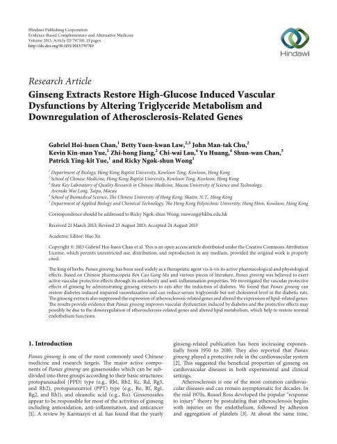
The contents of ginsenosides Rg1, Re, Rb1, Rc, Rb2, and<br />
Rd were 290.9, 339.6, 246.3, 231.3, 136.0, and 84.5 mg/g,<br />
respectively (Figure 1 and Table 1).<br />
2.3. Measurement of Contractile and Relaxant Responses<br />
in the Rat Aortic Rings. Similar procedures were followed<br />
according to the protocol as described by Chan and Fiscus<br />
2002 [10]. Briefly, thoracic aortae were isolated by cutting<br />
from the aortic arch to the diaphragm, resulting in a length<br />
of 30–40 mm tissue. In order to prevent physical damage<br />
of endothelium by forceps, the parts from the aortic arch<br />
were not used for experiment. Fat tissues were trimmed off<br />
from the aortae and before it was cut into 3 mm segments<br />
rings. The segments were then mounted carefully between<br />
two platinum hooks in 10 mL organ baths containing Krebs<br />
buffer (KRB) maintained at 37 ∘ Cbubbledwith95%O 2 –5%<br />
CO 2 continuously. Following a 30 min equilibration period of<br />
resting tension of 1 gram, cumulative doses of phenylephrine
Evidence-Based Complementary and Alternative Medicine 3<br />
(mAU)<br />
(mAU)<br />
700<br />
600<br />
500<br />
400<br />
300<br />
200<br />
100<br />
0<br />
300<br />
250<br />
200<br />
150<br />
100<br />
50<br />
0<br />
PPT- and PPD-type ginsenosides<br />
DAD1A, Sig = 203,8 Ref = 360,100 (PPT.D)<br />
Rg1<br />
Re<br />
0 10 20 30 40 50 60 70 80<br />
(min)<br />
(a)<br />
DAD1A, Sig = 203,8 Ref = 360,100 (PPD.D)<br />
0 10 20 30 40 50 60 70 80<br />
(min)<br />
(b)<br />
Figure 1: HPLC fingerprint of PPT-type (Panel (a)) and PPD-type (Panel (b)) ginseng extract used in current study. Instruments. An HP 1100<br />
system (Hewlett-Packard, Wilmington, DE) consisting of a G1312A binary pump, a G1329A automatic sample injector, and a G1315A diode<br />
array detector was used to perform HPLC analysis. Sample Preparation. Approximately 0.20 g powdered ginseng was accurately weighed into<br />
a 50 mL conical flask, and 10 mL 70% methanol was added. The suspension was sonicated for 30 min, and the sample solution was filtered<br />
through a 0.45 μm filter and used as the test solution for quantitative analysis of ginsenosides in Radix <strong>Ginseng</strong>. Chromatographic Conditions.<br />
HPLC analysis of Radix <strong>Ginseng</strong> was performed on an Alltima C 18 HPLC column (4.6 mm × 250 mm, 5 μm) at 25 ∘ C with a sample injection<br />
volume of 20 μL. The mobile phase was a gradient elution of KH2PO4 buffer (2 mmol/L, pH 6.8) and acetonitrile, starting isocratically with<br />
21% acetonitrile for 15 min and increasing to 38% acetonitrile over 55 min. The flow rate of the mobile phase was 1.0 mL/min, and the detector<br />
wavelength was 203 nm.<br />
Rb1<br />
Rc<br />
Rb2<br />
Rd<br />
(1 × 10 −9 Mto1× 10 −5 M) were added in each aortic ring. After<br />
the addition of phenylephrine, the aortic rings were washed<br />
with fresh and bubbled KRB solution every 10 minutes over<br />
a 30-minute period. A single dose of phenylephrine at 1 ×<br />
10 −7 M was added until the aortic rings maintained 50 percent<br />
of maximum tension. Doses of acetylcholine (1 ×10 −9 Mto<br />
1×10 −5 M) were added cumulatively to check the endothelial<br />
functions. All of the doses were added after the responses<br />
reached plateau.<br />
2.4. Blood Profile of the Experimental Rats. Serum tests<br />
for total cholesterol (TC), triglyceride (TG), low density<br />
lipoprotein (LDL), and high density lipoprotein (HDL) were<br />
conducted in The State Key laboratory for Chinese Medicine<br />
and Molecular Pharmacology of the Hong Kong Polytechnic<br />
University. Terminal colorimetric analysis method was used<br />
forquantificationofTC,TG,LDL,andHDL,respectively.The<br />
tests were conducted with ECHO automatic biochemistry<br />
analyzer (Logotech, Italy) and UV2800 spectrophotometer<br />
(Unico, Shanghai).<br />
Blood glucose level was measured using glucometer<br />
(Elite, Bayer Corporation, USA). Serum insulin and glycation<br />
endproductsweremeasuredusingkitspurchasedfromMillipore<br />
(EZRMI-13K) and Cusabio (CSB-E08140r), respectively.<br />
2.5. RT 2 Profiler Rat Atherosclerosis PCR Array Analysis.<br />
Aortic samples for PCR array analysis were used to obtain the<br />
total RNA by Qiagen RNeasy Mini Kit (Catalogue number:<br />
PARN-038A, Qiagen). This pathway specific RT-PCR array<br />
was used to evaluate the potential alterations of related genes<br />
after PPD-type and PPT-type treatments (30 mg/kg/day)<br />
in rats. The atherosclerosis array comprised of 87 genes<br />
selected based on their involvement in regulating vascular<br />
and endothelial cell homeostasis or inflammation. There<br />
were 5 housekeeping genes served as positive controls. Total<br />
RNA was reverse transcripted using the RT 2 First Strand<br />
Kit. Real-time PCR reactions were carried out on ABI 7500<br />
(Applied Biosystems) using the RT 2 SYBR Green qPCR Mastermix<br />
(Qiagen) according to manufacturer’s instructions.<br />
Data analysis was performed using the Qiagen’s integrated
4 Evidence-Based Complementary and Alternative Medicine<br />
Table 1: Contents of ginsenosides in the prepared PPT-type and PPD-type ginsenosides.<br />
Sample Ginsenosides Content (mg/g) Percentage<br />
Rg1 290.9 29.09%<br />
PPT-type ginsenosides<br />
Re 339.6 33.96%<br />
Rb1 246.3 24.63%<br />
Rc 231.30 23.13%<br />
PPD-type ginsenosides<br />
Rb2 136.0 13.61%<br />
Rd 84.5 8.45%<br />
web-based software package for the PCR Array System,<br />
which automatically performs all ΔΔCt based fold-change<br />
calculations from raw threshold cycle data.<br />
2.6. Statistical Analysis. All values are expressed as mean ±<br />
standarderrorofmean(SEM).Thesignificantdifferences<br />
between the young and aged groups in the isolated tissue<br />
experiments were analyzed using one way ANOVA with<br />
Newman-Keuls multiple comparison as post hoc test in the<br />
statistical package (Graphpad prism v6.0). A P value less<br />
than 0.05 was considered to be significant. The mean values<br />
were obtained from at least 5 animals or 3 DNA samples per<br />
treatment group.<br />
3. Results<br />
3.1. <strong>Ginseng</strong> <strong>Extracts</strong> <strong>Restore</strong> <strong>High</strong> <strong>Glucose</strong>-<strong>Induced</strong> Endothelial<br />
Dysfunction. Acetylcholine (ACh) causes vasodilation<br />
by activation of endothelial nitric oxide synthase and<br />
prostaglandin production. The aortic tissue was challenged<br />
with acetylcholine (1 × 10 −9 M–1 × 10 −5 M) and caused<br />
concentration-dependent relaxations in aortic rings from<br />
young rats. Normal rats showed 100% relaxation (restored<br />
the contracting state to resting state) at maximum dose 1×<br />
10 −5 M, while the response was only 62.5% of the relaxation<br />
in the diabetic rats (Figure 2(a)), showing an impairment<br />
of the endothelium. For positive control, diabetic rats were<br />
injected with insulin and the normal vasorelaxation was<br />
maintained (Figure 2(b)). After feeding PPD-type and PPTtype<br />
ginseng extracts for two weeks, the impaired vasorelaxationduetohighglucoselevelwasrestored(Figures<br />
2(c) to 2(f)), indicating that the endothelial functions were<br />
maintained under the diabetic conditions for the ginseng-fed<br />
groups.<br />
3.2. Blood Profile, Body Weight, Distribution of Visceral Adipose<br />
Tissue, and Organs Weight of the Experimental Rats. In<br />
this study, blood glucose, insulin, advanced glycation end<br />
products, serum total cholesterol, high density lipoprotein<br />
(HDL), low density lipoprotein (LDL), and triglyceride were<br />
examined. Except for normal and insulin injected positive<br />
control group, all of the diabetic groups were considered<br />
to be diabetic (blood glucose >16.7 mmol/dL) and with a<br />
significant reduction of insulin level (Figure 3(a)). There are<br />
no statistical differences between the ginseng-fed or nonfed<br />
diabetic groups for blood glucose, insulin, serum total cholesterol,<br />
HDL, and LDL (Figures 3 and 5(a)–5(c)), indicating<br />
the ginseng extracts have no improvement on hyperglycemic<br />
conditions or alternation of cholesterol levels. However, there<br />
is a slight decrease in the level of glycation end products<br />
(Figure 4), when the diabetic group was fed with PPT-type<br />
of ginseng at a dosage of 30 mg/kg/day. In addition, there<br />
was also significant decrease in serum triglyceride level for<br />
all ginseng-fed groups, which indicated that both PPD-type<br />
and PPT-type are effective in lowering serum triglyceride<br />
(Figure 5(d)). Visceral adipose tissue is associated with fatty<br />
acid metabolism. The distribution of visceral adipose tissue<br />
surrounding mesenteric arteries was shown in Figure 6.More<br />
visceraladiposetissuewasfoundincontrolgroupwhen<br />
comparedtothediabeticgroup.However,morevisceral<br />
adipose tissue was observed in diabetic rats after feeding with<br />
PPD-type and PPT-type of ginseng extracts. The body mass<br />
and organ mass are the health indicators for the experimental<br />
rats. Figure 7 showedthebodyandorganweightofthe<br />
experimental rats. The body weight of the insulin-injected<br />
diabetic groups is slightly larger than other groups. Among<br />
all organs measured (liver, pancreas, heart, adrenal gland, and<br />
kidneys), the PPD-type fed diabetic groups have significantly<br />
smaller adrenal glands than diabetic group (P < 0.05).<br />
3.3. <strong>Ginseng</strong> Extract Suppresses the Expression of Atherosclerosis-Related<br />
Genes. PCR array analysis showed the fold<br />
change of atherosclerosis-related gene expression (Figure 8<br />
and Table 2) for different treatment groups. When compared<br />
to normal control group, diabetic groups showed an upregulation<br />
on several atherosclerosis-related gene expressions,<br />
which indicate an increased risk of atherosclerosis. Besides,<br />
the gene expressions related to inflammations including<br />
adhesion molecules such as selectin (platelet) and ICAM1<br />
and macrophage activation including chemokine (C-C) motif<br />
ligand 2 (CCl-2), chemokine (C-X-C) motif ligand 1 (CxCl-<br />
1), interleukin 1 receptor 2 (IL1-R2), interleukins (IL3, IL4<br />
and IL5), and tumor necrosis factor-α (TNF-α) weredownregulated.<br />
Apart from genes related to inflammation, other<br />
genes involved in the development of atherosclerosis were<br />
also checked. Apoptotic genes, such as Bid as well as genes<br />
responsible for vascular endothelial cells and vascular smooth<br />
muscle cell proliferation and migration (including von Willebrand<br />
factor homolog, heparin-binding EGF-like growth<br />
factor, and thrombospondin 4), were also downregulated in<br />
ginseng-fed diabetic groups. On the other hand, lipid-related<br />
genes expression including apolipoprotein E (ApoE), lipase,<br />
and peroxisome proliferator-activated receptor (PPAR) γ<br />
were increased in the ginseng-fed groups.
Evidence-Based Complementary and Alternative Medicine 5<br />
Table2:PCRarrayanalysisofexpressionchangeinselectedatherosclerosis-relatedgenes.<br />
Fold change #<br />
Gene name<br />
Diabetes<br />
PPD-fed diabetic group<br />
PPT-fed diabetic group<br />
(30 mg/kg/day)<br />
(30 mg/kg/day)<br />
Adhesion molecules<br />
Selectin (platelet) +2.33 +1.18 +1.29<br />
Intercellular adhesion molecule 1 +2.06 +1.46 +1.46<br />
Macrophages<br />
Chemokine (C-C) motif ligand 2 +2.49 +0.56 +0.47<br />
Chemokine (C-X-C) motif ligand 1 +3.19 +0.89 +0.58<br />
Interleukin 1 receptor, type II +3.28 +1.22 +1.38<br />
Interleukin 3 +0.95 +0.48 +0.82<br />
Interleukin 4 +1.23 +0.53 +0.83<br />
Interleukin 5 +1.08 +0.78 +0.79<br />
Tumornecrosisfactor-α +1.56 +1.31 +1.03<br />
Lipid metabolism<br />
Apolipoprotein E +1.53 +2.95 +2.50<br />
Lipase +13.85 +24.93 +8.82<br />
Peroxisome proliferator-activated receptor-γ +2.54 +5.83 +2.06<br />
Cell growth and migration<br />
Fibrinogen beta chain +0.93 +0.43 +0.62<br />
von Willebrand factor homolog +4.45 +3.33 +2.99<br />
Heparin-binding EGF-like growth factor +2.53 +1.95 +2.04<br />
Laminin α 1 +0.93 +0.64 +0.61<br />
Extracellular matrix (ECM)<br />
Fibronectin +2.91 +1.94 +1.66<br />
Apoptosis<br />
Bcl2-like 1 +0.73 +0.75 +0.83<br />
BH3 interacting domain death agonist +1.45 +1.25 +1.23<br />
# Fold changes (comparing to control group, fold change = 1) are calculated according to manufacturer’s analysis software.<br />
In general, the ginseng-fed diabetic groups showed a<br />
decreased expression on atherosclerosis-related genes, which<br />
indicates the decreased risk of atherosclerosis after ginseng<br />
treatments.<br />
4. Discussion<br />
Endothelium controls vascular tone through the production<br />
of vasodilator mediators, endothelium-derived relaxing factors<br />
(EDRF), which act on vascular smooth muscle cells.<br />
The EDRF comprise nitric oxide (NO), prostacyclin, and an<br />
elusive endothelium-derived hyperpolarizing factor (EDHF).<br />
Multiple mechanisms lead to endothelial dysfunction [11,<br />
12], and endothelial dysfunction plays a key role in the<br />
pathogenesis of vascular diseases. Hyperglycemia is linked to<br />
the pathogenesis of diabetic complications involving alternations<br />
of intracellular metabolism and formation of advanced<br />
glycation end products.<br />
The attenuated endothelium-dependent vasodilations<br />
have been demonstrated in various vascular tissues of diabetic<br />
animal model [13]. In the present study, we examined<br />
the endothelial functions using the physiological isolated<br />
tissue bath setup and found that the high glucose-impaired<br />
vasodilations were restored after ginseng extracts treatment<br />
(Figure 2). The result indicates that ginseng extract plays a<br />
protective role in restoring normal endothelial functions in<br />
diabetic models. Different molecular mechanisms have been<br />
demonstratedtocausethevasculardysfunctions.Reports<br />
have suggested that hyperglycaemia-induced endothelial dysfunction<br />
is due to activation of protein kinase C (PKC)<br />
[14], inhibition of endothelial nitric oxide synthase [15, 16],<br />
early and advanced nonenzymatic glycation, and oxidative<br />
stress [17–19]. In atherosclerotic conditions, up-regulation<br />
of adhesion molecules, increased cytokine secretion, apoptosis,<br />
enhanced low-density lipoprotein oxidation, platelet<br />
activation, and vascular smooth muscle cell proliferation<br />
and migration are always observed [20–22]. Therefore, compounds<br />
that are able to modulate atherosclerosis and maintain<br />
normal endothelial functions are highly desirable.<br />
Although the levels of LDL, TC, HDL, and insulin in<br />
diabetic group were not significantly different when compared<br />
with the ginseng-fed groups, ginseng-fed diabetic
6 Evidence-Based Complementary and Alternative Medicine<br />
100<br />
100<br />
80<br />
80<br />
Relaxation (%)<br />
60<br />
40<br />
20<br />
0<br />
log[ACh]<br />
∗<br />
∗<br />
∗<br />
∗<br />
∗<br />
∗ P < 0.05<br />
−9 −8 −7 −6 −5<br />
∗<br />
∗<br />
∗<br />
Relaxation (%)<br />
60<br />
40<br />
20<br />
0<br />
−9 −8 −7 −6 −5<br />
log[ACh]<br />
Normal<br />
Diabetes<br />
Normal<br />
Insulin injected diabetic group<br />
(a)<br />
(b)<br />
100<br />
100<br />
80<br />
80<br />
Relaxation (%)<br />
60<br />
40<br />
Relaxation (%)<br />
60<br />
40<br />
20<br />
20<br />
0<br />
−9 −8 −7 −6 −5<br />
log[ACh]<br />
0<br />
−9 −8 −7 −6 −5<br />
log[ACh]<br />
Normal<br />
PPT-type (30 mg/kg/day)<br />
Normal<br />
PPT-type (10 mg/kg/day)<br />
(c)<br />
(d)<br />
100<br />
100<br />
80<br />
80<br />
Relaxation (%)<br />
60<br />
40<br />
Relaxation (%)<br />
60<br />
40<br />
20<br />
20<br />
0<br />
−9 −8 −7 −6 −5<br />
log[ACh]<br />
0<br />
−9 −8 −7 −6 −5<br />
log[ACh]<br />
Normal<br />
PPD-type (30 mg/kg/day)<br />
Normal<br />
PPD-type (10 mg/kg/day)<br />
(e)<br />
(f)<br />
Figure 2: <strong>Ginseng</strong> extracts restore acetylcholine-induced endothelium dependent vasorelaxation. After the addition of phenylephrine, doses<br />
of acetylcholine (1 × 10 −9 Mto1× 10 −5 M) were added cumulatively to check the endothelial functions. Control group showed an attenuation<br />
of acetylcholine-induced vasorelaxation (Panel (a)). The insulin injected diabetic group (Panel (b)), PPT-type (30 mg/kg/day) fed diabetic<br />
group (Panel (c)), PPT-type (10 mg/kg/day) fed diabetic group (Panel (d)), PPD-type (30 mg/kg/day) fed diabetic group (Panel (e)), and<br />
PPD-type (10 mg/kg/day) (Panel (f)) fed diabetic group showed restoration of the attenuated vasorelaxation. Results were expressed as the<br />
mean ± standard error; ∗ P < 0.05 for the indicated comparisons.
Evidence-Based Complementary and Alternative Medicine 7<br />
40<br />
4<br />
∗∗∗ P < 0.001<br />
∗ P < 0.05<br />
Blood glucose (mmol/dL)<br />
30<br />
20<br />
10<br />
∗∗∗<br />
Serum level of insulin (ng/mL)<br />
3<br />
2<br />
1<br />
∗<br />
0<br />
0<br />
Normal<br />
Diabetes<br />
Insulin injected control<br />
PPT-type 10 mg/kg/day<br />
PPT-type 30 mg/kg/day<br />
PPD-type 10 mg/kg/day<br />
PPD-type 30 mg/kg/day<br />
Normal<br />
Diabetes<br />
Insulin injected control<br />
PPT-type 10 mg/kg/day<br />
PPT-type 30 mg/kg/day<br />
PPD-type 10 mg/kg/day<br />
PPD-type 30 mg/kg/day<br />
(a)<br />
(b)<br />
Figure 3: Blood glucose level (Panel (a)) and serum insulin level (Panel (b)) of control, diabetic, and ginseng extract-fed diabetic groups. The<br />
bar indicates standard error; ∗ P < 0.05 for the indicated comparisons versus diabetic group.<br />
Level of advanced glycation end product (ng/mL)<br />
1500<br />
1000<br />
500<br />
0<br />
Normal<br />
Diabetes<br />
∗∗∗<br />
Insulin injected control<br />
∗ P < 0.05<br />
∗∗∗ P < 0.001<br />
Figure 4: Level of advanced glycation end product in serum of<br />
control, diabetic, and ginseng extract-fed diabetic groups. Results<br />
were expressed as the mean ± standard error; ∗ P < 0.05 for the<br />
indicated comparisons versus diabetic group.<br />
groups showed a decrease in serum triglyceride level after<br />
ginseng feeding (Figure 5(d)). Increased levels of serum<br />
triglycerideandfreefattyacidsarecommonfeaturesof<br />
PPT-type 10 mg/kg/day<br />
∗<br />
PPT-type 30 mg/kg/day<br />
PPD-type 10 mg/kg/day<br />
PPD-type 30 mg/kg/day<br />
diabetic dyslipidemia [23]. There are direct corelations of<br />
serum triglyceride with triglyceride-associated nonalcoholic<br />
fatty liver disease (NAFLD), which is a multifactorial syndrome<br />
linked with cardiovascular diseases [24]. To further<br />
investigate the underlying mechanisms of the altered<br />
triglyceride metabolism, we performed PCR array analysis<br />
to examine the changes of gene expressions in rat aorta<br />
after ginseng treatment. By comparing normal, diabetic, and<br />
ginseng-fed diabetic groups, we have studied the change of<br />
expression in 87 different atherosclerosis or lipid metabolism<br />
related genes. Several lipid metabolism related genes such<br />
as ApoE, lipase, and PPAR-γ areupregulatedintheaorta<br />
of ginseng extract-fed groups when compared to diabetic<br />
control group, showing the beneficial effects of ginseng. ApoE<br />
is responsible for catabolism of triglyceride-rich lipoprotein<br />
and cardiovascular diseases and was found to be related to<br />
proinflammatory cytokines [25]. On the other hand, upregulated<br />
gene expression of lipase leads to increase process<br />
of dietary lipids (e.g., triglyceride) which may explain the<br />
decreased triglyceride levels. PPAR-γ is up-regulated by PPD,<br />
and it is the target of thiazolidinediones, the drugs used in<br />
treatment of diabetes mellitus. The upregulations of these<br />
genes provide possible explanation to the lowered triglyceride<br />
levels.<br />
There is no statistically significant difference in body<br />
weight among the normal and the diabetic groups, possibly<br />
due to large variations of body weights of diabetic groups.<br />
Interestingly, the insulin-injected diabetic control group has<br />
significant weight gain (Figure 7(a)). The weight gain in the<br />
insulin-injected diabetic group has been reported by Jansen<br />
et al. in 2010 [26], which may be due to insulin therapy.
8 Evidence-Based Complementary and Alternative Medicine<br />
Serum level of total cholesterol (mmol/L)<br />
6<br />
4<br />
2<br />
0<br />
∗<br />
∗ P < 0.05<br />
Serum level of LDL (mmol/L)<br />
3<br />
2<br />
1<br />
0<br />
∗<br />
∗ P < 0.05<br />
Normal<br />
Diabetes<br />
Insulin injected control<br />
PPT-type 10 mg/kg/day<br />
PPT-type 30 mg/kg/day<br />
PPD-type 10 mg/kg/day<br />
PPD-type 30 mg/kg/day<br />
Normal<br />
Diabetes<br />
Insulin injected control<br />
PPT-type 10 mg/kg/day<br />
PPT-type 30 mg/kg/day<br />
PPD-type 10 mg/kg/day<br />
PPD-type 30 mg/kg/day<br />
3<br />
(a)<br />
(b)<br />
Serum level of HDL (mmol/L)<br />
2<br />
1<br />
0<br />
Serum level of triglyceride (mmol/L)<br />
8<br />
6<br />
4<br />
2<br />
0<br />
∗ P < 0.05<br />
∗ ∗ ∗ ∗<br />
Normal<br />
Diabetes<br />
Insulin injected control<br />
PPT-type 10 mg/kg/day<br />
PPT-type 30 mg/kg/day<br />
PPD-type 10 mg/kg/day<br />
PPD-type 30 mg/kg/day<br />
Normal<br />
Diabetes<br />
Insulin injected control<br />
PPT-type 10 mg/kg/day<br />
PPT-type 30 mg/kg/day<br />
PPD-type 10 mg/kg/day<br />
PPD-type 30 mg/kg/day<br />
(c)<br />
(d)<br />
Figure 5: Serum levels of total cholesterol (Panel (a)), LDL (Panel (b)), HDL (Panel (c)), and triglyceride (Panel (d)) in control, diabetic, and<br />
ginseng extract-fed diabetic groups. Results were expressed as the mean ± standard error; ∗ P < 0.05 for the indicated comparisons versus<br />
diabetic group.<br />
The weight of adrenal glands (Figure 7(b)) issignificantly<br />
smaller in the groups fed with PPD-type groups of ginseng<br />
extract except the liver (Figure 7(c)); other organs including<br />
pancreas, heart, and kidneys are not significantly different in<br />
weight among all diabetic groups (Figures 7(d)–7(f)).<br />
Known to be responsible for “fight-or-flight” response,<br />
the size of adrenal glands reflects adrenocorticoid secretion<br />
[27], and adrenal enlargement is directly related to stress [28]<br />
like diabetes mellitus [29]. Interestingly, though insulin therapy<br />
is the known most effective method for diabetes, it cannot<br />
reverse adrenal gland enlargement. This may due to intensive<br />
injection of insulin which imposed stress on the rats. However,<br />
PPD-type extract, at both dosages of 10 mg/kg/day and<br />
30 mg/kg/day, can reduce the size of enlarged adrenal glands<br />
in diabetic groups significantly (Figure 7(b)).<br />
It has been shown with evidence that endothelial apoptosis<br />
might be a major cause of plaque erosion [30]. If<br />
there is endothelial apoptosis, lipid-laden foam cells derived<br />
from macrophages produce phospholipid oxidation products<br />
(OX-PL) and play a role in atherosclerosis. There are two<br />
forms of atherosclerotic plaques, (1) stable plaque, which is<br />
made up of thick fibrous cap isolating small lipid core and
Evidence-Based Complementary and Alternative Medicine 9<br />
(a)<br />
(b)<br />
(c)<br />
(d)<br />
Figure 6: Distribution of visceral adipose tissue surrounding mesenteric arteries. The mesenteric bed from normal rats (Panel (a)) is<br />
surrounded by adipose tissue, whereas the mesenteric bed of diabetic rats (Panel (b)) is not surrounded by any adipose tissue. The PPDtype<br />
fed diabetic group (Panel (c)) and PPT-type fed diabetic group (Panel (d)) have comparatively more adipose tissue than the diabetic<br />
group.<br />
associated with low risk of thromboembolic complications,<br />
and (2) unstable plaque, which is a large lipid core covered<br />
by thin fibrous cap and prone to rupture and thrombus<br />
formation and associated with high risk of thromboembolic<br />
complications [31].Hence,inadditiontothedecreasein<br />
triglyceride levels and changes in lipid metabolism related<br />
genes expression, the decrease in apoptosis-related genes<br />
such as Bcl2-like 1 and Bid may also help to reduce risk<br />
of atherosclerosis and restore normal aorta vasorelaxation.<br />
Figure 6 shows that there is more visceral adipose tissue<br />
in the ginseng-fed groups, and the observation may be<br />
related to the altered lipid metabolism in diabetic conditions.<br />
It is known that visceral adipose tissue is linked to fatty<br />
acid metabolism [32]. However, as most of the researches<br />
focus on the adverse effects of visceral adipose tissue which<br />
is a common observation in obesity, we cannot find any<br />
evidence to explain the current phenomenon. However, it has<br />
been found that ginsenoside Rb1 promotes adipogenesis in<br />
3T3-L1 cells by enhancing PPAR-γ2 andC/EBP-α functions<br />
[33]. According to our present data on increased PPAR-γ<br />
expression, we believe that ginseng extract can modulate<br />
lipid metabolism in the diabetic condition through altered<br />
gene expression, which may result in an increased amount of<br />
visceral adipose tissue.<br />
Furthermore, similar observation has been reported by a<br />
recent paper published by Liu et al. [34], who showed that
10 Evidence-Based Complementary and Alternative Medicine<br />
0.15<br />
∗ P < 0.05<br />
400<br />
∗∗∗ ∗∗∗ P < 0.001<br />
Body weight (g)<br />
300<br />
200<br />
100<br />
Weight of adrenal glands (g)<br />
0.10<br />
0.05<br />
∗<br />
∗<br />
0<br />
0.00<br />
(a)<br />
(b)<br />
20<br />
∗<br />
∗ P < 0.05<br />
1.5<br />
Weight of liver (g)<br />
15<br />
10<br />
5<br />
Weight of pancreas (g)<br />
1.0<br />
0.5<br />
0<br />
0.0<br />
(c)<br />
(d)<br />
1.5<br />
4<br />
Weight of heart (g)<br />
1.0<br />
0.5<br />
3<br />
2<br />
1<br />
0.0<br />
0<br />
Normal<br />
Diabetes<br />
Insulin injected control<br />
PPT-type 10 mg/kg/day<br />
PPT-type 30 mg/kg/day<br />
PPD-type 10 mg/kg/day<br />
PPD-type 30 mg/kg/day<br />
Normal<br />
Diabetes<br />
Insulin injected control<br />
PPT-type 10 mg/kg/day<br />
PPT-type 30 mg/kg/day<br />
Weight of kidneys (g)<br />
PPD-type 10 mg/kg/day<br />
PPD-type 30 mg/kg/day<br />
(e)<br />
(f)<br />
Figure 7: Weights of rats (Panel (a)) and weights of adrenal gland (Panel (b)), liver (Panel (c)), pancreas (Panel (d)), hearts (Panel (e)), and<br />
kidneys (Panel (f)). The insulin injected control group is slightly heavier than other groups. Results were expressed as the mean ± standard<br />
error; ∗ P < 0.05 fortheindicatedcomparisonsversusdiabeticgroup.
Evidence-Based Complementary and Alternative Medicine 11<br />
4<br />
6<br />
Fold change<br />
3<br />
2<br />
1<br />
Fold change<br />
4<br />
2<br />
0<br />
Selectin (platelet)<br />
ICAM1<br />
0<br />
Ccl2 Cxcl1 IL1 R2 IL3 IL4 IL5 TNF-α<br />
(a)<br />
(b)<br />
40<br />
6<br />
Fold change<br />
30<br />
20<br />
10<br />
Fold change<br />
4<br />
2<br />
0<br />
ApoE Lipase PPAR-γ<br />
0<br />
Fibrinogen β chain<br />
von Willebrand factor<br />
homolog<br />
Heparin-binding EGF<br />
-like growth factor<br />
Laminin α1<br />
(c)<br />
(d)<br />
4<br />
2.0<br />
3<br />
1.5<br />
Fold change<br />
2<br />
Fold change<br />
1.0<br />
1<br />
0.5<br />
0<br />
Fibronectin<br />
0.0<br />
Bcl2l1<br />
Bid<br />
Diabetes<br />
PPD<br />
PPT<br />
Diabetes<br />
PPD<br />
PPT<br />
(e)<br />
(f)<br />
Figure 8: Comparison of different atherosclerosis related-gene expressions on adhesion molecules (Panel (a)), macrophages (Panel (b)), lipid<br />
metabolism (Panel (c)), smooth muscle cells proliferation and migration (Panel (d)), extracellular matrix (Panel (e)), and apoptosis (Panel<br />
(f)) by PCR array analysis. The PPD and PPT groups were fed with PPD-type and PPT-type of ginseng extract at dosage of 30/mg/kg/day,<br />
respectively. The fold change for normal control was set at 1.
12 Evidence-Based Complementary and Alternative Medicine<br />
ginsenosides can lower both triglyceride and total cholesterol<br />
level. In contrast, our results demonstrated a decrease in<br />
only the triglyceride level by using a lower dosage of ginseng<br />
extract. Therefore, we believe that ginseng may have potential<br />
therapeutic effects on elevated lipid levels and this effect may<br />
be dose dependent.<br />
Although glucose level is not lowered in all ginseng-fed<br />
diabetic groups, PPT-type ginseng extract fed at a dose of<br />
30 mg/kg/day decreased the level of glycation end products in<br />
diabetic rats (Figure 4).Glycationend-productcanbeformed<br />
exogenously by heating or cooking or endogenously through<br />
normal aging or accelerated formation under diabetic conditions.<br />
The glycation process yields two different products:<br />
early and advanced glycation endproducts (AGEs). Recent<br />
finding shows advanced glycation end products formed<br />
on haemoglobin and HbA1c, which is a well-established<br />
important indicator for glycaemia monitoring. The advanced<br />
glycation end products that accumulate in vascular tissues<br />
are likely related to alterations in the connective tissue<br />
composition of the microvascular wall, which results in<br />
increased tissue rigidity [11]. During the pathogenesis of<br />
diabetes, endothelial cells intake more glucose [35, 36] and<br />
in turn increase the proton gradient and eventually produce<br />
reactive oxygen species and damaged DNA and more glycation<br />
end products will be produced. In other words, lower<br />
levels of advanced glycation end products usually show less<br />
hyperglycemic damages.<br />
Biomarkers for accurately predicting clinical outcome<br />
and assessing disease risk and progression would greatly<br />
facilitate cardiovascular disease diagnosis and therapy. Finding<br />
the right balance between safety and efficacy of therapeutic<br />
methods probably requires assessing a variety of<br />
anti-inflammatory mechanisms and so forth [37]. Figure 8<br />
showed a panel of atherosclerosis genes (including genes<br />
related to adhesion molecules, inflammation, vascular cell<br />
proliferation, and migration) which are downregulated. The<br />
findings may bring beneficial therapeutic implications to the<br />
vascular complications in diabetes. However, the underlying<br />
mechanisms, for example, increased lipid metabolism<br />
but observable increased amount of visceral adipose tissue,<br />
remain to be determined.<br />
5. Conclusion<br />
Ancient pieces of literature Shennong Ben Cao Jing and Kai<br />
Bao Ben Cao have mentioned the potential antiobesity and<br />
antidiabetic effects of Panax ginseng. Here, our present study<br />
proves that the endothelium-dependent relaxation can be<br />
impaired by diabetes mellitus and the damage can be protected<br />
by feeding with ginseng extracts (both PPD-type and<br />
PPT-type). Furthermore, the PCR array result reveals that<br />
ginseng may exert endothelial protection effect by downregulating<br />
the gene expressions of adhesion molecules, inflammatory<br />
cytokines, and chemokines. The protective mechanisms<br />
may partially due to lowering of serum triglyceride levels<br />
or alternating atherosclerosis-related and lipid-related gene<br />
expressions, which may result in anti-inflammation and<br />
endothelial cell protection. Therefore, further studies would<br />
be required to differentiate the protective mechanisms by<br />
individual ginsenosides in the future.<br />
Conflict of Interests<br />
The authors declare that they have no conflict of interests.<br />
Authors’ Contribution<br />
Gabriel Hoi-huen Chan and Betty Yuen-kwan Law contributed<br />
equally to this work and should be considered cofirst<br />
authors.<br />
Acknowledgments<br />
This work was supported by Strategic Development Fund of<br />
HKBU (SDF 11-0117-P05) awarded to Professor Ricky NgokshunWong.Theauthorswouldliketogivespecialthanksto<br />
Dr. AE James, Director of the Laboratory Animal Services<br />
Center at The Chinese University of Hong Kong, Mr. L. W.<br />
Lam, and Dr. T. W. C. Lo for their technical assistance.<br />
References<br />
[1] J.-M. Lü, Q. Yao, and C. Chen, “<strong>Ginseng</strong> compounds: an update<br />
on their molecular mechanisms and medical applications,”<br />
Current Vascular Pharmacology,vol.7,no.3,pp.293–302,2009.<br />
[2] M. Karmazyn, M. Moey, and X. T. Gan, “Therapeutic potential<br />
of ginseng in the management of cardiovascular disorders,”<br />
Drugs, vol. 71, no. 15, pp. 1989–2008, 2011.<br />
[3] R. Ross and J. A. Glomset, “The pathogenesis of atherosclerosis,”<br />
New England Journal of Medicine, vol.295,no.7,pp.369–377,<br />
1976.<br />
[4]R.F.FurchgottandJ.V.Zawadzki,“Theobligatoryroleof<br />
endothelial cells in the relaxation of arterial smooth muscle by<br />
acetylcholine,” Nature, vol. 288, no. 5789, pp. 373–376, 1980.<br />
[5] A. Faggiotto, R. Ross, and L. Harker, “Studies of hypercholesterolemia<br />
in the nonhuman primate—I. Changes that lead to<br />
fatty streak formation,” Arteriosclerosis, vol.4,no.4,pp.323–<br />
340, 1984.<br />
[6] G. K. Hansson and A. Hermansson, “The immune system in<br />
atherosclerosis,” Nature Immunology,vol.12,no.3,pp.204–212,<br />
2011.<br />
[7]P.Libby,P.M.Ridker,andG.K.Hansson,“Progressand<br />
challenges in translating the biology of atherosclerosis,” Nature,<br />
vol. 473, no. 7347, pp. 317–325, 2011.<br />
[8] H.-K. Kuo, P.-C. Wu, C.-N. Kuo, and Y.-H. Chen, “Effect of<br />
insulin on the expression of intraocular vascular endothelial<br />
growth factor in diabetic rats,” Chang Gung Medical Journal,vol.<br />
29, no. 6, pp. 555–560, 2006.<br />
[9] G.-Y. Zhu, Y.-W. Li, D. K.-P. Hau, Z.-H. Jiang, Z.-L. Yu, and W.-<br />
F. Fong, “Protopanaxatriol-type ginsenosides from the root of<br />
Panax ginseng,” Journal of Agricultural and Food Chemistry,vol.<br />
59, no. 1, pp. 200–205, 2011.<br />
[10] G. H. H. Chan and R. R. Fiscus, “Severe impairment of CGRPinduced<br />
hypotension in vivo and vasorelaxation in vitro in<br />
elderly rats,” European Journal of Pharmacology,vol.434,no.3,<br />
pp.133–139,2002.
Evidence-Based Complementary and Alternative Medicine 13<br />
[11] G. M. Rubanyi, “Endothelium-derived relaxing and contracting<br />
factors,” Journal of Cellular Biochemistry, vol.46,no.1,pp.27–<br />
36, 1991.<br />
[12] A. S. De Vriese, T. J. Verbeuren, J. Van De Voorde, N. H.<br />
Lameire, and P. M. Vanhoutte, “Endothelial dysfunction in<br />
diabetes,” British Journal of Pharmacology, vol.130,no.5,pp.<br />
963–974, 2000.<br />
[13] C. H. Leo, J. L. Hart, and O. L. Woodman, “Impairment<br />
of both nitric oxide-mediated and EDHF-type relaxation in<br />
small mesenteric arteries from rats with streptozotocin-induced<br />
diabetes,” British Journal of Pharmacology, vol.162,no.2,pp.<br />
365–377, 2011.<br />
[14] K. Taguchi, T. Kobayashi, T. Matsumoto, and K. Kamata,<br />
“Dysfunction of endothelium-dependent relaxation to insulin<br />
via PKC-mediated GRK2/Akt activation in aortas of ob/ob<br />
mice,” American Journal of Physiology: Heart and Circulatory<br />
Physiology,vol.301,no.2,pp.H571–H583,2011.<br />
[15] E. Linden, W. Cai, J. C. He et al., “Endothelial dysfunction<br />
in patients with chronic kidney disease results from advanced<br />
glycation end products (AGE)-mediated inhibition of endothelial<br />
nitric oxide synthase through RAGE activation,” Clinical<br />
Journal of the American Society of Nephrology, vol.3,no.3,pp.<br />
691–698, 2008.<br />
[16] D. N. Atochin and P. L. Huang, “Endothelial nitric oxide synthase<br />
transgenic models of endothelial dysfunction,” Pflugers<br />
Archiv European Journal of Physiology,vol.460,no.6,pp.965–<br />
974, 2010.<br />
[17] S.-I. Yamagishi and T. Matsui, “Advanced glycation end products,<br />
oxidative stress and diabetic nephropathy,” Oxidative<br />
Medicine and Cellular Longevity,vol.3,no.2,pp.101–108,2010.<br />
[18] Z. Hegab, S. Gibbons, L. Neyses, and M. A. Mamas, “Role of<br />
advanced glycation end products in cardiovascular disease,”<br />
World Journal of Cardiology,vol.4,no.4,pp.90–102,2012.<br />
[19] C. G. Schalkwijk and T. Miyata, “Early- and advanced nonenzymatic<br />
glycation in diabetic vascular complications: the<br />
search for therapeutics,” Amino Acids, vol.42,no.4,pp.1193–<br />
1204, 2012.<br />
[20] J. K. Liao, “Endothelium and acute coronary syndromes,”<br />
Clinical Chemistry, vol. 44, no. 8, pp. 1799–1808, 1998.<br />
[21] A. N. N. Mertens and P. Holvoet, “Oxidized LDL and HDL:<br />
antagonists in atherothrombosis,” FASEB Journal,vol.15,no.12,<br />
pp. 2073–2084, 2001.<br />
[22] P. L. Faries, D. I. Rohan, M. C. Wyers et al., “Vascular<br />
smooth muscle cells derived from atherosclerotic human arteries<br />
exhibit greater adhesion, migration, and proliferation than<br />
venous cells,” Journal of Surgical <strong>Research</strong>,vol.104,no.1,pp.22–<br />
28, 2002.<br />
[23] L. Rossetti and I. J. Goldberg, “A new piece in the diabetes<br />
puzzle,” Nature Medicine,vol.8,no.2,pp.112–114,2002.<br />
[24] K.Hosoyamada,H.Uto,andY.Imamura,“Fattyliverinmenis<br />
associated with high serum levels of small, dense low-density<br />
lipoprotein cholesterol,” Diabetology & Metabolic Syndrome,<br />
vol.4,no.1,article34,2012.<br />
[25] L. Liu, O. Aboud, R. A. Jones, R. E. Mrak, W. S. T. Griffin,<br />
and S. W. Barger, “Apolipoprotein E expression is elevated by<br />
interleukin 1 and other interleukin 1-induced factors,” Journal<br />
of Neuroinflammation,vol.8,article175,2011.<br />
[26] H. J. Jansen, G. Vervoort, M. Van der Graaf, and C. J. Tack,<br />
“Pronounced weight gain in insulin-treated patients with type<br />
2diabetesmellitusisassociatedwithanunfavourablecardiometabolic<br />
risk profle,” Netherlands Journal of Medicine, vol.<br />
68,no.11,pp.359–366,2010.<br />
[27] L. Adams and S. Hane, “Adrenal gland size as an index of<br />
adrenocortical secretion rate in the California ground squirrel,”<br />
Journal of Wildlife Diseases,vol.8,no.1,pp.19–23,1972.<br />
[28] P. Du Ruisseau, Y. Taché, and H. Selye, “Effects of chronic<br />
stress on pituitary hormone release induced by combined<br />
hemi extirpation of the thyroid, adrenal and ovary in rats,”<br />
Neuroendocrinology,vol.24,no.3-4,pp.169–182,1977.<br />
[29] P. Naeser, “Adrenal function in the diabetic mutant mouse (gene<br />
symbol dbm),” Acta Physiologica Scandinavica,vol.98,no.4,pp.<br />
395–399, 1976.<br />
[30] E. Durand, A. Scoazec, A. Lafont et al., “In vivo induction of<br />
endothelial apoptosis leads to vessel thrombosis and endothelial<br />
denudation: a clue to the understanding of the mechanisms<br />
of thrombotic plaque erosion,” Circulation, vol.109,no.21,pp.<br />
2503–2506, 2004.<br />
[31]V.Fuster,Z.A.Fayad,andJ.J.Badimon,“Acutecoronary<br />
syndromes: biology,” The Lancet,vol.353,no.2,pp.5–9,1999.<br />
[32] J. O. Ebbert and M. D. Jensen, “Fat depots, free fatty acids, and<br />
dyslipidemia,” Nutrients, vol. 5, no. 2, pp. 498–508, 2013.<br />
[33] W. Shang, Y. Yang, B. Jiang et al., “Ginsenoside Rb1 promotes<br />
adipogenesis in 3T3-L1 cells by enhancing PPARγ2andC/EBPα<br />
gene expression,” Life Sciences, vol. 80, no. 7, pp. 618–625, 2007.<br />
[34] Z. Liu, W. Li, X. Li et al., “Antidiabetic effects of malonyl<br />
ginsenosides from Panax ginseng on type 2 diabetic rats induced<br />
by high-fat diet and streptozotocin,” Journal of Ethnopharmacology,vol.145,no.1,pp.233–240,2013.<br />
[35] M. H. Dominiczak, “Obesity, glucose intolerance and diabetes<br />
and their links to cardiovascular disease. Implications for laboratory<br />
medicine,” Clinical Chemistry and Laboratory Medicine,<br />
vol.41,no.9,pp.1266–1278,2003.<br />
[36] M. Brownlee, “The pathobiology of diabetic complications: a<br />
unifying mechanism,” Diabetes, vol.54,no.6,pp.1615–1625,<br />
2005.<br />
[37] I. F. Charo and R. Taub, “Anti-inflammatory therapeutics for the<br />
treatment of atherosclerosis,” Nature Reviews Drug Discovery,<br />
vol.10,no.5,pp.365–376,2011.
Hindawi Publishing Corporation<br />
http://www.hindawi.com<br />
Volume 2013<br />
Hindawi Publishing Corporation<br />
http://www.hindawi.com Volume 2013<br />
Hindawi Publishing Corporation<br />
http://www.hindawi.com Volume 2013<br />
Hindawi Publishing Corporation<br />
http://www.hindawi.com Volume 2013<br />
Hindawi Publishing Corporation<br />
http://www.hindawi.com Volume 2013<br />
Hindawi Publishing Corporation<br />
http://www.hindawi.com Volume 2013<br />
Hindawi Publishing Corporation<br />
http://www.hindawi.com Volume 2013<br />
Hindawi Publishing Corporation<br />
http://www.hindawi.com Volume 2013<br />
Journal of<br />
Obesity<br />
Journal of<br />
Oncology<br />
Hindawi Publishing Corporation<br />
http://www.hindawi.com Volume 2013<br />
Gastroenterology<br />
<strong>Research</strong> and Practice<br />
Hindawi Publishing Corporation<br />
http://www.hindawi.com Volume 2013<br />
The Scientific<br />
World Journal<br />
Journal of<br />
Diabetes <strong>Research</strong><br />
Hindawi Publishing Corporation<br />
http://www.hindawi.com<br />
Volume 2013<br />
International Journal of<br />
Endocrinology<br />
Hindawi Publishing Corporation<br />
http://www.hindawi.com<br />
Volume 2013<br />
Evidence-Based<br />
Complementary and<br />
Alternative Medicine<br />
Hindawi Publishing Corporation<br />
http://www.hindawi.com<br />
Volume 2013<br />
BioMed <strong>Research</strong><br />
International<br />
Submit your manuscripts at<br />
http://www.hindawi.com<br />
PPAR<br />
<strong>Research</strong><br />
Hindawi Publishing Corporation<br />
http://www.hindawi.com Volume 2013<br />
Hindawi Publishing Corporation<br />
http://www.hindawi.com Volume 2013<br />
Clinical &<br />
Developmental<br />
Immunology<br />
MEDIATORS<br />
of<br />
INFLAMMATION<br />
Oxidative Medicine and<br />
Cellular Longevity<br />
Hindawi Publishing Corporation<br />
http://www.hindawi.com Volume 2013<br />
Hindawi Publishing Corporation<br />
http://www.hindawi.com Volume 2013<br />
Computational and<br />
Mathematical Methods<br />
in Medicine<br />
Hindawi Publishing Corporation<br />
http://www.hindawi.com Volume 2013<br />
Journal of<br />
Ophthalmology<br />
Hindawi Publishing Corporation<br />
http://www.hindawi.com Volume 2013<br />
ISRN<br />
AIDS<br />
ISRN<br />
Biomarkers<br />
ISRN<br />
Addiction<br />
ISRN<br />
Anesthesiology<br />
ISRN<br />
Allergy