Case Report Lower lid retraction surgical treatment
Case Report Lower lid retraction surgical treatment
Case Report Lower lid retraction surgical treatment

Create successful ePaper yourself
Turn your PDF publications into a flip-book with our unique Google optimized e-Paper software.
ACTA MEDICA LITUANICA. 2005. VOLUME 12 No. 2. P. 42–44<br />
© Lietuvos mokslø akademija, 2005<br />
42<br />
Raimonda Piðkinienë<br />
© Lietuvos mokslø akademijos leidykla, 2005<br />
<strong>Case</strong> <strong>Report</strong><br />
<strong>Lower</strong> <strong>lid</strong> <strong>retraction</strong> <strong>surgical</strong> <strong>treatment</strong><br />
Raimonda Piðkinienë<br />
Eye Clinic,<br />
Kaunas University of Medicine,<br />
Eiveniø 2, Kaunas, Lithuania,<br />
E-mail: irpec@yahoo.com<br />
The purpose of our work was to present a case report of lower <strong>lid</strong> <strong>retraction</strong><br />
after squint surgery and the result of <strong>lid</strong> <strong>retraction</strong> <strong>surgical</strong> <strong>treatment</strong>.<br />
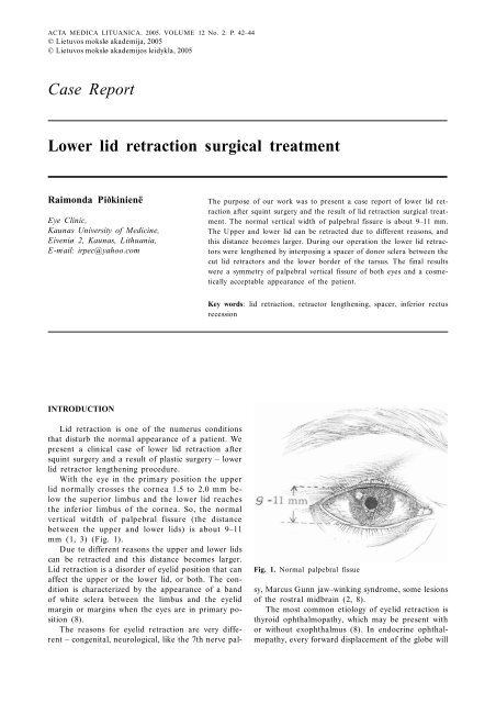
The normal vertical width of palpebral fissure is about 9–11 mm.<br />
The Upper and lower <strong>lid</strong> can be retracted due to different reasons, and<br />
this distance becomes larger. During our operation the lower <strong>lid</strong> retractors<br />
were lengthened by interposing a spacer of donor sclera between the<br />
cut <strong>lid</strong> retractors and the lower border of the tarsus. The final results<br />
were a symmetry of palpebral vertical fissure of both eyes and a cosmetically<br />
acceptable appearance of the patient.<br />
Key words: <strong>lid</strong> <strong>retraction</strong>, retractor lengthening, spacer, inferior rectus<br />
recession<br />
INTRODUCTION<br />
Fig. 1. Normal palpebral fissue<br />
Lid <strong>retraction</strong> is one of the numerus conditions<br />
that disturb the normal appearance of a patient. We<br />
present a clinical case of lower <strong>lid</strong> <strong>retraction</strong> after<br />
squint surgery and a result of plastic surgery – lower<br />
<strong>lid</strong> retractor lengthening procedure.<br />
With the eye in the primary position the upper<br />
<strong>lid</strong> normally crosses the cornea 1.5 to 2.0 mm below<br />
the superior limbus and the lower <strong>lid</strong> reaches<br />
the inferior limbus of the cornea. So, the normal<br />
vertical witdth of palpebral fissure (the distance<br />
between the upper and lower <strong>lid</strong>s) is about 9–11<br />
mm (1, 3) (Fig. 1).<br />
Due to different reasons the upper and lower <strong>lid</strong>s<br />
can be retracted and this distance becomes larger.<br />
Lid <strong>retraction</strong> is a disorder of eye<strong>lid</strong> position that can<br />
affect the upper or the lower <strong>lid</strong>, or both. The condition<br />
is characterized by the appearance of a band<br />
of white sclera between the limbus and the eye<strong>lid</strong><br />
margin or margins when the eyes are in primary position<br />
(8).<br />
The reasons for eye<strong>lid</strong> <strong>retraction</strong> are very different<br />
– congenital, neurological, like the 7th nerve palsy,<br />
Marcus Gunn jaw–winking syndrome, some lesions<br />
of the rostral midbrain (2, 8).<br />
The most common etiology of eye<strong>lid</strong> <strong>retraction</strong> is<br />
thyroid ophthalmopathy, which may be present with<br />
or without exophthalmus (8). In endocrine ophthalmopathy,<br />
every forward displacement of the globe will
<strong>Lower</strong> <strong>lid</strong> <strong>retraction</strong> <strong>surgical</strong> <strong>treatment</strong> 43<br />
lead to a widening of the palpebral fissure above the<br />
normal upper value. Proptosis and <strong>retraction</strong> of the<br />
upper <strong>lid</strong> are to be ascribed to the shortening of the<br />
contractile substance of the levator palpebrae, inhibiting<br />
the orbicularis oculi, as well as an increased sympathetic<br />
stimulation of Mueller’s muscle, as well as<br />
its fibrous contraction. Retraction of the lower <strong>lid</strong><br />
also occurs because of proptosis, but is frequently amplified<br />
due to the shortening of the <strong>lid</strong> retractors (3).<br />
The mechanical factors that cause <strong>lid</strong> <strong>retraction</strong><br />
are trauma, scars, tissue loss, posterior or middle <strong>lid</strong><br />
lamella shortening, complications of other surgery such<br />
as blepharoplasty, overcorrection of ptosis repair,<br />
blow-out fracture repair, large recession of the inferior<br />
rectus (2, 8).<br />
The anatomic basis of lower <strong>lid</strong> <strong>retraction</strong> in inferior<br />
rectus recession involves anterior extensions of<br />
Tenon’s capsule which surrounds the inferior rectus<br />
and the inferior oblique, thus forming Lockwood’s<br />
ligament. Lockwood’s ligament is the origin of the<br />
capsulopalpebral fascia, which is inserted at the lower<br />
tarsal border (8).<br />
Excessive recession of the inferior rectus causes<br />
lower <strong>lid</strong> <strong>retraction</strong> through the check ligaments between<br />
the inferior rectus and the tarsus of the lower<br />
<strong>lid</strong> (4, 5). Retraction frequently occurs even in spite<br />
of severing the connections between the inferior rectus<br />
and the lower eye<strong>lid</strong> (6).<br />
If <strong>lid</strong> <strong>retraction</strong> occurs despite this procedure, a<br />
recession of the <strong>lid</strong> retractors can be done (8).<br />
MATERIALS AND METHODS<br />
A 28-year-old female underwent strabismus surgery of<br />
the left eye. Recession of the inferior rectus was done.<br />
We saw the patient first six months after surgery.<br />
The vertical distance between the upper and the lower<br />
eye<strong>lid</strong> was 8 mm in the right eye and 12 mm in<br />
the left eye. The distance between the inferior limbus<br />
of the cornea and the inferior eye<strong>lid</strong> was 0.5 mm in<br />
the right and 3 mm in the left eye (Fig. 2).<br />
The patient was operated on the lower <strong>lid</strong> retractor<br />
lengthening procedure was done, interposing a<br />
graft of donor sclera between the cut <strong>lid</strong> retractors<br />
and the lower border of the tarsus (12).<br />
Freeing of the inferior eye<strong>lid</strong> retractors is anatomically<br />
equivalent to levator weakening in the upper eye<strong>lid</strong>,<br />
however, it does not elevate the lower <strong>lid</strong> because of<br />
the gravity vector with which the surgeon must contend.<br />
Thus, a spacer is the inevitable requirement for<br />
an effective lower eye<strong>lid</strong> elevation (7). Sometimes <strong>lid</strong>s<br />
receiving scleral implants become thickened and edematous<br />
postoperatively and the sclera resorbs in an<br />
unpredictable manner in many cases. Therefore, scleral<br />
grafts may end up less than cosmetically acceptable<br />
and require further surgery (9, 11). So, we demonstrate<br />
a realy successful case.<br />
Alternatively, the hammock sling procedure (10, 11)<br />
or different types of aloplastic spacers can be used.<br />
References<br />
Received 10 January 2005<br />
Accepted 15 March 2005<br />
1. Iliff CE, Iliff WJ. Oculoplastic Surgery. 1979: 35.<br />
2. Collin JRO. A Manual of Systematic Eye<strong>lid</strong> Surgery.<br />
2nd edition. 1989: 144.<br />
3. Olivari N. Endocrine Ophthalmolopathy. Kaden Verlag,<br />
Heidelberg, Germany 2001: 11.<br />
4. Roth A, Speeg-Shatz. Eye Muscle Surgery. English edition<br />
Swets & Zeitlinger b.v., Lisse 2001: 277.<br />
5. Huber A. Electrophysiology of the <strong>retraction</strong> syndromes.<br />
Brit J Ophthalmol 1974; 5: 293–300.<br />
6. Richards R. Strabismus Surgery. First edition 1991, Williams<br />
& Wilkins: 143.<br />
Fig. 2. Patient after an inferior rectus recession and before<br />
a lower <strong>lid</strong> surgery<br />
RESULTS<br />
The final results were a cosmetically acceptable appearance<br />
of the patient, a symmetry of palpebral vertical<br />
fissure in the right and left eyes, and a great satisfaction<br />
of the patient and the surgeon (Fig. 3).<br />
DISCUSSION<br />
Whereas the surgeon is able to apply several techniques<br />
for lowering an affected upper eye<strong>lid</strong>, correction of<br />
lower eye<strong>lid</strong> <strong>retraction</strong> does not permit such flexibility.<br />
Fig. 3. The same patient after a lower <strong>lid</strong> lengthening<br />
procedure
44<br />
Raimonda Piðkinienë<br />
7. Beyer-Machule ChK. Surgical <strong>treatment</strong> of thyroid-related<br />
eye<strong>lid</strong> <strong>retraction</strong>. International Ophthalmology Clinics<br />
1989; 29(4): 233.<br />
8. Heffernan JT, Tenzel RR. Lid <strong>retraction</strong>. Current Ocular<br />
Therapy. USA: W.B. Sounders Company 1995: 583.<br />
9. Dryden RM, Soll DB. The use of scleral transplantation<br />
in cicatricial entropion and eye<strong>lid</strong> <strong>retraction</strong>. Trans<br />
Am Acad Ophthalmol Otolaryngol 1971; 83: 669.<br />
10. Heffernan JT, Tenzel RR. Correction of vertical <strong>retraction</strong><br />
of the lower <strong>lid</strong> with an orbicularis hammock.<br />
Ophthalmic Plast Reconstr Surg 1989; 5: 92–8.<br />
11. Heffernan JT, Tenzel RR. Lid <strong>retraction</strong> current ocular<br />
therapy. USA: W.B. Sounders Company 1995: 584.<br />
12. Collin JRO. A manual of systematc eye<strong>lid</strong> surgery, 2nd<br />
edition 1989: 146.<br />
13. Olivari N. Endocrine Ophthalmopathy. Kaden Verlag,<br />
Heidelberg, 2001: 12.<br />
Raimonda Piðkinienë<br />
APATINIO VOKO RETRAKCIJOS CHIRURGINIS<br />
GYDYMAS<br />
Santrauka<br />
Normalus vertikalus voko plyðio matmuo yra apie 9–11 mm.<br />
Dël ávairiø prieþasèiø, sukelianèiø virðutinio ar apatinio voko<br />
retrakcijà, ðis matmuo gali padidëti. Ðiame darbe pateikiame<br />
sëkmingà apatinio voko retrakcijos, atsiradusios po apatinio<br />
tiesiojo raumens recesijos, chirurginio gydymo atvejá.<br />
Operacijos metu apatinis vokas pailgintas, tarp jo apatinio<br />
kremzlës kraðto ir voko retraktoriø ásiuvant donorinës skleros<br />
intarpà. Operacijos rezultatas: simetrija tarp abiejø akiø<br />
vokø plyðiø ir geras kosmetinis efektas.<br />
Raktaþodþiai: vokø retrakcija, retraktoriø pailginimas,<br />
intarpas, apatinio tiesiojo raumens recesija