Anthropological description of skeletons from graves no. 123, 124 ...
Anthropological description of skeletons from graves no. 123, 124 ...
Anthropological description of skeletons from graves no. 123, 124 ...

Create successful ePaper yourself
Turn your PDF publications into a flip-book with our unique Google optimized e-Paper software.
This picture <strong>of</strong> extreme stress on the upper limbs can be reasonably explained by the<br />
fracture <strong>of</strong> the arch <strong>of</strong> second lumbar vertebra (see below) which undoubtedly determined the<br />
complete flaccid paralysis <strong>of</strong> both lower limbs. The woman survived the severe trauma but<br />
could <strong>no</strong>t walk any more, <strong>no</strong>t even on crutches, therefore we can presume that she moved on<br />
a kind <strong>of</strong> low wheeled cart, driving it by arms, possibly holding a facility tool with her hands.<br />
On the other hand, muscle enthesopaties are observable also on femurs (iliopsoas, adductor<br />
magnus, gluteus maximus, vastus medialis) and tibiae (soleus), leading to suppose alternative<br />
hypotheses <strong>of</strong> a sort <strong>of</strong> polyenthesopathy syndrome or an intense overall activity, previous the<br />
accident.<br />
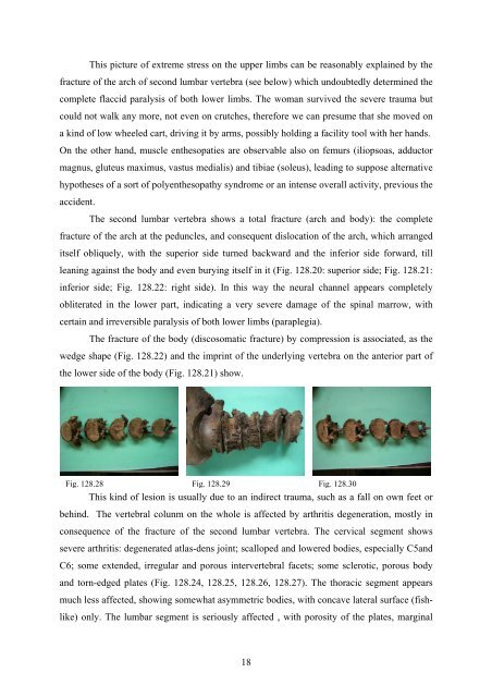
The second lumbar vertebra shows a total fracture (arch and body): the complete<br />
fracture <strong>of</strong> the arch at the peduncles, and consequent dislocation <strong>of</strong> the arch, which arranged<br />
itself obliquely, with the superior side turned backward and the inferior side forward, till<br />
leaning against the body and even burying itself in it (Fig. 128.20: superior side; Fig. 128.21:<br />
inferior side; Fig. 128.22: right side). In this way the neural channel appears completely<br />
obliterated in the lower part, indicating a very severe damage <strong>of</strong> the spinal marrow, with<br />
certain and irreversible paralysis <strong>of</strong> both lower limbs (paraplegia).<br />
The fracture <strong>of</strong> the body (discosomatic fracture) by compression is associated, as the<br />
wedge shape (Fig. 128.22) and the imprint <strong>of</strong> the underlying vertebra on the anterior part <strong>of</strong><br />
the lower side <strong>of</strong> the body (Fig. 128.21) show.<br />
Fig. 128.28 Fig. 128.29 Fig. 128.30<br />
This kind <strong>of</strong> lesion is usually due to an indirect trauma, such as a fall on own feet or<br />
behind. The vertebral colunm on the whole is affected by arthritis degeneration, mostly in<br />
consequence <strong>of</strong> the fracture <strong>of</strong> the second lumbar vertebra. The cervical segment shows<br />
severe arthritis: degenerated atlas-dens joint; scalloped and lowered bodies, especially C5and<br />
C6; some extended, irregular and porous intervertebral facets; some sclerotic, porous body<br />
and torn-edged plates (Fig. 128.24, 128.25, 128.26, 128.27). The thoracic segment appears<br />
much less affected, showing somewhat asymmetric bodies, with concave lateral surface (fishlike)<br />
only. The lumbar segment is seriously affected , with porosity <strong>of</strong> the plates, marginal<br />
18