Anthropological description of skeletons from graves no. 123, 124 ...
Anthropological description of skeletons from graves no. 123, 124 ...
Anthropological description of skeletons from graves no. 123, 124 ...

Create successful ePaper yourself
Turn your PDF publications into a flip-book with our unique Google optimized e-Paper software.
Elsa Pacciani<br />
<strong>Anthropological</strong> <strong>description</strong><br />
<strong>of</strong> <strong>skeletons</strong> <strong>from</strong> <strong>graves</strong> <strong>no</strong>. <strong>123</strong>, <strong>124</strong>, 125,<br />
126, 127, 128, 129, 130, 131, 132, 133, 134,<br />
135, 136, 141, 142, 143, 145 and 146 at<br />
Skriðuklaustur Monastery<br />
Skýrslur Skriðuklaustursrannsókna XXVIII
Elsa Pacciani<br />
<strong>Anthropological</strong> <strong>description</strong><br />
<strong>of</strong> <strong>skeletons</strong> <strong>from</strong> <strong>graves</strong> <strong>no</strong>. <strong>123</strong>, <strong>124</strong>, 125,<br />
126, 127, 128, 129, 130, 131, 132, 133, 134,<br />
135, 136, 141, 142, 143, 145 and 146 at<br />
Skriðuklaustur Monastery<br />
© Elsa Pacciani 2010<br />
<strong>Anthropological</strong> <strong>description</strong><br />
<strong>of</strong> <strong>skeletons</strong> <strong>from</strong> <strong>graves</strong> <strong>no</strong>. <strong>123</strong>, <strong>124</strong>, 125, 126, 127, 128, 129, 130, 131, 132,<br />
133, 134, 135, 136, 141, 142, 143, 145 and 146 at Skriðuklaustur Monastery<br />
Vala Gunnarsdóttir bjó til prentunar<br />
Skýrslur Skriðusklaustursrannsókna XXVIII<br />
Útgefandi: Skriðuklaustursrannsóknir og Soprintendenza per i Beni<br />
Archeologici della Toscana, Italy<br />
Útgáfustaður: Reykjavík<br />
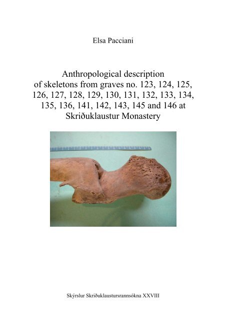
Forsíðumynd: A femur <strong>from</strong> grave 130. Shows a healed fracture.<br />
ISBN 978-9979-9970-6-1<br />
ISSN 1670-7982<br />
2
Table <strong>of</strong> contents<br />
INTRODUCTION ................................................................................................................................. 4<br />
ANTHROPOLOGICAL DESCRIPTION 2009.................................................................................. 6<br />
GRAVE <strong>123</strong>.......................................................................................................................................... 6<br />
GRAVE <strong>124</strong>.......................................................................................................................................... 8<br />
GRAVE 125.......................................................................................................................................... 9<br />
GRAVE 126........................................................................................................................................ 11<br />
LOOSE BONES IN GRAVE 126:............................................................................................................. 14<br />
GRAVE 127........................................................................................................................................ 14<br />
GRAVE 128........................................................................................................................................ 15<br />
GRAVE 129........................................................................................................................................ 19<br />
GRAVE 130........................................................................................................................................ 23<br />
GRAVE 131........................................................................................................................................ 31<br />
GRAVE 132........................................................................................................................................ 33<br />
GRAVE 133........................................................................................................................................ 34<br />
GRAVE 134........................................................................................................................................ 35<br />
GRAVE 135........................................................................................................................................ 37<br />
GRAVE 136........................................................................................................................................ 38<br />
GRAVE 141........................................................................................................................................ 40<br />
GRAVE 142........................................................................................................................................ 40<br />
GRAVE 143........................................................................................................................................ 41<br />
GRAVE 145........................................................................................................................................ 45<br />
GRAVE 146........................................................................................................................................ 50<br />
REFERENCES .................................................................................................................................... 52<br />
3
Introduction<br />
This <strong>description</strong> was made “on the field”, soon after the excavation, the preliminary cleaning<br />
and restoration. Thus, its aim is mostly to record the identifying data and some relevant<br />
observations, thereby <strong>of</strong>fering cues, research lines and suggestions for widening <strong>of</strong> particular<br />
aspects.<br />
So there is <strong>no</strong> pretence <strong>of</strong> exhaustiveness, as the anthropological study requires more<br />
time and the simultaneous disposability <strong>of</strong> the whole sample, for screening and comparisons<br />
about the various characters. Moreover many features need to be examined by lab specific<br />
tools and equipping. However some generalities, observations and statements can be related<br />
here:<br />
A marked sexual dimorphism characterizes this sample and makes the sex diag<strong>no</strong>sis<br />
relatively easy, together with the good state <strong>of</strong> preservation; very few cases raised some<br />
uncertainty. So a morphological diag<strong>no</strong>sis was performed, on the basis <strong>of</strong> the most<br />
discriminant hip bone and skull features, and taking into account also the other bones. No sex<br />
diag<strong>no</strong>sis was attempted on subadults.<br />
About the age-at–death diag<strong>no</strong>sis I decided to avoid a subdivision <strong>of</strong> adult individuals<br />
in small age classes, because <strong>of</strong> the weakness <strong>of</strong> all the aging indicators due to the high<br />
individual and population variability. For this reason I adopted a gross subdivision in three<br />
classes: young adult (conventionally beginning <strong>from</strong> the sphe<strong>no</strong>-occipital suture closure or<br />
the third molars eruption), mature adult and old adult, on the basis <strong>of</strong> a complex <strong>of</strong> traits<br />
appearance, such as pubic symphysis, auricular surface, dental wear and pathology, cranial<br />
suture closure, joint degeneration, spongy bone rarefaction etc.. A more precise diag<strong>no</strong>sis will<br />
be possible when the whole sample will be examined in order to detect the “population”<br />
aging rate, or /and other traits will be examined, such as dental cement anulation, pulp/tooth<br />
ratio etc.<br />
For dental wear quantification I used the Lovejoy 1985 graphic scheme which<br />
represents phases <strong>of</strong> maxilla and mandible wear, but without attributing the specimens to the<br />
associated age classes, because I have found, in my previous methodological research, a huge<br />
divergence between the Lovejoy age classes and the real age. As aging methods I adopted:<br />
- for the pubic symphysis, Brooks and Suchey 1990 scale<br />
- for the auricular surface, Lovejoy et al 1985 scale<br />
- for the sternal end <strong>of</strong> the ribs, Iscan et al 1984 scale<br />
Subadult age-at–death diag<strong>no</strong>sis was made according the Ubelaker (1989) dental<br />
development standard.<br />
4
A restricted selection <strong>of</strong> measurements was made with a purely identifying aim<br />
regarding the skull, and with the purpose <strong>of</strong> underscoring some stress indicators and<br />
anthropological conditions (stature, robusticy, platymeria, platicnemia) regarding the<br />
postcranial bones. In measuring pair bones, I chose the best preserved side; when sides were<br />
equally preserved, I chose the left one; when I detected a clear asymmetry, I reported it in the<br />
text.<br />
Of course it will be possible and advisable to take a much larger amount <strong>of</strong><br />
measurements in the anthropology laboratory, where having the availability <strong>of</strong> all the<br />
necessary anthropometric instruments and, above all, having specific finalities in an organic<br />
research project.<br />
The dental formula is presented for each individual in a table, whose legend is the<br />
following:<br />
P = present<br />
AM = lost ante mortem<br />
PM = lost postmortem<br />
AG = genetically absent<br />
R = root only<br />
NE = <strong>no</strong>t erupted<br />
- - = <strong>no</strong>t detectable<br />
The stature was calculated by the formulas <strong>of</strong> Olivier et al. 1978, based on the<br />
physiologic lenght <strong>of</strong> the femur (n. 2 according Martin and Saller).<br />
Degree <strong>of</strong> resorption <strong>of</strong> alveolar bone, due to periodontal disease is attributed<br />
according to the simple scale <strong>of</strong> Brothwell 1981 (No alveolar destruction; Slight, Medium,<br />
Considerable).<br />
The same author was followed for the degree <strong>of</strong> calculus formation (Slight, Medium,<br />
Considerable). Diastema is a gap or space between two teeth. It happens when there is an<br />
unequal relationship between the size <strong>of</strong> the teeth and the jaw.<br />
A very peculiar and interesting pathologic condition is recurrent in the sample and<br />
requires an accurate analysis and a differential diag<strong>no</strong>sis. Even 5 individuals (see the single<br />
<strong>description</strong>s below), out <strong>of</strong> 13 adults in this group exhibit the characteristic signs: the distal<br />
epiphyses <strong>of</strong> some long bones appear “swollen” and show porosity, erosion, pitted surface,<br />
new bone apposition, while other long bones appear <strong>no</strong>rmal. Hand bones are the most<br />
affected, with a “swollen” look and lytic lesions: holes, erosions and pits. Also the joint<br />
surfaces appear altered, with erosion areas, deformation and osteophytosis.<br />
A<strong>no</strong>ther group characteristic is the high frequency <strong>of</strong> Echi<strong>no</strong>coccus cysts: even 4<br />
individuals are affected.<br />
5
<strong>Anthropological</strong> <strong>description</strong> 2009<br />
Grave <strong>123</strong><br />
Sex: Uncertain. Some features are<br />
masculine: inion, absence <strong>of</strong> frontal<br />
eminences, mastoid process, square<br />
chin, ulna and fibula well shaped for<br />
muscle attachments; other feature are<br />
feminine: supraorbital edge, gonion,<br />
glabella, mandibular condyle.<br />
Age at death: Mature adult.<br />
Dental wear is low: degree E for<br />
posterior teeth; degree B2 for anterior<br />
Fig. <strong>123</strong>.1<br />
ones. But cranial sutures are partly<br />
closed: lateral parts <strong>of</strong> the coronal, almost all the sagittal, some portion <strong>of</strong> the lambdoid.<br />
Stature: Not detectable.<br />
Dental characteristics:<br />
Right<br />
Left<br />
M3 M2 M1 P2 P1 C I2 I1 I1 I2 C P1 P2 M1 M2 M3<br />
Maxilla -- -- -- -- -- -- -- -- -- -- -- -- -- -- -- --<br />
Mandibula P P P P P P P P P P P P P P P P<br />
- Malocclusion: left P2 dislocated lingually.<br />
- Caries <strong>no</strong>.<br />
- Periodontitis (phase: Medium) (Fig. <strong>123</strong>.2).<br />
- Calculus (phase: Medium).<br />
Fig. <strong>123</strong>.2 Fig. <strong>123</strong>.3<br />
6
Occupational stress indicators and pathological aspects:<br />
The few bones better preserved show signs <strong>of</strong> activity: ulna diaphysis, fibula, metacarpals.<br />
The first right metacarpal shows enthesopathy <strong>of</strong> the muscle opponens pollicis.<br />
Occipital condyles are affected by marginal osteophytosis. Besides they are<br />
asymmetric: the left one is flattened, the right one shows an accessory facet in lateralposterior<br />
position (Fig.<strong>123</strong>.3). Moreover, the occipital foramen has an asymmetric shape.<br />
Atlas can give an explanation about that: it is lacking <strong>of</strong> the right arm <strong>of</strong> the posterior arch,<br />
because <strong>of</strong> a congenital defect (Fig.<strong>123</strong>.4). The a<strong>no</strong>maly has repercussions on C2 and C3,<br />
which show asymmetric shape <strong>of</strong> the intervertebral facets and anterior shortening <strong>of</strong> the<br />
bodies (Fig.<strong>123</strong>.5, <strong>123</strong>.6).<br />
Fig. <strong>123</strong>.4 Fig. <strong>123</strong>.5 Fig. <strong>123</strong>.6<br />
A<strong>no</strong>ther consequence is the temporo-mandibular arthritis, bilateral and asymmetric:<br />
on the right side, flattening and enlargement <strong>of</strong> the fossa mandibularis and an area <strong>of</strong> bony<br />
buildup (Fig.<strong>123</strong>.7); on the left side, the formation <strong>of</strong> an accessory facet (Fig.<strong>123</strong>.8).<br />
Other observations: trace <strong>of</strong> metopism: a little (1 cm) segment close to the naso-frontal<br />
suture.<br />
Os acromiale <strong>no</strong>t detectable for incompleteness.<br />
Fig. <strong>123</strong>.7 Fig. <strong>123</strong>.8<br />
7
Grave <strong>124</strong><br />
Sex: Female (small mandible, sharp<br />
supraorbital edge, small humerus<br />
dimensions).<br />
Age at death: Adult (weared M3) young<br />
(frontal suture completely open, dental<br />
wear: maxillary degree C; mandibular<br />
degree D).<br />
Dental characteristics:<br />
Fig. <strong>124</strong>.1<br />
Right<br />
Left<br />
M3 M2 M1 P2 P1 C I2 I1 I1 I2 C P1 P2 M1 M2 M3<br />
Maxilla P P P -- -- P P P P P P P P P P --<br />
Mandibula P P P P P P P P PM P P P P P P PM<br />
- Hypoplasia striae on central incisors, canines and premolars.<br />
- No shovel shape incisors.<br />
- No caries.<br />
- Calculus: Medium.<br />
- Periodontitis: absent.<br />
- Incisors have fractured edge (Fig.<strong>124</strong>.2, <strong>124</strong>.3); upper right M1 has fractured disto-lingual<br />
cusp (Fig.<strong>124</strong>.4).<br />
Fig. <strong>124</strong>.2 Fig. <strong>124</strong>.3 Fig. <strong>124</strong>.4<br />
8
Grave 125<br />
Only lower limbs are present.<br />
Sex: Male (femur head and<br />
condyles dimensions).<br />
Age at death: Mature adult<br />
(slight arthritis signs at the at the<br />
femur head and knee joints).<br />
Stature: Cm 160.<br />
Occupational stress indicators<br />
and pathological aspects:<br />
On femurs: enthesopathies <strong>of</strong> the<br />
ligament <strong>of</strong> the head (Fig.125.2)<br />
Fig. 125.1<br />
and gluteus maximus (Fig.125.3).<br />
Arthritis signs at the head: marginal osteophytosis and endoarticular neoapposition<br />
(Fig. 125.2) and at the knee joint. Platimeria.<br />
Both tibiae show periostitis all over the diaphysis, and vascular imprints on the<br />
antero-lateral face (Fig.125.4). Also the fibulae are periostitic.<br />
The right fibula is<br />
curved, with medial<br />
convessity (Fig.125.5,<br />
125.6) causing<br />
twisted relations and<br />
osteophytosis at the<br />
ankle joint (Fig<br />
Fig. 125.2 Fig. 125.3<br />
125.7), and osteophytosis at the foot joints, in particular<br />
the right first metatarsal (Fig 125.8, 125.9) and first<br />
phalanx (Fig. 125.10). The whole foot trim is altered.<br />
Also the soleus muscle attachment on the right tibia is<br />
more expressed than left, indicating an a<strong>no</strong>malous<br />
deambulation (Fig.125.11).<br />
Both patellae show the quadriceps tendon<br />
enthesopathy (Fig. 125.12).<br />
Fig. 125.4<br />
9
A scattered right femur and rotula are included in the box, <strong>no</strong>t belonging to this<br />
individual (the excavator says they could be matched with the articulated individual <strong>of</strong> grave<br />
140). It is well preserved, except some exfoliation <strong>of</strong> the diaphysis and extremities erosion. It<br />
shows a slight marginal osteophytosis at the knee articulation (Fig.125.13).<br />
The stature <strong>of</strong> the individual is cm 173.<br />
Fig. 125.7<br />
Fig. 125.5 Fig. 125.6<br />
Fig. 125.8 Fig. 125.9 Fig. 125.10<br />
Fig. 125.12 Fig. 125.13<br />
Fig. 125.11<br />
10
Grave 126<br />
Fig. 126.1 Fig. 126.2<br />
Sex: Female (morphological diag<strong>no</strong>sis, on the basis <strong>of</strong> the most discriminant cranial and hip<br />
bone features).<br />
Age at death: Mature, on the basis <strong>of</strong> dental wear (phase G on maxilla, H on mandible),<br />
according to the scale <strong>of</strong> Lovejoy, 1985), auricular surface (degree 7) sternal ends <strong>of</strong> the ribs<br />
and arthritis signs; even if cranial sutures are completely open.<br />
Stature: Cm 154.<br />
Dental characteristics:<br />
Right<br />
Left<br />
M3 M2 M1 P2 P1 C I2 I1 I1 I2 C P1 P2 M1 M2 M3<br />
Maxilla P AM P P R P P P P P P P P P P P<br />
Mandibula P P P P P P P AM R P P P P P P P<br />
An abscess is located in correspondence with upper right<br />
M2, lost. A second one is in correspondence with lower<br />
right M1, which is fractured in the proximal side (Fig.<br />
126.3, 126.4).<br />
Periodontitis : Considerable.<br />
Calculus: Considerable.<br />
Fig. 126.3<br />
Caries absent.<br />
A diastema is located<br />
between upper left M3<br />
and M2.<br />
Malocclusion absent.<br />
Shovel shaped incisors.<br />
Fig. 126.4<br />
Fig. 126.5<br />
11
Occupational stress indicators and pathological aspects:<br />
The deltoid muscle attachments are enthesopathic on both clavicles and very developed on<br />
both humeri (Fig.126.5); also the the pectoralis major (Fig 126.6) and teres major (Fig. 126.7)<br />
are very developed and enthesopathic as well as the brachiradialis (Fig. 126.8).<br />
The ulnae and radii show marked insertions <strong>of</strong> flexors and extensors on the diaphysis,<br />
besides the biceps. Hands show sharp edges on metacarpals for dorsal and palmar interossei,<br />
and exceptionally developed insertions for flexor digitorum muscles (Fig. 126.9). As this<br />
complex <strong>of</strong> stress indicators is bilateral and involves the deltoid (abductor muscle) and the<br />
flexion/extension muscles, we can imagine an explaining activity such as laundress. The right<br />
ulna shows also enthesopathic insertions <strong>of</strong> brachialis and pronator quadratus.<br />
Fig. 126.6 Fig. 126.7 Fig. 126.8<br />
Arthritis signs (marginal osteophytosis ) are visible on humeral head (Fig. 126.10),<br />
elbow, wristle, carpals and metacarpals , confirming a stressing activity <strong>of</strong> upper limbs.<br />
Platimeria.<br />
Bilateral acetabulum arthritis.<br />
Marked bilateral superior tibi<strong>of</strong>ibular joint arthritis (Fig. 126.11).<br />
“Squatting” facets on the anterior edge <strong>of</strong> the ankle joint.<br />
Ankylosis between the first and second phalanx <strong>of</strong> the third finger, <strong>of</strong> traumatic origin. (Fig.<br />
126.12).<br />
Dorsal extension <strong>of</strong> the first metatarsal distal joints (Fig. 126.13). Severe arthritis<br />
(porosity, enlargement) on the left intervertebral facets <strong>of</strong> C4-C5 and C7-T1 (Fig 126.14,<br />
126.15). Slight arthritis on the cervical and thoracic segments <strong>of</strong> the column.<br />
Radio and ulna lengths de<strong>no</strong>te asymmetry <strong>of</strong> the upper limbs (radio max. length: right mm<br />
215, left mm 211; ulna max. length: right mm 231, left mm 227). Left humerus is <strong>no</strong>t<br />
measurable.<br />
A deep preauricular sulcus indicates pregnancies.<br />
Echi<strong>no</strong>coccus cyst (a parasitic infection which affects people who herd sheep using<br />
dogs) <strong>of</strong> huge dimensions was found in the liver region (Fig.126.16).<br />
Other observations: Os acromiale <strong>no</strong>t detectable. Presence <strong>of</strong> torus palatinus.<br />
12
Fig. 126.9 Fig. 126.10<br />
Fig. 126.11 Fig. 126.12<br />
Fig. 126.13 Fig. 126.14<br />
Fig. 126.15 Fig. 126.16<br />
13
Loose bones in grave 126:<br />
Some skull fragments<br />
1 mandible, lacking teeth (most <strong>of</strong> them lost in life, at least all molars) and rami.<br />
2 controlateral humerus diaphyses<br />
2 controlateral ulna diaphyses<br />
2 controlateral mastoid processes<br />
2 controlateral femur diaphyses<br />
2 controlateral tibia diaphyses<br />
fragment <strong>of</strong> scapula, hip bone, ribs, hands and feet<br />
All the bones can belong to a female individual, characterized by little build, and rather old<br />
age.<br />
Grave 127<br />
Fig. 127.1 Fig. 127.2<br />
Age at death: Newborn, on the basis <strong>of</strong> teeth development (Fig. 127.2).<br />
Very fragmentary remains. The skull (squama fragments, pyramids, left orbit, basis) and most<br />
<strong>of</strong> the postacranial bones are represented: right clavicle, right and left ribs, vertebrae, humeri,<br />
forearms, hip bones, femurs, tibiae, fibulae, right hand (10 elements), left hand (9 elements,<br />
right foot (5 elements), left foot (2 elements). The bones are incomplete and curled up. The<br />
presence <strong>of</strong> some tooth crowns allows us to determinate the age at death, coincident with the<br />
birth.<br />
Dental characteristics:<br />
right<br />
left<br />
m2 m1 c i2 i1 i1 i2 c m1 m2<br />
Maxilla P -- -- P P P P P P P<br />
Mandibula -- -- -- -- -- -- -- -- -- --<br />
14
Grave 128<br />
Fig. 128.1 Fig. 128.2<br />
Sex: Female (morphological diag<strong>no</strong>sis, on the basis <strong>of</strong> the most discriminant cranial and hip<br />
bone features).<br />
Age at death: Old:<br />
Advanced dental wear, exceeding the maximum degree <strong>of</strong> the Lovejoy scale.<br />
Cranial sutures in advanced closure.<br />
Presence <strong>of</strong> osteoporosis.<br />
Presence <strong>of</strong> diffuse arthritis.<br />
Pubis symphysis: degree 4.<br />
Auricular surface: very porotic, irregular edges, retroauricular activity traces (phase 8).<br />
Dental characteristics:<br />
Right<br />
Left<br />
M3 M2 M1 P2 P1 C I2 I1 I1 I2 C P1 P2 M1 M2 M3<br />
Maxilla AG P P P P P P P P P P P P P P AG<br />
Mandibula AG P P P AM P P AM AM P PM R AM P AM AG<br />
Periodontal disease: considerable.<br />
Calculus: considerable.<br />
4 diastemas on maxilla: right P1-C; right I2-I1; I1-I1; left I1-I2 (Fig. 128.3).<br />
Dental wear <strong>of</strong> incisors is huge, almost reaching the roots (Fig. 128.3).<br />
Buccal abscess <strong>of</strong> upper right M1.<br />
Fig. 128.3 Fig. 128.4 Fig. 128.5<br />
15
Occupational stress indicators and pathological aspects:<br />
Enthesopaties: severe on deltoid attachments on both clavicles and both humeri (Fig. 128.4,<br />
128.5); infraspinatus and teres mi<strong>no</strong>r on both scapulae (Fig. 128.6) and humeri (Fig. 128.7),<br />
but mainly on the left side; triceps on scapulae (Fig. 128.8); pectoralis major and teres major<br />
are well developed on humeri, mainly on the left side.<br />
Common extensor and extensor carpi radialis longus muscles are enthesopatic on<br />
humeri, bilaterally (Fig. 128.9). Common flexor attachments are very developed on both<br />
humeri.<br />
Fig. 128.6 Fig. 128.7 Fig. 128.8<br />
Particularly <strong>no</strong>ticeable are the enthesopaties <strong>of</strong> biceps tuberosity on both radii (Fig.<br />
128.10, 128.11); <strong>of</strong> brachialis and the muscles attaching on the postero-lateral face <strong>of</strong> ulnae<br />
(abductor pollicis longus; extensor pollicis longus; extensor indicis) (Fig. 128.12, 128.13,<br />
128.14); <strong>of</strong> supinator on radii (Fig. 128.15, see the grooving imprints and entesophyties); <strong>of</strong><br />
teres pronator on radii (Fig. 128.16); <strong>of</strong> pronator quadratus on right ulna (Fig. 128.17).<br />
Finally the flexor digitorum muscle attachments on the anterior diaphyseal surfaces <strong>of</strong> radii<br />
and ulnae are very marked (Fig. 128.10), as well as the crests on the palmar surface <strong>of</strong> the<br />
phalangeal diaphyses (Fig. 128.18).<br />
Fig. 128.9 Fig. 128.10 Fig. 128.11<br />
Generally, all the main arm muscles involved in abdution/addution <strong>of</strong> the arm,<br />
flexion/extension <strong>of</strong> the forearm; pro<strong>no</strong>/supination <strong>of</strong> the forearm and hand grip appear<br />
subject to strong and continue stress forces.<br />
Both scapulae show gle<strong>no</strong>id arthritis, with marginal osteophytosis and, on the right<br />
side, a forward extension <strong>of</strong> the articular surface (Fig. 128.19).<br />
16
Fig. 128.12 Fig. 128.13 Fig. 128.14<br />
Fig. 128.15 Fig. 128.16 Fig. 128.17<br />
Fig. 128.18<br />
Fig. 128.19<br />
Fig. 128.20<br />
Fig. 128.21 Fig. 128.22 Fig. 128.24<br />
Fig. 128.25 Fig. 128.26 Fig. 128.27<br />
17
This picture <strong>of</strong> extreme stress on the upper limbs can be reasonably explained by the<br />
fracture <strong>of</strong> the arch <strong>of</strong> second lumbar vertebra (see below) which undoubtedly determined the<br />
complete flaccid paralysis <strong>of</strong> both lower limbs. The woman survived the severe trauma but<br />
could <strong>no</strong>t walk any more, <strong>no</strong>t even on crutches, therefore we can presume that she moved on<br />
a kind <strong>of</strong> low wheeled cart, driving it by arms, possibly holding a facility tool with her hands.<br />
On the other hand, muscle enthesopaties are observable also on femurs (iliopsoas, adductor<br />
magnus, gluteus maximus, vastus medialis) and tibiae (soleus), leading to suppose alternative<br />
hypotheses <strong>of</strong> a sort <strong>of</strong> polyenthesopathy syndrome or an intense overall activity, previous the<br />
accident.<br />
The second lumbar vertebra shows a total fracture (arch and body): the complete<br />
fracture <strong>of</strong> the arch at the peduncles, and consequent dislocation <strong>of</strong> the arch, which arranged<br />
itself obliquely, with the superior side turned backward and the inferior side forward, till<br />
leaning against the body and even burying itself in it (Fig. 128.20: superior side; Fig. 128.21:<br />
inferior side; Fig. 128.22: right side). In this way the neural channel appears completely<br />
obliterated in the lower part, indicating a very severe damage <strong>of</strong> the spinal marrow, with<br />
certain and irreversible paralysis <strong>of</strong> both lower limbs (paraplegia).<br />
The fracture <strong>of</strong> the body (discosomatic fracture) by compression is associated, as the<br />
wedge shape (Fig. 128.22) and the imprint <strong>of</strong> the underlying vertebra on the anterior part <strong>of</strong><br />
the lower side <strong>of</strong> the body (Fig. 128.21) show.<br />
Fig. 128.28 Fig. 128.29 Fig. 128.30<br />
This kind <strong>of</strong> lesion is usually due to an indirect trauma, such as a fall on own feet or<br />
behind. The vertebral colunm on the whole is affected by arthritis degeneration, mostly in<br />
consequence <strong>of</strong> the fracture <strong>of</strong> the second lumbar vertebra. The cervical segment shows<br />
severe arthritis: degenerated atlas-dens joint; scalloped and lowered bodies, especially C5and<br />
C6; some extended, irregular and porous intervertebral facets; some sclerotic, porous body<br />
and torn-edged plates (Fig. 128.24, 128.25, 128.26, 128.27). The thoracic segment appears<br />
much less affected, showing somewhat asymmetric bodies, with concave lateral surface (fishlike)<br />
only. The lumbar segment is seriously affected , with porosity <strong>of</strong> the plates, marginal<br />
18
osteophytosis and lowered bodies (except L1, almost <strong>no</strong>rmal and L5, with porous upper plate<br />
only) (Fig. 128.28, 128.29, 128.30).<br />
Degenerative arthritis signs are detectable on occipital condyles, temporo-mandibular<br />
joints, elbow, carpus, metacarpus.<br />
Hand bones have a “swollen” look and lytic lesions: holes, erosions, pits (Fig. 128.31,<br />
128.32), as other individuals <strong>of</strong> the group (see individuals N.129 ; N.130; N.143; N.145).<br />
Other observations: Os acromiale absent, bilaterally. Metopism: initial and final short<br />
segments. Presence <strong>of</strong> torus palatinus. Presence <strong>of</strong> some small button osteomas (benign<br />
tumors) on the skull vault (Fig. 128.33). Presence <strong>of</strong> preauricular sulcus, bilaterally.<br />
Platymeria.<br />
Bilateral asymmetry is detectable in the humerus lengths (right mm<br />
306, left mm 295).<br />
Fig. 128.31 Fig. 128.32 Fig. 128.33<br />
Grave 129<br />
Sex: Male (morphological diag<strong>no</strong>sis, on the basis <strong>of</strong> the most discriminant cranial and hip<br />
bone features).<br />
Age at death: old:<br />
Advanced dental wear, exceeding the maximum degree <strong>of</strong> the Lovejoy scale; even, the whole<br />
crown is worn in almost all the maxillary teeth (Fig. 129.4, 129.5, 129.6).<br />
Cranial sutures are open except the lateral segments <strong>of</strong> the coronal.<br />
Auricular surface is porotic, with irregular edges and moderate traces <strong>of</strong> retroauricular<br />
activity (phase 7).<br />
Fig. 129.1 Fig. 129.2 Fig. 129.3<br />
19
Stature: Cm 160.<br />
Dental characteristics:<br />
Right<br />
Left<br />
M3 M2 M1 P2 P1 C I2 I1 I1 I2 C P1 P2 M1 M2 M3<br />
Maxilla PM AM R P P P AM P P P P P P R P P<br />
Mandibula P AM P P P P P P P P P P P P AM P<br />
Periodontal disease: considerable.<br />
Six abscesses can be counted, all with a vestibular fistula:<br />
1 – the cause <strong>of</strong> the loss <strong>of</strong> right I2 in a short time before death (alveolar edges are still<br />
sharp),<br />
2 - a sole large abscess in correspondence <strong>of</strong> upper right M2 and M3,<br />
3 - in correspondence <strong>of</strong> upper right M1; this tooth rotated on its lingual root (working as a<br />
swivel) and occupied the space <strong>of</strong> M2 (lost AM) with its vestibular posterior root completely<br />
raised out <strong>of</strong> the alveolus, while the vestibular anterior one is lacking (Fig. 129.7).<br />
4 - in correspondence <strong>of</strong> upper right I2<br />
5 - in correspondence <strong>of</strong> lower left M1; only the lingual root remains.<br />
6 - in correspondence <strong>of</strong> lower left M2<br />
Calculus: considerable (Fig.129.8).<br />
Malocclusion: lower anterior teeth crowded, with vestibular dislocation <strong>of</strong> left I2 and lingual<br />
dislocation <strong>of</strong> left I1.<br />
Fig. 129.4<br />
Fig. 129.5 Fig. 129.6<br />
Shovel shape incisors <strong>no</strong>t detectable.<br />
Hypoplasia <strong>no</strong>t detectable.<br />
Occupational stress indicators and pathological aspects:<br />
Pathological aspect <strong>of</strong> the scapular gle<strong>no</strong>ids, mostly on the left side: marginal osteophytes,<br />
porosity and sclerosis <strong>of</strong> the articular surface (Fig. 129.9); these signs can be associated to an<br />
impingement syndrome resulted <strong>from</strong> the presence <strong>of</strong> os acromiale (see below). An erosive<br />
area is present on the right side at the origin <strong>of</strong> the biceps tendon (Fig. 129.10); a similar<br />
erosive area is also at the sternal end <strong>of</strong> the clavicles (Fig. 129.11).<br />
20
Fig. 129.7 Fig. 129.8 Fig. 129.9<br />
Fig. 129.10 Fig. 129.11 Fig. 129.12<br />
Fig. 129.13 Fig. 129.14 Fig. 129.15<br />
Fig. 129.16 Fig. 129.16bis Fig. 129.17<br />
Fig. 129.18 Fig. 129.19<br />
Fig. 129.20<br />
21
Both humeri show evident insertions for the pectoralis major and deltoid muscles, and<br />
enthesopathies <strong>of</strong> the teres major and rotator cuff (Fig. 129.12, 129.13).<br />
Advanced degenerative arthritis at the right elbow (Fig. 129.14). An erosive area is<br />
present on the right olecra<strong>no</strong>n apex (Fig. 129.15, 129.16), on the left radial tuberosity, on the<br />
back side <strong>of</strong> both rotulae (Fig. 129.16 bis).<br />
Fig. 129.21 Fig. 129.22 Fig. 129.23<br />
The distal epiphyses <strong>of</strong> the right radio and ulna appear “swollen” (Fig. 129.17) and the<br />
joint surfaces appear altered too (Fig.129.18); the left one is <strong>no</strong>rmal.<br />
The same “swollen” appearance can be found on<br />
the hands: the right one, mostly at the proximal end <strong>of</strong> the<br />
second metacarpal and at the distal end <strong>of</strong> the third one<br />
(Fig. 129.19, 129.20, 129.21); the left one, even more<br />
damaged, where the carpal bones are more affected,<br />
besides the distal end <strong>of</strong> all the metacarpals and the<br />
proximal phalanges <strong>of</strong> the second and third finger (Fig.<br />
Fig. 129.24<br />
129.22, 129.23, 129.24);. These “swollen” areas show<br />
also porosity, erosion, pitted surface, new bone<br />
apposition. This pathological condition is very similar to<br />
the one found on Individuals N. 128 and N.130.<br />
Finally, an Echi<strong>no</strong>coccus cyst was found in the right side<br />
<strong>of</strong> the abdomen(Fig. 129.25).<br />
Other observations: Os acromiale present bilaterally, but<br />
the <strong>no</strong>n-fusion involves different ossification nuclei, so<br />
that the right acromion is shorter the the left one.<br />
Torus palatinus absent.<br />
Fig. 129.25<br />
22
Grave 130<br />
Fig. 130.1 Fig. 130.2 Fig. 130.3<br />
Sex: Male (morphological diag<strong>no</strong>sis, on the basis <strong>of</strong> the most discriminant cranial and hip<br />
bone features).<br />
Age at death: Mature adult.<br />
- Cranial sutures open except the lateral segments <strong>of</strong> the coronal.<br />
- Dental wear advanced, exceeding the Lovejoy scale (Fig. 130.4, 130.5).<br />
- Sternal ends <strong>of</strong> ribs in Phase 3 (Iscan et al. 1984) (Fig.130.6).<br />
- Auricular surface <strong>of</strong> ilium: phase 5.<br />
Stature: Cm 163.<br />
Dental characteristics:<br />
Right<br />
Left<br />
M3 M2 M1 P2 P1 C I2 I1 I1 I2 C P1 P2 M1 M2 M3<br />
Maxilla P P P P P P P p P P P AM P P P P<br />
Mandibula P P P P P P P P P P P P P P P P<br />
Fig. 130.4 Fig. 130.5 Fig. 130.6<br />
Caries absent.<br />
Periodontal disease: medium.<br />
Abscess with buccal fistula in correspondence <strong>of</strong> upper right M1; buccal periodontal pocket<br />
in correspondence <strong>of</strong> upper right M2; buccal and lingual periodontal pockets in<br />
correspondence <strong>of</strong> upper left P1; large lingual abscess in correspondence <strong>of</strong> upper left M2.<br />
Calculus: considerable.<br />
3 diastemas on maxilla: right I2-I1; I1-I1; left I2-I1 (Fig. 130.4).<br />
Malocclusion: slight rotation <strong>of</strong> lower canines.<br />
23
Severe hypoplasia on lower canines; presence <strong>of</strong> striae also on other teeth, detectable<br />
despite <strong>of</strong> the abundant calculus.<br />
Dental wear is particularly advanced on upper anterior teeth, where it is oriented<br />
obliquely, reaching the root on the lingual side.<br />
Occupational stress indicators and pathological aspects:<br />
The cranial vault shows multiple lesions, probably due to tertiary syphilis: gummatous pits <strong>of</strong><br />
different width, surrounded by hyperostotic porous bone, and erosive or sclerotic areas. Four<br />
<strong>of</strong> these lesions on the frontal bone (Fig. 130.7, 130.8, 130.9) (n.1 and n.2 on the left anterior<br />
part <strong>of</strong> the squama behind the left superciliary arch, small and close to each other, oval, with<br />
the major axis in sagittal direction,; n.3, behind the right superciliary arch, larger and flatter,<br />
quadrangular shaped; n.4 on the posterior part <strong>of</strong> the squama, before the bregma, slightly on<br />
the left, grossly rounded, large and flat); four on the left parietal (Fig. 130.10, 130.11, 130.12)<br />
(n.5, the most anterior, in the corner between the coronal and sagittal sutures, is an initial<br />
erosion area; n.6, soon behind it, with irregular shape and bottom, and transversal major axis;<br />
n.7, rather flat erosion area behind n.6, close to the sagittal suture, with irregular shape and<br />
oblique major axis; n. 8, the posterior one, larger and deeper, oval, with rounded edges, rather<br />
smooth and undulated surface, transverse major axis); one on the right parietal: n.9,<br />
(Fig.130.11) the largest one, oval, sagittal major axis, rather smooth and pitted bottom,<br />
rounded edges.<br />
The left femur distal diaphysis shows signs <strong>of</strong> gummatous and <strong>no</strong>ngummatous<br />
osteoperiostitis: a large thickened area (10 cm long), mostly on the lateral side and extending<br />
to the anterior and posterior faces. The surface is rough, sieve or sclerotic, following a bony<br />
buildup process (Fig. 130.13, 130.14). In the antero-lateral side there are three grouped<br />
irregular cavities, furrowed by grooves and surrounded by porotic bone, probably<br />
corresponding to the location <strong>of</strong> gummas (Fig. 130.15, 130.16).<br />
Tibiae are both affected by severe <strong>no</strong>ngummatous osteoperiostitis. The right one<br />
shows two osteoperiostitis areas: 1) a huge thickened area with the maximum bulge mostly<br />
on the anterior margin at half diaphysis but extending to the antero-lateral and antero-medial<br />
faces on almost the whole diaphysis (Fig. 130.17, 130.18, 130.19). A core <strong>of</strong> swelling (9 cm<br />
long) with clear edge is observable at the centre <strong>of</strong> the thickened area, surrounded by a<br />
periostitis area (irregular and remodelled surface, porous or sclerotic aspect due to bony<br />
neoapposition, hypervascular imprints. 2) a thickened area focusing on the medial margin (8<br />
cm long), in the distal half <strong>of</strong> the diaphysis, extending on the antero-medial and posterior<br />
faces (Fig. 130.20, 130.21, 130.22).<br />
24
Fig. 130.7 Fig. 130.8 Fig. 130.9<br />
Fig. 130.10 Fig. 130.11 Fig. 130.12<br />
Fig. 130.13 Fig. 130.14 Fig. 130.15<br />
Fig. 130.16 Fig. 130.17 Fig. 130.18<br />
Fig. 130.19 Fig. 130.20 Fig. 130.21<br />
25
The left tibia shows an extensive thickning (7 cm long) in the distal half <strong>of</strong> the<br />
diaphysis, mostly on the lateral margin, but extending on the antero-lateral and posterior<br />
faces, with large areas <strong>of</strong> roughness, macroporosity or sclerotic neoapposition and with rather<br />
clear edges, surrounded by limited periostitis areas (Fig. 130.23, 130.24, 130.25, 130.26,<br />
130.27). Fibulae appear untouched.<br />
Fig. 130.22 Fig. 130.23 Fig. 130.24<br />
Other bones affected:<br />
- Both clavicles, mostly the left one: sternal ends are deeply degenerated with porosity and<br />
irregular and widen out edges (Fig. 130.28) The left one shows two gummatous cavities<br />
close to the articular surface and opening on this one (Fig. 130.29, 130.30).<br />
- Mandible right condyle (Fig. 130.31, 130.32 ) has twisted and flattened shape , with porous<br />
surface, that does <strong>no</strong>t seem a temporomandibular joint arthritis.<br />
Fig. 130.25 Fig. 130.26 Fig. 130.27<br />
- The distal end <strong>of</strong> both radii, but mostly the right one, are “swollen” and cribrose , with<br />
osteomyelitic appearance ; the distal diaphysis is grooved by vascular imprints (Fig.130.33).<br />
On the right one the articular surface also is altered (Fig. 130.34).<br />
- The distal end <strong>of</strong> the right ulna shows a similar osteomyelitic swelling and alteration (Fig.<br />
130.35). Also the right olecra<strong>no</strong>n (the only observable one) is grossly altered, with irregular<br />
surface, deep grooves and porosity (Fig. 130.36, 130.37). Instead, the distal end <strong>of</strong> the right<br />
humerus is undamaged.<br />
26
Fig. 130.28<br />
Fig. 130.29<br />
Fig. 130.30<br />
Fig. 130.31<br />
Fig. 130.32<br />
Fig. 130.33<br />
Fig. 130.34 Fig. 130.35<br />
Fig. 130.36<br />
Fig. 130.37 Fig. 130.38 Fig. 130.39<br />
Fig. 130.40 Fig. 130.41<br />
Fig. 130.42<br />
27
- The right carpal bones, as well as the proximal end <strong>of</strong> the second metacarpal are also<br />
“swollen”, festooned, porous; phalanges are undamaged. (Fig. 130.38, 130.39. 130.40,<br />
130.41); the left hand has a similar appearance (Fig. 130.42), but also the proximal end <strong>of</strong> the<br />
first phalanx is affected (Fig. 130.43).<br />
- The left talus and calcaneum (articular surface) (Fig. 130.44, 130.45) and the distal end <strong>of</strong><br />
the first foot phalanx (Fig. 130.46) are affected, whereas the right foot is undamaged.<br />
- The left auricular surface <strong>of</strong> ilium has an eroded area (macroporosity) at the angle<br />
(Fig.130.47).<br />
In addition to multiple syphilitic lesions, we can observe other pathological features:<br />
The cervical vertebrae show arthritis signs both on bodies (festooned edges and porous<br />
plates) (Fig. 130.48, 130.49) and on intervertebral facets (some <strong>of</strong> them are porous and<br />
irregularly enlarged) (Fig.130.50, 130.51). The high toracic vertebrae are also affected.<br />
The right femur exhibits the result <strong>of</strong> a healed fracture <strong>of</strong> the distal diaphysis, at about<br />
10 cm <strong>from</strong> the half (Fig.130.52, 130.53, 130.54), with marked longitudinal displacement <strong>of</strong><br />
the broken ends (about 4 cm overlapping) and angulation on the sagittal plane (the angle is<br />
about 30°, convex in front) but very slight mediolateral displacement (angle less than 10<br />
°convex laterally). Callus and post-traumatic ossification are <strong>no</strong>t particularly abundant<br />
(Fig.130.55, 130.56, 130.57, 130.58).<br />
Two bony thorns are observable on the lateral and medial side, close to the fracture<br />
area, probably due to tendon ossifications. The callus is spangled by a dozen <strong>of</strong> large holes.<br />
Misalignment caused a shortening and a change in the axis <strong>of</strong> the bone which certainly made<br />
trouble in the mechanical function <strong>of</strong> the lower limb, like uneven locomotion and joint stress.<br />
However joints do <strong>no</strong>t have a degenerated look and the left femur does <strong>no</strong>t show<br />
compensative enthesopathy <strong>no</strong>r marked signs <strong>of</strong> secondary arthritis at the hip or the knee<br />
extremities. This could mean that the individual did <strong>no</strong>t survive for a long time after the<br />
trauma. On the other hand, conspicuous development or slight enthesopathy can be observed<br />
on muscle insertions <strong>of</strong> humeri (deltoid, pectoralis major and teres major) (Fig. 130.59) left<br />
humerus), radii (biceps and pronator teres) and ulnae (pronator quadratus), besides arthritis<br />
signs on gle<strong>no</strong>ids <strong>of</strong> scapulae, indicating that at least for a period the individual tried to walk<br />
on crutches.<br />
Other observations Os acromiale <strong>no</strong>t detectable.<br />
Bilateral asymmetry is detectable in the humerus lengths (right mm 316, left mm 311).<br />
28
Fig. 130.43 Fig. 130.44<br />
Fig. 130.45<br />
Fig. 130.46<br />
Fig. 130.47<br />
Fig. 130.48<br />
Fig. 130.49 Fig. 130.50 Fig. 130.51<br />
Fig. 130.52 Fig. 130.53 Fig. 130.54<br />
29
Fig. 130.55 Fig. 130.56<br />
Fig. 130.57 Fig. 130.58<br />
Fig. 130.59<br />
30
Grave 131<br />
Fig. 131.1 Fig. 131.2 Fig. 131.3<br />
Sex: Male (morphological diag<strong>no</strong>sis, on the basis <strong>of</strong> the most discriminant cranial and hip<br />
bone features).<br />
Age at death: Mature adult.<br />
Dental wear (maxilla phase H; mandible phase G).<br />
Sutures: coronal open except lateral segments; sagittal in advanced closure, lambdoid in<br />
initial closure).<br />
but the pubis symphysis has still a billowing surface (Phase 4) (Fig. 131.4).<br />
Auricular joint has degenerated, sclerotic surface and warped edges (Phase 6) (Fig.131.5).<br />
Stature: Cm 173.<br />
Dental characteristics:<br />
Right<br />
Left<br />
M3 M2 M1 P2 P1 C I2 I1 I1 I2 C P1 P2 M1 M2 M3<br />
Maxilla P P P P P P P P P P P P P P P AM<br />
Mandibula P P P P P P P P P P P P P P P P<br />
1 small caries on right lower M2 (occlusal, cusp) (Fig.131.6).<br />
Malocclusion: left upper I2 rotation.<br />
Periodontal disease: Slight.<br />
Calculus: Medium.<br />
Shovel shape absent.<br />
Fig. 131.4<br />
Fig. 131.5<br />
Fig. 131.6<br />
31
Occupational stress indicators and pathological aspects:<br />
Claviculae: entesophatic deltoid; well developed pectoralis major ; severely entesophatic<br />
costoclavicular ligament.<br />
Only the right scapula shows evident insertion lines <strong>of</strong> subscapularis muscle (Fig.<br />
131.7).<br />
Humeri: teres mi<strong>no</strong>r entesophatic bilaterally but especially on the left side; teres major<br />
bilaterally; pectoralis major on the right side. All the muscle attachments are well developed.<br />
Forearm: muscle attachments are well developed, especially pronator quadratus on<br />
ulnae (Fig. 131.8). The right ulna and radio have larger diaphysis then the left ones.<br />
Moreover, bilateral asymmetry in upper limb length (humerus max. length: right mm<br />
351, left mm 345; radio max. length: right mm 247, left mm 244; ulna max. length: right mm<br />
262, left mm 260).<br />
Both radio diaphyses are very curved (lateral convexity) (Look for a possible<br />
biomechanical reason).<br />
Hands show sharp edges on metacarpals and well developed insertions for the fibrous<br />
flexor sheaths.<br />
All bones have a particularly high weight and density, indicating a considerable bone<br />
mass.<br />
Fig. 131.7 Fig. 131.8 Fig. 131.9<br />
Fig. 131.10<br />
Fig. 131.11<br />
32
Femurs are platimeric; tibiae are platicnemic. Lower limbs are well shaped, show well<br />
developed attachments, <strong>no</strong> enthesopathy, <strong>no</strong> arthritis signs.<br />
Some small Echi<strong>no</strong>coccus cysts come <strong>from</strong> the grave, but the provenance district is unk<strong>no</strong>wn<br />
(Fig. 131.9).<br />
The calcification <strong>of</strong> the longitudinal anterior ligament is observable like a bridge<br />
between L1 and L2, as consequence <strong>of</strong> vertebral arthritis (Fig. 131.10). Vertebral bodies are<br />
well separated and osteophytosis free; osteophytosis is abundant and festooned between L2<br />
and L3; L3 and L4, mi<strong>no</strong>r between L4 and L5. This appearance is only involving the superior<br />
plates <strong>of</strong> the vertebrae (Fig. 131.10).<br />
Other observations: Os acromiale absent on both sides.<br />
Many lambdoid ossicles (Fig. 131.11).<br />
Lime deposits are present in form <strong>of</strong> lumps on the right ribs.<br />
Grave 132<br />
Fig. 132.1 Fig. 132.2<br />
Age at death: Six months, on the basis <strong>of</strong> teeth development (Fig. 132.2).<br />
Very fragmentary remains. The skull (squama fragments, pyramids), the mandibula and most<br />
<strong>of</strong> the postacranial bones are represented: clavicles, scapulae, right and left ribs, vertebrae,<br />
humeri, forearms, hip bones, femurs, tibiae, fibulae, right hand (11 elements), left hand (9<br />
elements). The bones are incomplete and curled up.<br />
Dental characteristics:<br />
Right<br />
Left<br />
m2 m1 c i2 i1 i1 i2 c m1 m2<br />
Maxilla -- P P P -- -- -- -- -- --<br />
Mandibula -- P P P P -- P P P P<br />
33
Grave 133<br />
Fig. 133.1<br />
Dental characteristics:<br />
Sex: Female (morphological diag<strong>no</strong>sis, on the<br />
basis <strong>of</strong> the most discriminant cranial<br />
features).<br />
Age at death: Mature adult. The evaluation<br />
tries to balance the following contradictory<br />
features:<br />
dental wear is very advanced, exceeding the<br />
Lovejoy scale, but sutures are open and<br />
vertebral degenerative arthritis signs are<br />
absent.<br />
Right<br />
Left<br />
M3 M2 M1 P2 P1 C I2 I1 I1 I2 C P1 P2 M1 M2 M3<br />
Maxilla AG R P AM AM AM P PM PM PM P AM P P P AG<br />
Mandibula AG P P AM P P P AM AM AM R P P P AM AG<br />
Vestibular abscesses: deep alveolar resorption at upper left M2 (Fig. 133.2) and lower right<br />
M1 (Fig. 133.3) and fistula at upper right M1.<br />
Periodontal disease: Medium.<br />
Calculus: Abundant (Fig. 133.3).<br />
Hypoplasia on the canine.<br />
Shovel shape <strong>no</strong>t detectable.<br />
Fig. 133.2 Fig. 133.3 Fig. 133.4<br />
Fig. 133.5 Fig. 133.6 Fig. 133.7<br />
34
Occupational stress indicators and pathological aspects:<br />
Clavicles are small but the pectoralis major insertion area is very defined and even<br />
enthesopathic (Fig.133.4), as well as the deltoid one Fig.133.5).<br />
Humeri have a very large and robust appearance, with deltoid tuberosities highly<br />
prominent (Fig. 133.6). Also the pectoralis major insertion is well developed. Slight<br />
degenerative arthritis signs (lipping) are detectable on the scapular gle<strong>no</strong>ids (Fig. 133.7).<br />
Other observations: Os acromiale <strong>no</strong>t detectable<br />
Grave 134<br />
Fig. 134.1 Fig. 134.2<br />
Sex: Female (morphological diag<strong>no</strong>sis, on the basis <strong>of</strong> the most discriminant cranial and hip<br />
bone features).<br />
Age at death: Old.<br />
Dental wear <strong>of</strong> mandibular posterior teeth is very advanced, exceeding the Lovejoy scale;<br />
dental wear <strong>of</strong> mandibular anterior teeth is phase D. Vertebrae show arthritis signs.<br />
Stature: Not detectable.<br />
Dental characteristics:<br />
Right<br />
Left<br />
M3 M2 M1 P2 P1 C I2 I1 I1 I2 C P1 P2 M1 M2 M3<br />
Maxilla AG P P AM AM P P -- P -- P P PM PM P P<br />
Mandibula P P P P P P P P P P P P P P P P<br />
35
Fig. 134.3 Fig. 134.4 Fig. 134.5<br />
Periodontal disease: Considerable (Fig. 134.3). Alveolar resorption at lower right M2 (Fig.<br />
134.4) and left M2 (Fig. 134.5).<br />
Calculus: Abundant (Fig. 134.3).<br />
Malocclusion: lower anterior teeth crowded, with vestibular dislocation <strong>of</strong> right P1 and left<br />
I1, and rotation <strong>of</strong> right I2 (Fig. 134.6).<br />
Shovel shape <strong>of</strong> Upper I1.<br />
Occupational stress indicators and pathological aspects:<br />
The advanced bone erosion prevents to detect most <strong>of</strong> the indicators. Yet, the forearm muscle<br />
insertions appear markedly developed (Fig. 134.7).<br />
Despite <strong>of</strong> the bad preservation <strong>of</strong> the column we can observe that 2 cervical vertebrae<br />
are affected by the ankylosis <strong>of</strong> the arches (Fig 134.8).<br />
The cervical vertebrae show arthritis signs, such as porous surface <strong>of</strong> the<br />
intervertebral facets (Fig.134.9, 134.10), besides C5 body is very shortened.<br />
Other observations: Presence <strong>of</strong> Torus palatinus. Os acromiale <strong>no</strong>t detectable.<br />
Fig. 134.6 Fig. 134.7 Fig. 134.8<br />
Fig. 134.9 Fig. 134.10<br />
36
Grave 135<br />
Sex: Male: cranial morphological characters;<br />
incisura ischiadica major, general robustness<br />
and great dimensions.<br />
Age at death: Mature adult. The fine femoral<br />
head spongy bone indicates a <strong>no</strong>t old<br />
individual, as well as the completely open<br />
cranial sutures. But dental wear is quite<br />
advanced (maxilla phase H; mandible phase<br />
H).<br />
Dental characteristics:<br />
Fig. 135.1<br />
Right<br />
Left<br />
M3 M2 M1 P2 P1 C I2 I1 I1 I2 C P1 P2 M1 M2 M3<br />
Maxilla P P P P P P P P PM PM P PM P P P P<br />
Mandibula P P P PM P P P P P P P P P P P P<br />
Caries absent.<br />
Malocclusion: crowding: left lower P2 buccal dislocation; left lowerP1 lingual dislocation<br />
(Fig. 135.2).<br />
Periodontal disease: Slight.<br />
Calculus: Slight.<br />
Shovel shape: <strong>no</strong>t detectable (Fig. 135.3).<br />
Fig. 135.2 Fig. 135.3 Fig. 135.4<br />
Fig. 135.5<br />
Fig. 135.6 Fig. 135.7<br />
37
Occupational stress indicators and pathological aspects:<br />
The upper limbs are well shaped, with robust appearance. The right humerus shows a severe<br />
enthesopathy <strong>of</strong> the pectoralis major, while the left one has only a marked attachment <strong>of</strong> the<br />
muscle(Fig. 135.4). Both humeri show enthesopathies <strong>of</strong> the teres major (Fig. 135.5) and<br />
deltoid.<br />
Femurs are platimeric (Index = 82.6) and show marked linea aspra but <strong>no</strong><br />
enthesopathies. Tibiae show diffuse periostitis on diaphyses (Fig. 135.6). Besides the left one<br />
shows a swollen area on the upper half <strong>of</strong> the medial diaphyseal face, due to osteitis (Fig.<br />
135.7) A X ray would be useful to check the medullary cavity.<br />
Other observations: complete metopism. Presence <strong>of</strong> Torus palatinus. Os acromiale <strong>no</strong>t<br />
detectable<br />
Grave 136<br />
Fig. 136.1 Fig. 136.2 Fig. 136.3<br />
Age at death: 10 years, on the basis <strong>of</strong> teeth development.<br />
Dental characteristics:<br />
Right<br />
Left<br />
M3 M2 M1 m2 P1 C I2 I1 I1 I2 C P1 m2 M1 M2 M3<br />
Maxilla NE P P P P P P P P P P P P P P NE<br />
Mandibula NE P P P P P P P P P P P P P P NE<br />
Fig. 136.4 Fig. 136.5 Fig. 136.6<br />
Caries absent.<br />
Calculus: Medium (Fig. 136.4).<br />
Hypoplasia: slight stria at the base <strong>of</strong> the canine.<br />
38
Shovel shape: right and left upper I2 (Fig 136.5, 136.6).<br />
Occupational stress indicators and pathological aspects:<br />
Severe enthesopathies can be detected <strong>of</strong> the deltoid muscle on both clavicles Fig. 136.7) and<br />
humeri (Fig. 136.8), <strong>of</strong> the costoclavicular ligament on the right one (the left one is <strong>no</strong>t<br />
detectable) (Fig. 136.9), <strong>of</strong> the teres major and pectoralis major on both humeri (Fig.136.10),<br />
<strong>of</strong> the biceps on both radii (Fig. 136.11). The complex <strong>of</strong> these observations indicates a<br />
protracted effort on shoulders and arms, probably due to a working activity, e.g. carrying<br />
heavy loads (pack-saddle).<br />
Fig. 136.7 Fig. 136.8 Fig. 136.9<br />
Fig. 136.10 Fig. 136.11<br />
The right femur is more curved (the diaphysis is concave behind) and more<br />
platymeric than the left one (platymeric index = 82 vs 86).<br />
39
Grave 141<br />
Fig. 141.1 Fig. 141.2<br />
Not yet cleaned/restored<br />
Age at death: 5 years, on the basis <strong>of</strong> teeth development (Fig. 141.2).<br />
Only the skull (fragmentary), the mandible and scattered teeth are present.<br />
Dental characteristics:<br />
Permanent Right Left<br />
M3 M2 M1 P2 P1 C I2 I1 I1 I2 C P1 P2 M1 M2 M3<br />
Maxilla -- NE -- NE NE NE NE NE NE NE NE NE NE NE -- --<br />
Mandibula -- NE NE NE NE NE NE NE NE NE NE NE NE NE NE --<br />
Deciduous Right Left<br />
m2 m1 c i2 i1 i1 i2 c m1 m2<br />
Maxilla P P P P P P P P P P<br />
Mandibula P P P P P P P P P P<br />
Grave 142<br />
Age at death: Nine months, on the basis <strong>of</strong><br />
teeth development.<br />
Teeth are the only findings.<br />
Dental characteristics:<br />
Fig. 142.1<br />
Right<br />
Left<br />
M1 m2 m1 c i2 i1 i1 i2 c m1 m2 M1<br />
Maxilla NE P P P P P P P P P P NE<br />
Mandibula -- P P P P P P P P P P NE<br />
40
Grave 143<br />
Fig. 143.1 Fig. 143.2 Fig. 143.3<br />
Sex: Female, cranial morphological characters; incisura ischiadica major.<br />
Age at death: Mature-old adult. Dental wear is very advanced on molars (exceeding the<br />
Lovejoy scale), while it is slight on anterior teeth (phase D on maxilla; phase C on mandible).<br />
Advanced vertebral degenerative arthritis and diffuse degenerative arthritis.<br />
Stature: Not detectable.<br />
Dental characteristics:<br />
Right<br />
Left<br />
M3 M2 M1 P2 P1 C I2 I1 I1 I2 C P1 P2 M1 M2 M3<br />
Maxilla AG AM P P P AM P P AM AM P P P P P AG<br />
Mandibula P P P P P P P P P P P P P P P P<br />
Caries absent.<br />
Periodontal disease: Considerable Fig. 143.4), with particularly advanced alveolar resorption<br />
at upper left C, upper right and left M1 and left M2 (Fig. 143.4, 143.5) , lower right I1, lower<br />
left M1(Fig. 143.6).<br />
Calculus: Abundant (Fig. 143.4, 143.6).<br />
Malocclusion: Absent.<br />
Shovel shape: Upper I1 and I2.<br />
Fig. 143.4 Fig. 143.5 Fig. 143.6<br />
41
Occupational stress indicators and pathological aspects:<br />
(Unfortunately diagenetic erosion limits greatly the possibility to detect features). Muscle<br />
insertions appear well developed (pectoralis major, dorsalis major and deltoid on right<br />
humerus (Fig. 143.7, 143.8); biceps, abductor pollicis longus and pronator teres on right<br />
radius (Fig 143.9, 143.10); brachialis and pronator quadratus on ulna. On the femurs,<br />
insertions <strong>of</strong> vastus medialis and linea aspra are enthesopathic (Fig.143.11, 143.12). On the<br />
tibiae, insertions <strong>of</strong> soleus (Fig 143.13).<br />
Vertebral arthritis: in the cervical segment the bodies are severely affected by<br />
shortening, porosity <strong>of</strong> the plates, marginal lipping, exuberant new bone formation along the<br />
walls; some diartrodial facets are affected by porosity and irregular enlargement (Fig. 143.14,<br />
143.15); the thoracic segment is <strong>no</strong>t detectable; the 3 lumbar vertebrae are severely affected<br />
by osteophytosis and porosity <strong>of</strong> the plates (Fig. 143.16, 143.17).<br />
Arthritis signs are detectable at the elbows (Fig143.18. 143.19), at the right wrist (Fig.<br />
143.20)<br />
A number <strong>of</strong> pathologic characters seems referable to the same syndrome as N.145<br />
and N.130, a chronic inflammatory condition, even if to a less extent:<br />
- the distal end <strong>of</strong> the right ulna shows the periarticular surface grossly porous and swollen<br />
(Fig. 143.21), somewhat similar to the individual N.145; also some carpal bones, mostly the<br />
scaphoid and capitate <strong>of</strong> the left side, resemble those <strong>of</strong> N.145: swollen, perforated, porous,<br />
osteophytic and deformed (Fig. 143.22, 143.23);<br />
- the femur head shows a very altered surface, with gross porosity, cavities, sclerosis,<br />
remodelling (Fig. 143.24);<br />
- the same appearance can be seen on the humerus head, despite <strong>of</strong> its incompleteness;<br />
- the right radio shows, on the proximal radio-ulnar joint, a depressed, eroded area (Fig.<br />
143.25);<br />
- the right rotula articular surface shows gross porosity (Fig.143.26);<br />
- finally, the first phalanx <strong>of</strong> the left foot shows an oval pit with blunted margin, surrounded<br />
by remodelled surface, on the proximal joint (Fig. 143.27)<br />
Both tibiae are affected by advanced periostitis on the whole antero-medial face <strong>of</strong> the<br />
diaphysis, with apposition <strong>of</strong> new bone (Fig. 143.28)<br />
Several ossified Echi<strong>no</strong>coccus cysts and attendant fragments are present, <strong>of</strong> different<br />
dimensions (e.g. Fig. 143.29)<br />
42
Fig. 143.7 Fig. 143.8 Fig. 143.9<br />
Fig. 143.10 Fig. 143.11 Fig. 143.12<br />
Fig. 143.13 Fig. 143.14 Fig. 143.15<br />
Fig. 143.16 Fig. 143.17 Fig. 143.18<br />
Fig. 143.19 Fig. 143.20 Fig. 143.21<br />
43
Other observations: Os acromiale <strong>no</strong>t detectable. Presence <strong>of</strong> Torus palatinus. Four small<br />
rounded bumps are detectable on the lingual side <strong>of</strong> the mandible body, aligned in the space<br />
between the right and left premolars Fig. 143.30). The meaning is <strong>no</strong>t clear.<br />
Fig. 143.22 Fig. 143.23 Fig. 143.24<br />
Fig. 143.26 Fig. 143.27<br />
Fig. 143.25<br />
Fig. 143.28 Fig. 143.29 Fig. 143.30<br />
44
Grave 145<br />
Fig. 145.1 Fig. 145.2<br />
Fig. 145.3 Fig. 145.4 Fig. 145.5<br />
Sex: Male (morphological diag<strong>no</strong>sis, on the basis <strong>of</strong> the most discriminant cranial and hip<br />
bone features).<br />
Age at death: Mature-old<br />
adult. Pubic symphysis has a<br />
rather juvenile appearance<br />
(Phase 3), but dental wear is<br />
advanced: degree more than<br />
H for the posterior maxillary<br />
Fig. 145.6 Fig. 145.7<br />
teeth; degree H for the<br />
anterior ones. Auricular surfaces show granularity but also erosive areas and marginal lipping<br />
(Fig. 145.6). Cranial sutures are: the coronal completely closed, the sagittal mostly closed, the<br />
lambdoid partly closed. Advanced and diffuse degenerative arthritis is observable. Tyroid<br />
cartilage is ossified.<br />
Stature: Cm 161.<br />
45
Dental characteristics:<br />
Right<br />
Left<br />
M3 M2 M1 P2 P1 C I2 I1 I1 I2 C P1 P2 M1 M2 M3<br />
Maxilla P P P P P P P P P P P P P P P PM<br />
Mandibula AG P P - P - P P P P PM AM AM P P P<br />
Upper incisors have fractured edge, moreover they are worn almost until the roots.<br />
Periodontal disease: considerable.<br />
Calculus: Abundant (Fig.145.7).<br />
Occupational stress indicators and pathological aspects:<br />
Bones are robust and exhibit scarce enthesopathy. Exceptions are the severely degenerated<br />
attachments <strong>of</strong> subscapularis muscle (more generally the rotator cuff) on both humeri but<br />
more evident on the left one (Fig. 145.8), <strong>of</strong> the biceps on both radii (Fig. 145.9, 145.10).<br />
Vertebral arthritis: in the cervical segment the bodies are affected <strong>from</strong> C3 to C6,<br />
mostly C5, with shortening, porosity and lipping (Fig. 145.11, 145.12); in the thoracic<br />
segment the body shortening is asymmetric (T3 on the right side; T5 and T6 on the left side;<br />
T7 and T8 on the front part); the T8-T11 piece is the most affected (Fig. 145.13); also the<br />
costovertebral joint are affected; the lumbar segment is severely affected, with marginal<br />
lipping, erosion, osteophytes development, festooned appearance, longitudinal ligament<br />
ossification, body shortening, porotic and broaden diarthrodial facets; the anterior erosion <strong>of</strong><br />
the adjacent edges <strong>of</strong> the bodies indicates probably a bony reaction to an anterior herniation<br />
<strong>of</strong> the disk (Fig. 145.14, 145.15). Also the sacrum right diarthrodial facet is sclerotic, porous<br />
and broaden (Fig. 145.16).<br />
Many joints are affected by arthritis, more or less severe:<br />
- left elbow shows exuberant marginal lipping and, besides, erosion areas on the articular<br />
surfaces <strong>of</strong> the radio head and humerus capitulum and trochlea (Fig 145.17); the right elbow<br />
is <strong>no</strong>rmal except the marginal lipping on ulna.<br />
- wrists are also severely affected; the appearance <strong>of</strong> the periarticular surface is grossly<br />
porous and swollen; the joints show marginal lipping , porous or eburnated surfaces and<br />
deformation (Fig. 145.18, 145.19, 145.20).<br />
- similar aspects can be observed on left acromion-clavicle joint (Fig.145.21, 145.22), while<br />
the gle<strong>no</strong>id joints <strong>of</strong> the scapulas are almost undamaged<br />
- hands are both affected: carpal bones have a swollen , perforated, porous, osteophytic and<br />
deformed appearance, as so as the metacarpal ends and the proximal end <strong>of</strong> the proximal<br />
phalanges (Fig. 145.23, 145.24, 145.25, 145.26, 145.27).<br />
46
Fig. 145.8 Fig. 145.9<br />
Fig. 145.10<br />
Fig. 145.11<br />
Fig. 145.12<br />
Fig. 145.13<br />
Fig. 145.14 Fig. 145.15<br />
Fig. 145.16<br />
Fig. 145.17 Fig. 145.18 Fig. 145.19<br />
Fig. 145.20 Fig. 145.21 Fig. 145.22<br />
47
- hip joints show little marginal lipping but endoarticular signs <strong>of</strong> cartilage degeneration<br />
(porosity areas) (Fig.145.28);<br />
- knees (mostly the left one) show exuberant marginal lipping <strong>of</strong> femur and tibia;<br />
endoarticular new bone apposition on femur; erosive and eburnated areas on tibia<br />
(Fig.145.29, 145.30, 145.31);<br />
- tibias show in the metaphysisl medial side, in the area <strong>of</strong> ligamentum tibialis collateralis<br />
attachment, an erosive depression, rounded by a porotic area , whose meaning in <strong>no</strong>t clear<br />
(Fig.145.32); the distal epiphyses appear porotic and slightly bulging in the medial side.<br />
- feet are both affected: talus, calcaneum, other tarsal bones, and, on the left side, distal end <strong>of</strong><br />
the first metacarpus and proximal end <strong>of</strong> the first phalanx.<br />
On the skull vault an eroded area can be observed on the left parietal bone, 1.5 cm to<br />
the sagittal suture and 9 cm to the lambdoid one, with oval shape (cm 1.5x1.0) and sagittal<br />
major axis (Fig.145.33). The hole can be <strong>of</strong> diagenetic meaning, but the photograph taken<br />
<strong>from</strong> inside (Fig. 145.34) let us see a thinning <strong>of</strong> the bone, <strong>no</strong>t only around the hole but also<br />
in other adjacent areas.<br />
A little depressed area is observable close to the left frontal bossing, with irregular<br />
margins and pitted bottom (Fig.145.35).<br />
The mandible body is shortened because <strong>of</strong> the resorption <strong>of</strong> the alveolar process. A<br />
huge and protracted inflammatory process caused resorption, remodelling and swelling,<br />
besides the enlargement <strong>of</strong> the mental foramen (Fig.145.36, 145.37).<br />
Some general considerations can be done about the arthritis disease: it does <strong>no</strong>t seem<br />
<strong>of</strong> degenerative origin, but rather the result <strong>of</strong> a chronic inflammatory condition, which<br />
caused a considerable disruption <strong>of</strong> many articular surfaces, periarticular reaction and some<br />
muscle attachments degeneration.<br />
About the burial deposition in a short c<strong>of</strong>fin, we can<strong>no</strong>t see a clear anatomical reason,<br />
but we can suppose that this may be evidence <strong>of</strong> rheumatoid deformity without marked bone<br />
changes.<br />
Other observations Os acromiale absent on both sides. Lower right M3 agenesis. Lower left<br />
M3 reduced. Shovel shape: <strong>no</strong>t detectable.<br />
Bilateral asymmetry is detectable in the humerus lengths (right mm 325, left mm 322).<br />
48
Fig. 145.23 Fig. 145.24 Fig. 145.25<br />
Fig. 145.26 Fig. 145.27 Fig. 145.28<br />
Fig. 145.29 Fig. 145.30 Fig. 145.31<br />
Fig. 145.32 Fig. 145.33 Fig. 145.34<br />
Fig. 145.35 Fig. 145.36 Fig. 145.37<br />
49
Grave 146<br />
Fig. 146.1 Fig. 146.2<br />
Not yet cleaned/restored<br />
Age at death: Newborn, on the basis <strong>of</strong> teeth development (Fig. 146.3, 146.4) and some bone<br />
length measurements e.g.<br />
Humerus: Diaphyseal length mm57<br />
Transverse diameter at half diaphysis mm 4.5<br />
Transverse width <strong>of</strong> proximal epiphysis mm 10<br />
Transverse width <strong>of</strong> distal epiphysis mm 14<br />
Femur: Diaphyseal length mm 66.5<br />
Transverse diameter at half diaphysis mm 6.5<br />
Transverse width <strong>of</strong> proximal epiphysis mm 15<br />
Transverse width <strong>of</strong> distal epiphysis mm 19<br />
Fig. 146.3 Fig. 146.4<br />
50
Some anthropometric measurements in mm (code numbers according Martin and<br />
Saller):<br />
125<br />
125<br />
scatt.<br />
126<br />
M M F F M M M M F M<br />
1- maximum cranial - - 176 173 192.5 172 196 178 181.5 185<br />
lenght<br />
8- maximum cranial - - 132 128 147 131 140 138 - 142<br />
breadth<br />
17- basion/bregma - - 126.5 127 137 131 132 135 133 132<br />
height<br />
1- maximum humerus - - 301 306 313 316 351 - - 325<br />
lenght<br />
2- total humerus lenght - - 295 300 309 307 345 - - 322<br />
7- minimum hum. - - 60 60 66 63 67 70 65 68<br />
circumference<br />
1- maximum radius - - 211 207 227 231 247 - 235 242<br />
lenght<br />
3- minimum radius - - 41 39 45 42 47 46 43.5 47<br />
circumference<br />
1- maximum ulna lenght - - 227 224 242 256 262 - 256 254<br />
3- minimum ulna - - 37.5 34 36 - 40 47 37.5 40<br />
circumference<br />
1- maximum femur 420 476 405 - 427 433.5 480 - - 426<br />
lenght<br />
2- physiological femur 418 474 402.5 - 420 430 475 - - 425<br />
lenght<br />
6- femur sagittal 27 31 25 27 30 27 31 34.5 29 32.5<br />
diameter in middle<br />
7- femur transv. 30 30 28 28 30 26.5 32 29.5 30 31<br />
diameter in middle<br />
9- femur superior transv. 34 32.5 34.5 29.5 31 32 37 34.5 34 33<br />
diameter<br />
10- femur superior 26 30 22 23.5 27.5 24.5 28 28.5 29 29<br />
sagittal diameter<br />
1- total tibia lenght 333 - 326 328 334 338 370 - 343 326<br />
8 a – tibia sagittal 34.5 - 31.5 33 35 32 38 38.5 32.5 36<br />
diameter for nutr<br />
9 a – tibia transv. 25 - 24 22 27.5 27.5 26 27 28 29<br />
diameter for nutr<br />
128<br />
129<br />
130<br />
131<br />
135<br />
143<br />
145<br />
51
References<br />
Brooks S. and Suchey J.M., 1990. “Skeletal age determination based on the os pubis: a<br />
comparison <strong>of</strong> the Acsádi-Nemeskéri and Suchey-Brooks methods.” Human Evolution,<br />
5:227-238.<br />
Iscan M.Y., Loth S.R., Wright R.K., 1984. “Age estimation <strong>from</strong> the rib by Phase analysis:<br />
white males.” Journal <strong>of</strong> Forensic Sciences, 29, 1094-1104.<br />
Iscan M.Y., Loth S.R., Wright R.K., 1985. “Age estimation <strong>from</strong> the rib by Phase analysis:<br />
white females.” Journal <strong>of</strong> Forensic Sciences, 30, 853-863.<br />
Lovejoy C.O., Meindl R.S., Pryzbeck T.R., Mensforth R., 1985. “Chro<strong>no</strong>logical<br />
metamorphosis <strong>of</strong> the auricular surface <strong>of</strong> the ilium: a new method for the determination <strong>of</strong><br />
adult skeletal age at death”. American Journal <strong>of</strong> Physical Anthropology, 68:15-28.<br />
Lovejoy C.O., 1985. “Dental wear in the Libben population: its functional pattern and role in<br />
the determination <strong>of</strong> adult skeletal age at death.” American Journal <strong>of</strong> Physical Anthropology,<br />
68: 47-56.<br />
52
Skýrslur Skriðuklaustursrannsókna<br />
I. Steinunn Kristjánsdóttir 2003: Skriðuklaustur –<br />
híbýli helgra manna. Áfangaskýrsla fornleifarannsókna<br />
2002. Reykjavík: Skriðuklaustursrannsóknir.<br />
II. Magnús Sigurgeirsson 2003: Skriðuklaustur í<br />
Fljótsdal – fornleifarannsókn 2002. Gjóskulagagreining.<br />
Reykjavík:<br />
Skriðuklaustursrannsóknir.<br />
III. Jonathan Møller 2003: Identification <strong>of</strong> Skriðuklaustur´s<br />
animal bones 2002. Reykjavík:<br />
Skriðuklaustursrannsóknir.<br />
IV. Steinunn Kristjánsdóttir 2004: Skriðuklaustur –<br />
híbýli helgra manna. Áfangaskýrsla fornleifarannsókna<br />
2003. Reykjavík: Skriðuklaustursrannsóknir.<br />
V. Giuseppe Venturini 2004: Preservation<br />
Condition <strong>of</strong> Metal Objects From Skriðuklaustur<br />
Excavation 2003. Reykjavík: Skriðuklaustursrannsóknir.<br />
VI. Hákon Jensson 2004: Garðrækt í Skriðuklaustri.<br />
Verkefni Nýsköpunarsjóðs námsmanna.<br />
Reykjavík: Skriðuklaustursrannsóknir.<br />
VII. Albína Hulda Pálsdóttir 2004: Bókagerð í miðaldaklaustrinu<br />
á Skriðu í Fljótsdal. Verkefni<br />
Nýsköpunarsjóðs námsmanna. Reykjavík:<br />
Skriðuklaustursrannsóknir.<br />
VIII. Macchioni, Nicola og Lazzeri, Simonia 2004:<br />
Anatomical identification <strong>of</strong> the wooden<br />
samples <strong>from</strong> the Skriðuklaustur excavation<br />
(samples collection summer 2003). Reykjavík:<br />
Skriðuklaustursrannsóknir.<br />
IX. Steinunn Kristjánsdóttir 2005: Skriðuklaustur –<br />
híbýli helgra manna. Áfangaskýrsla Skriðuklaustursrannsókna<br />
2004. Reykjavík: Skriðuklaustursrannsóknir.<br />
X. Ragnheiður Gló Gylfadóttir 2005: Miðaldaklaustrið<br />
á Skriðu. Leirker. Reykjavík: Skriðuklaustursrannsóknir.<br />
XI. Steinunn Kristjánsdóttir 2006: Skriðuklaustur –<br />
híbýli helgra manna. Áfangaskýrsla Skriðuklaustursrannsókna<br />
2005. Reykjavík: Skriðuklaustursrannsóknir.<br />
XII. Þóra Pétursdóttir 2006: Sjónarhólsk<strong>of</strong>i á Múlaafrétti.<br />
– rannsókn og uppgröftur 8.‐15. ágúst<br />
2006. Reykjavík: Skriðuklaustursrannsóknir.<br />
XIII. Dagný Arnarsdóttir 2006: Miðaldaklaustrið á<br />
Skriðu – gerðir líkkistna. Reykjavík: Skriðuklaustursrannsóknir.<br />
XIV. Pacciani, Elsa 2006: <strong>Anthropological</strong> <strong>description</strong> <strong>of</strong><br />
<strong>skeletons</strong> <strong>from</strong> <strong>graves</strong> <strong>no</strong>. 4, 62, 63, 65, 66, 67 and 68<br />
at Skriðuklaustur Monastery. Reykjavík:<br />
Skriðuklaustursrannsóknir.<br />
XV. Steinunn Kristjánsdóttir 2007: Skriðuklaustur – híbýli<br />
helgra manna. Áfangaskýrsla fornleifarannsókna<br />
2006. Reykjavík: Skriðuklaustursrannsóknir.<br />
XVI. Davíð Bragi Konráðsson 2007: Bygging Skriðuklausturs.<br />
Verkefni Nýsköpunarsjóðs námsmanna.<br />
Reykjavík: Skriðuklaustursrannsóknir.<br />
XVII. Steinunn Kristjánsdóttir 2008: Skriðuklaustur – híbýli<br />
helgra manna. Áfangaskýrsla fornleifarannsókna<br />
2007. Reykjavík: Skriðuklaustursrannsóknir.<br />
XVIII. Pacciani, Elsa 2008: <strong>Anthropological</strong> <strong>description</strong> <strong>of</strong><br />
<strong>skeletons</strong> <strong>from</strong> <strong>graves</strong> <strong>no</strong>. 5, 17, 27, 34, 54, 74 and 75<br />
at Skriðuklaustur Monastery. Reykjavík: Skriðuklaustursrannsóknir.<br />
XIX. Ragnheiður Gló Gylfadóttir 2008: Skrá yfir leirkerabrot<br />
fundin á Skriðuklaustri 2002‐2007. Reykjavík:<br />
Skriðuklaustursrannsóknir.<br />
XX. Hrönn Konráðsdóttir 2008: An Archaeoentomological<br />
Research <strong>of</strong> Skriðuklaustur Samples I.<br />
XXI. Hrönn Konráðsdóttir 2009: Archaeoentomological<br />
analysis <strong>of</strong> samples <strong>from</strong> the 2008 season <strong>of</strong> Skriðuklaustur<br />
excavation. Reykjavík: Skriðuklaustursrannsóknir.<br />
XXII. Pacciani, Elsa 2009: <strong>Anthropological</strong> <strong>description</strong> <strong>of</strong><br />
<strong>skeletons</strong> <strong>from</strong> <strong>graves</strong> <strong>no</strong>. 83, 84, 85, 87, 88, 95, 96,<br />
97 and 99 at Skriðuklaustur Monastery. Reykjavík:<br />
Skriðuklaustursrannsóknir.<br />
XXIII. Steinunn Kristjánsdóttir 2009: Skriðuklaustur – híbýli<br />
helgra manna. Áfangaskýrsla fornleifarannsókna<br />
2008. Reykjavík: Skriðuklaustursrannsóknir.<br />
XXIV. Steinunn Kristjánsdóttir og Margrét Valmundsdóttir<br />
2009: Kirkja staðarhaldara og sýslumanna á Skriðuklaustri<br />
1554‐1793. Áfangaskýrsla um kirkju, grafir<br />
og grafsiði. Reykjavík: Skriðuklaustursrannsóknir.<br />
XXV. Steinunn Kristjánsdóttir 2010: Skriðuklaustur – híbýli<br />
helgra manna. Áfangaskýrsla fornleifarannsókna<br />
2009. Reykjavík: Skriðuklaustursrannsóknir.<br />
XXVI. Hamilton‐Dyer, Sheila 2010: Skriðuklaustur<br />
monastery, Iceland. Animal bones 2003‐2007.<br />
Reykjavík: Skriðuklaustursrannsóknir.<br />
XXVII. Collins, Cecilia 2010: An Osteological Analysis <strong>of</strong> the<br />
Human Remains <strong>from</strong> the 2009 Excavation Season at<br />
Skriðuklaustur, East Iceland. Reykjavík:<br />
Skriðuklaustursrannsóknir.<br />
53