

HTRF® package insert Human Interleukin 17A ... - Cisbio Bioassays
HTRF® package insert Human Interleukin 17A ... - Cisbio Bioassays
HTRF® package insert Human Interleukin 17A ... - Cisbio Bioassays

You also want an ePaper? Increase the reach of your titles
YUMPU automatically turns print PDFs into web optimized ePapers that Google loves.
Headquarters & Europe Office<br />
<strong>Cisbio</strong> <strong>Bioassays</strong><br />
Phone: +33 (0)4 66 79 67 05<br />
Fax: +33 (0)4 66 79 19 20<br />
bioassays@cisbio.com<br />
USA Office<br />
<strong>Cisbio</strong> US, Inc.<br />
Phone: +1 888 963 4567<br />
Fax: +1 781 687 1500<br />
htrfinfo@cisbio.us<br />
China Office<br />
IBA China<br />
Phone: +86 10 8080 9288<br />
Fax: +86 10 8080 9299<br />
htrfinfo@iba-group.com<br />
Japan Office<br />
Sceti Medical Labo K.K.<br />
Phone: +81 (0)3 5510 2932<br />
Fax: +81 (0)3 5510 0130<br />
reagent@scetimedilabo.co.jp<br />
<strong>Human</strong> <strong>Interleukin</strong> <strong>17A</strong><br />
20,000 tests<br />
For in vitro research use only<br />
Reagent storage temperature: -20°C or below<br />
www.htrf.com<br />
HTRF ® <strong>package</strong> <strong>insert</strong><br />
Document reference : 64H17PEC rev 10 (August 2012)<br />
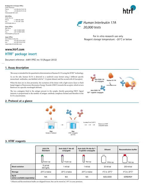
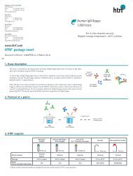
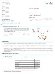
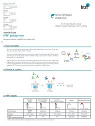
1. Assay description<br />
This assay is intended for the quantitative determination of human IL<strong>17A</strong> using the HTRF® technology.<br />
As on the side, human IL<strong>17A</strong> is detected in a sandwich assay format using 2 different specific<br />
monoclonal antibodies, one labelled with Eu 3+ -Cryptate (donor) and the second with d2 (acceptor).<br />
When the dyes are in close proximity, the excitation of the donor with a light source (laser or flash<br />
lamp) triggers a Fluorescence Resonance Energy Transfer (FRET) towards the acceptor, which in turn<br />
fluoresces at a specific wavelength (665nm).<br />
The two conjugates bind to the antigen present in the sample, thereby generating FRET. Signal<br />
intensity is proportional to the number of antigen–antibody complexes formed and therefore to the<br />
IL<strong>17A</strong> concentration.<br />
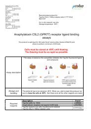
2. Protocol at a glance<br />
Anti-(h)IL<strong>17A</strong><br />
d2 conjugate<br />
Anti-(h)IL<strong>17A</strong><br />
EU 3+ cryptate conjugate<br />
<strong>Human</strong> IL<strong>17A</strong><br />
Incubate 5h at 18-25°C<br />
Read on an HTRF<br />
compatible reader<br />
10µL<br />
Sample or Calibrator<br />
10µL<br />
HTRF conjugates<br />
(pre-mix)<br />
3. HTRF reagents<br />
(h)IL<strong>17A</strong><br />
Standard<br />
Anti-(h)IL17 Ab-d2<br />
conjugate<br />
Anti-(h)IL<strong>17A</strong> Ab-Eu 3+ -<br />
Cryptate-conjugate<br />
Diluent<br />
Reconstitution buffer<br />
Green cap Purple cap Red cap White cap Red cap<br />
Stock solution<br />
10 µl/vial<br />
300 ng/mL<br />
1 ml/vial 1 ml/vial 20 ml/vial 200 ml/vial<br />
Storage -20°C or below -20°C or below -20°C or below 4°C to -20°C* 4°C to -20°C*<br />
Ref #<br />
(when available separately)<br />
N/A N/A N/A 62DL3DDD 62RB3RDF<br />
* Diluent and Reconstitution buffer are shipped frozen, but can be stored at 2-8°C in your premises.
4. Reagent preparation<br />
HTRF® reagent concentrations have been set for optimal assay performances. Note that any dilution or improper use of the d2 and Cryptate-conjugates will impair the assay<br />
quality.<br />
For an accurate quantitative determination of sample, dilution must be carried out with the medium used for preparing the samples (i.e. diluent, culture medium or any other<br />
compatible medium).<br />
Conjugates may be frozen and thawed once: to avoid freeze/thaw cycles it is recommended to dispense remaining stock solutions of conjugates into disposable plastic vials for<br />
storage at –20°C or below.<br />
Be careful, working solution preparation may differ between the 1,000 and the 20,000 data point kits.<br />
• Thaw all reagents at room temperature, allow them to warm up.<br />
• Prepare the working solutions from stock solutions (§3) by following the instructions below.<br />
4.1. Preparation of working solutions of conjugates<br />
Determine the amounts of conjugates needed for the experiment. Each well requires 5µL of each conjugates. In practice:<br />
Anti-(h)IL<strong>17A</strong>-d2 conjugate<br />
Anti- (h)IL<strong>17A</strong>-Eu 3+ -Cryptate-conjugate<br />
1 volume<br />
99 volumes 1 volume<br />
99 volumes<br />
Purple cap<br />
Red cap<br />
Red cap<br />
Red cap<br />
Prepare a 100X diluted solution using the reconstitution buffer: e.g. take 50 µL of conjugate<br />
stock solution and add it to 4950 µL of reconstitution buffer<br />
Prepare a 100X diluted solution using the reconstitution buffer: e.g. take 50 µL of conjugate<br />
stock solution and add it to 4950 µL of reconstitution buffer.<br />
4.2. Standard curve preparation<br />
Determine how many standard levels and replicates will be tested. Each well requires 10µl of standard.<br />
A recommended standard dilution procedure is listed and illustrated below.<br />
Standards Working concentration (pg/mL) Preparation<br />
Std 9 5000 200 µl working solution<br />
Std 8 2500 100 µl Std 9 + 100 µl diluent<br />
Std 7 1250 100 µl Std 8 + 100 µl diluent<br />
Std 6 625 100 µl Std 7 + 100 µl diluent<br />
Std 5 312.5 100 µl Std 6 + 100 µl diluent<br />
Std 4 156.25 100 µl Std 5 + 100 µl diluent<br />
Std 3 78.12 100 µl Std 4 + 100 µl diluent<br />
Std 2 39 100 µl Std 3 + 100 µl diluent<br />
Std 1 19.5 100 µl Std 2 + 100 µl diluent<br />
Std 0 0 100 µl diluent<br />
→ Dilute the standard stock solution 60-fold with diluent; this yields the high standard (Std 9: 5000 pg/mL) for the top of the curve.<br />
In practice:<br />
• e.g. take 5µL of standard stock solution and add it to 295 µL of diluent. Mix gently.<br />
→ Use the high standard (Std 9) to prepare the standard curve using 1/2 serial dilutions as follows:<br />
• Dispense 100µL of diluent in each vial from Std 8 to Std 0.<br />
• Add 100µL of standard to 100µL of diluent, mix gently and repeat the 1/2 serial dilution to make standard solutions: 2500, 1250, 625, 312.5, 156.25, 78.12, 39, 19.5 pg/<br />
mL. This will create 9 standards for the analyte. Std 0 (Positive control) is diluent alone.<br />
Step1: Dispense diluent in each vial<br />
100µL 100µL 100µL<br />
295µL<br />
100µL 100µL 100µL 100µL 100µL 100µL<br />
Step 2: Standards dilution<br />
5µL 100µL 100µL 100µL 100µL 100µL 100µL 100µL 100µL<br />
Diluent<br />
(white cap)<br />
Std 9 Std 8 Std 7 Std 6 Std 5 Std 4 Std 3 Std 2 Std 1<br />
Std 0<br />
Standard stock<br />
solution<br />
(green cap)<br />
Std 9<br />
Std 8 Std 7<br />
Std 6 Std 5 Std 4 Std 3 Std 2 Std 1
5. Assay protocol<br />
Dispense the reagents in the following order:<br />
10µL<br />
standard or sample<br />
5µL<br />
anti-(h)IL<strong>17A</strong>-d2 conjugate<br />
5µL<br />
anti-(h)IL<strong>17A</strong>-Eu 3+ cryptate conjugate<br />
It is possible to pre-mix the two conjugates just before dispensing and add 10 µl of this mix.<br />
→ Cover the plate with a plate sealer.<br />
→ Incubate at room temperature for 5 hours.<br />
→ Remove the plate sealer and,<br />
→ Read the fluorescence emission at two different wavelengths (665nm and 620nm) on a compatible HTRF® reader.<br />
For more information about HTRF compatible readers, please visit our website at: htrf.com/technology/htrfmeasurement/compatible_readers/<br />
Assay controls<br />
Negative control Cryptate control Buffer control Sample / Std<br />
Used to calculate the<br />
delta F%<br />
Used to check the Cryptate signal<br />
at 620 nm<br />
used to check background<br />
fluorescence<br />
Sample / Std - - - 10µL<br />
Diluent 10µL 10µL 10µL -<br />
Anti-(h)IL<strong>17A</strong>-d2 conjugate 5µL - - 5µL<br />
Anti-(h)IL<strong>17A</strong>-Eu 3+ -Cryptate conjugate 5µL 5µL - 5µL<br />
Reconstitution buffer - 5µL 10µL -<br />
6. Data reduction<br />
This data must not be substituted for that obtained in the laboratory and should be considered only as an example (readouts on PHERAstar plus ). Results may vary from one HTRF<br />
compatible reader to another.<br />
The assay standard curve is drawn up by plotting delta F% versus the analyte concentration, after an overnight incubation.<br />
Standard – pg/mL Ratio (1) CV % (2) Delta F % (3)<br />
Std 0 - Negative control 514 2 0<br />
Std 1 - 19.5 543 5 6<br />
Std 2 - 39 592 0 15<br />
Std 3 - 78.1 631 1 23<br />
Std 4 - 156.2 771 0 50<br />
Std 5 - 312.5 1000 2 95<br />
Std 6 - 625 1566 1 205<br />
Std 7 - 1250 2826 2 450<br />
Std 8 - 2500 5344 2 939<br />
Std 9 - 5000 11028 4 2045<br />
DeltaF%<br />
2500<br />
2000<br />
1500<br />
1000<br />
<strong>Human</strong> IL<strong>17A</strong> standard curve<br />
Incubation at RT<br />
5h incubation<br />
500<br />
Overnight incubation<br />
0<br />
0 1000 2000 3000 4000 5000 6000<br />
[<strong>Human</strong> IL17] pg/mL<br />
Ratio (1)<br />
Signal 665nm<br />
------------------- x 10 4<br />
Signal 620nm<br />
Ratio must be calculated for each individual well.<br />
CV% (2)<br />
Standard deviation<br />
-------------------------- x 100<br />
Mean ratio<br />
The mean and standard deviation can then be worked out from ratio replicates.<br />
DeltaF % (3)<br />
Ratio standard or sample<br />
– Ratio Negative control<br />
---------------------------------------------- x 100<br />
Ratio Negative control<br />
Reflects the signal to background of the assay. The negative control plays the role of an<br />
internal assay control.<br />
For more information about data reduction, please visit our website at: htrf.com/technology/htrfmeasurement/ratio_data_reduction/<br />
To obtain additional information or support, please contact your technical support team (htrfservices@cisbio.com).
7. Assay characteristics<br />
Detection limit (dose of mean zero + 3SD)<br />
Linear range study<br />
Incubation time<br />
In diluent<br />
28 pg/ml (ON)<br />
19.5 to 5000 pg/ml<br />
5H at RT<br />
Copyright © 2012 <strong>Cisbio</strong> <strong>Bioassays</strong>, France<br />
HTRF®, TRACE®, and the HTRF logo are trademarks belonging to <strong>Cisbio</strong> <strong>Bioassays</strong>.
![HTRF meeting_130426_villa_pour pdf [Mode de compatibilité]](https://img.yumpu.com/22345646/1/190x135/htrf-meeting-130426-villa-pour-pdf-mode-de-compatibilitac.jpg?quality=85)