Acaulospora rehmii and Gigaspora margarita, arbuscular ...
Acaulospora rehmii and Gigaspora margarita, arbuscular ...
Acaulospora rehmii and Gigaspora margarita, arbuscular ...

Create successful ePaper yourself
Turn your PDF publications into a flip-book with our unique Google optimized e-Paper software.
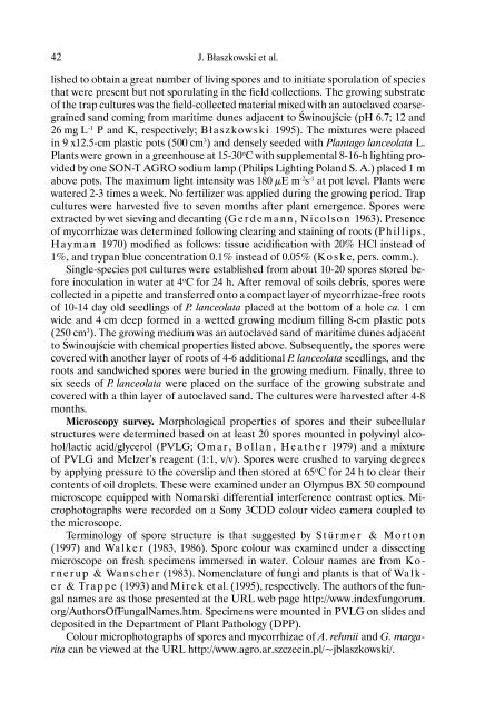
42 J. Błaszkowski et al.<br />
lished to obtain a great number of living spores <strong>and</strong> to initiate sporulation of species<br />
that were present but not sporulating in the field collections. The growing substrate<br />
of the trap cultures was the field-collected material mixed with an autoclaved coarsegrained<br />
s<strong>and</strong> coming from maritime dunes adjacent to Świnoujście (pH 6.7; 12 <strong>and</strong><br />
26 mg L -1 P <strong>and</strong> K, respectively; Błaszkowski 1995). The mixtures were placed<br />
in 9 x12.5-cm plastic pots (500 cm 3 ) <strong>and</strong> densely seeded with Plantago lanceolata L.<br />
Plants were grown in a greenhouse at 15-30 o C with supplemental 8-16-h lighting provided<br />
by one SON-T AGRO sodium lamp (Philips Lighting Pol<strong>and</strong> S. A.) placed 1 m<br />
above pots. The maximum light intensity was 180 μE m -2 s -1 at pot level. Plants were<br />
watered 2-3 times a week. No fertilizer was applied during the growing period. Trap<br />
cultures were harvested five to seven months after plant emergence. Spores were<br />
extracted by wet sieving <strong>and</strong> decanting (Gerdemann, Nicolson 1963). Presence<br />
of mycorrhizae was determined following clearing <strong>and</strong> staining of roots (Phillips,<br />
Hayman 1970) modified as follows: tissue acidification with 20% HCl instead of<br />
1%, <strong>and</strong> trypan blue concentration 0.1% instead of 0.05% (Koske, pers. comm.).<br />
Single-species pot cultures were established from about 10-20 spores stored before<br />
inoculation in water at 4 o C for 24 h. After removal of soils debris, spores were<br />
collected in a pipette <strong>and</strong> transferred onto a compact layer of mycorrhizae-free roots<br />
of 10-14 day old seedlings of P. lanceolata placed at the bottom of a hole ca. 1 cm<br />
wide <strong>and</strong> 4 cm deep formed in a wetted growing medium filling 8-cm plastic pots<br />
(250 cm 3 ). The growing medium was an autoclaved s<strong>and</strong> of maritime dunes adjacent<br />
to Świnoujście with chemical properties listed above. Subsequently, the spores were<br />
covered with another layer of roots of 4-6 additional P. lanceolata seedlings, <strong>and</strong> the<br />
roots <strong>and</strong> s<strong>and</strong>wiched spores were buried in the growing medium. Finally, three to<br />
six seeds of P. lanceolata were placed on the surface of the growing substrate <strong>and</strong><br />
covered with a thin layer of autoclaved s<strong>and</strong>. The cultures were harvested after 4-8<br />
months.<br />
Microscopy survey. Morphological properties of spores <strong>and</strong> their subcellular<br />
structures were determined based on at least 20 spores mounted in polyvinyl alcohol/lactic<br />
acid/glycerol (PVLG; Omar, Bollan, Heather 1979) <strong>and</strong> a mixture<br />
of PVLG <strong>and</strong> Melzer’s reagent (1:1, v/v). Spores were crushed to varying degrees<br />
by applying pressure to the coverslip <strong>and</strong> then stored at 65 o C for 24 h to clear their<br />
contents of oil droplets. These were examined under an Olympus BX 50 compound<br />
microscope equipped with Nomarski differential interference contrast optics. Microphotographs<br />
were recorded on a Sony 3CDD colour video camera coupled to<br />
the microscope.<br />
Terminology of spore structure is that suggested by Stürmer & Morton<br />
(1997) <strong>and</strong> Walker (1983, 1986). Spore colour was examined under a dissecting<br />
microscope on fresh specimens immersed in water. Colour names are from Kornerup<br />
& Wanscher (1983). Nomenclature of fungi <strong>and</strong> plants is that of Walker<br />
& Trappe (1993) <strong>and</strong> Mirek et al. (1995), respectively. The authors of the fungal<br />
names are as those presented at the URL web page http://www.indexfungorum.<br />
org/AuthorsOfFungalNames.htm. Specimens were mounted in PVLG on slides <strong>and</strong><br />
deposited in the Department of Plant Pathology (DPP).<br />
Colour microphotographs of spores <strong>and</strong> mycorrhizae of A. <strong>rehmii</strong> <strong>and</strong> G. <strong>margarita</strong><br />
can be viewed at the URL http://www.agro.ar.szczecin.pl/~jblaszkowski/.