Acaulospora rehmii and Gigaspora margarita, arbuscular ...
Acaulospora rehmii and Gigaspora margarita, arbuscular ...
Acaulospora rehmii and Gigaspora margarita, arbuscular ...
You also want an ePaper? Increase the reach of your titles
YUMPU automatically turns print PDFs into web optimized ePapers that Google loves.
ACTA MYCOLOGICA<br />
Vol. 41 (1): 41-48<br />
2006<br />
Dedicated to Professor Alina Skirgiełło<br />
on the occasion of her ninety-fifth birthday<br />
<strong>Acaulospora</strong> <strong>rehmii</strong> <strong>and</strong> <strong>Gigaspora</strong> <strong>margarita</strong>, <strong>arbuscular</strong> mycorrhizal<br />
fungi (Glomeromycota) new for Europe <strong>and</strong> Pol<strong>and</strong>, respectively<br />
JANUSZ BŁASZKOWSKI, SŁAWOMIR KOWALCZYK <strong>and</strong> BEATA CZERNIAWSKA<br />
Department of Plant Pathology, University of Agriculture<br />
Słowackiego 17, PL-71-434 Szczecin, jblaszkowski@agro.ar.szczecin.pl<br />
Błaszkowski J., Kowalczyk S., Czerniawska B.: <strong>Acaulospora</strong> <strong>rehmii</strong> <strong>and</strong><br />
<strong>Gigaspora</strong> <strong>margarita</strong>, <strong>arbuscular</strong> mycorrhizal fungi (Glomeromycota) new for Europe <strong>and</strong><br />
Pol<strong>and</strong>, respectively. Acta Mycol. 41 (1): 41-48, 2006.<br />
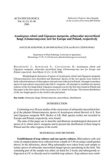
Morphological characters of spores of <strong>Acaulospora</strong> <strong>rehmii</strong> <strong>and</strong> <strong>Gigaspora</strong> <strong>margarita</strong><br />
(Glomeromycota) were described <strong>and</strong> illustrated. Spores of the two species were found in<br />
field-collected mixtures of rhizosphere soil <strong>and</strong> roots collected in Pol<strong>and</strong>. Attempts to produce<br />
spores in trap cultures succeeded only with G. <strong>margarita</strong>. All attempts to establish one-species<br />
cultures of the two fungi failed. <strong>Gigaspora</strong> <strong>margarita</strong> was for the first time found in Pol<strong>and</strong> <strong>and</strong><br />
this paper is the first report of the occurrence of A. <strong>rehmii</strong> in Europe. The known distribution<br />
of the two fungal species in the world is also presented.<br />
Key words: <strong>arbuscular</strong> fungi, Glomeromycota, mycorrhizae, distribution<br />
INTRODUCTION<br />
Continuing over 20-year studies of the occurrence of <strong>arbuscular</strong> mycorrhizal fungi<br />
of the phylum Glomeromycota, spores of <strong>Acaulospora</strong> <strong>rehmii</strong> Sieverd. et S. Toro<br />
<strong>and</strong> <strong>Gigaspora</strong> <strong>margarita</strong> W.N. Becker et I.R. Hall, species earlier not recorded in<br />
Europe <strong>and</strong> Pol<strong>and</strong>, respectively, were found.<br />
The aims of this paper are to describe <strong>and</strong> illustrate morphological characters of<br />
spores of A. <strong>rehmii</strong> <strong>and</strong> G. <strong>margarita</strong> <strong>and</strong> to present the distribution of these fungi in<br />
Pol<strong>and</strong> <strong>and</strong> the other regions of the world.<br />
MATERIALS AND METHODS<br />
Establishment of trap cultures <strong>and</strong> one-species cultures. Rhizosphere soils <strong>and</strong><br />
roots of sampled plants were collected from a depth of 5-30 cm using a small garden<br />
shovel. In the laboratory, about 100-g subsamples were taken from each sample to<br />
isolate spores of <strong>arbuscular</strong> mycorrhizal fungal species sporulating in the field. The<br />
remaining part of the sample was either air dried for 2 weeks <strong>and</strong> subsequently refrigerated<br />
at 4 o C or directly used to establish trap cultures. Trap cultures were estab-
42 J. Błaszkowski et al.<br />
lished to obtain a great number of living spores <strong>and</strong> to initiate sporulation of species<br />
that were present but not sporulating in the field collections. The growing substrate<br />
of the trap cultures was the field-collected material mixed with an autoclaved coarsegrained<br />
s<strong>and</strong> coming from maritime dunes adjacent to Świnoujście (pH 6.7; 12 <strong>and</strong><br />
26 mg L -1 P <strong>and</strong> K, respectively; Błaszkowski 1995). The mixtures were placed<br />
in 9 x12.5-cm plastic pots (500 cm 3 ) <strong>and</strong> densely seeded with Plantago lanceolata L.<br />
Plants were grown in a greenhouse at 15-30 o C with supplemental 8-16-h lighting provided<br />
by one SON-T AGRO sodium lamp (Philips Lighting Pol<strong>and</strong> S. A.) placed 1 m<br />
above pots. The maximum light intensity was 180 μE m -2 s -1 at pot level. Plants were<br />
watered 2-3 times a week. No fertilizer was applied during the growing period. Trap<br />
cultures were harvested five to seven months after plant emergence. Spores were<br />
extracted by wet sieving <strong>and</strong> decanting (Gerdemann, Nicolson 1963). Presence<br />
of mycorrhizae was determined following clearing <strong>and</strong> staining of roots (Phillips,<br />
Hayman 1970) modified as follows: tissue acidification with 20% HCl instead of<br />
1%, <strong>and</strong> trypan blue concentration 0.1% instead of 0.05% (Koske, pers. comm.).<br />
Single-species pot cultures were established from about 10-20 spores stored before<br />
inoculation in water at 4 o C for 24 h. After removal of soils debris, spores were<br />
collected in a pipette <strong>and</strong> transferred onto a compact layer of mycorrhizae-free roots<br />
of 10-14 day old seedlings of P. lanceolata placed at the bottom of a hole ca. 1 cm<br />
wide <strong>and</strong> 4 cm deep formed in a wetted growing medium filling 8-cm plastic pots<br />
(250 cm 3 ). The growing medium was an autoclaved s<strong>and</strong> of maritime dunes adjacent<br />
to Świnoujście with chemical properties listed above. Subsequently, the spores were<br />
covered with another layer of roots of 4-6 additional P. lanceolata seedlings, <strong>and</strong> the<br />
roots <strong>and</strong> s<strong>and</strong>wiched spores were buried in the growing medium. Finally, three to<br />
six seeds of P. lanceolata were placed on the surface of the growing substrate <strong>and</strong><br />
covered with a thin layer of autoclaved s<strong>and</strong>. The cultures were harvested after 4-8<br />
months.<br />
Microscopy survey. Morphological properties of spores <strong>and</strong> their subcellular<br />
structures were determined based on at least 20 spores mounted in polyvinyl alcohol/lactic<br />
acid/glycerol (PVLG; Omar, Bollan, Heather 1979) <strong>and</strong> a mixture<br />
of PVLG <strong>and</strong> Melzer’s reagent (1:1, v/v). Spores were crushed to varying degrees<br />
by applying pressure to the coverslip <strong>and</strong> then stored at 65 o C for 24 h to clear their<br />
contents of oil droplets. These were examined under an Olympus BX 50 compound<br />
microscope equipped with Nomarski differential interference contrast optics. Microphotographs<br />
were recorded on a Sony 3CDD colour video camera coupled to<br />
the microscope.<br />
Terminology of spore structure is that suggested by Stürmer & Morton<br />
(1997) <strong>and</strong> Walker (1983, 1986). Spore colour was examined under a dissecting<br />
microscope on fresh specimens immersed in water. Colour names are from Kornerup<br />
& Wanscher (1983). Nomenclature of fungi <strong>and</strong> plants is that of Walker<br />
& Trappe (1993) <strong>and</strong> Mirek et al. (1995), respectively. The authors of the fungal<br />
names are as those presented at the URL web page http://www.indexfungorum.<br />
org/AuthorsOfFungalNames.htm. Specimens were mounted in PVLG on slides <strong>and</strong><br />
deposited in the Department of Plant Pathology (DPP).<br />
Colour microphotographs of spores <strong>and</strong> mycorrhizae of A. <strong>rehmii</strong> <strong>and</strong> G. <strong>margarita</strong><br />
can be viewed at the URL http://www.agro.ar.szczecin.pl/~jblaszkowski/.
<strong>Acaulospora</strong> <strong>rehmii</strong> <strong>and</strong> <strong>Gigaspora</strong> <strong>margarita</strong> 43<br />
DESCRIPTIONS OF THE SPECIES<br />
<strong>Acaulospora</strong> <strong>rehmii</strong> Sieverd. et S. Toro<br />
Spores single in the soil; develop laterally on the neck of a sporiferous saccule.<br />
Spores dull yellow (3B3) to Nankeen yellow (3B7); globose to subglobose;<br />
(90-)140(-165) μm diam. Subcellular structure of spores consists of a spore wall <strong>and</strong><br />
two inner germination walls (Fig. 1). Spore wall composed of three layers (layers<br />
1-3). Layer 1 evanescent, hyaline, (0.8-)1.0(-1.2) μm thick (Fig. 2), continuous with<br />
the wall of a sporiferous saccule, rarely present in mature spores. Layer 2 laminate,<br />
dull yellow (3B3) to Nankeen yellow (3B7), (4.5-)10.0(-12.0) μm thick, ornamented<br />
with labyrinthine folds, 1.0-3.5 μm wide <strong>and</strong> 1.0-4.5 μm high when seen in a<br />
plane view <strong>and</strong> a cross view, respectively (Figs 1-3). Layer 3 semi-flexible, hyaline,<br />
(0.5-)0.7(-0.8) μm thick, rarely separable from layer 2 (Fig. 2). Germination wall<br />
1 consists of two tightly adherent hyaline, semi-flexible layers, each 0.5-0.8 μm thick;<br />
these layers are tightly adherent to each other <strong>and</strong>, hence, difficult to see (Figs 1, 2,<br />
4, 5). Germination wall 2 composed of two adherent layers (layers 1 <strong>and</strong> 2; Figs 1 <strong>and</strong><br />
2). Layer 1 flexible, hyaline, (0.5-)0.7(-1.0) μm thick, covered with small,
44 J. Błaszkowski et al.<br />
A. <strong>rehmii</strong>, those of A. foveata are much greater [(240-)289(-300) vs. (82-)141(-175)<br />
μm diam in A. <strong>rehmii</strong>] <strong>and</strong> darker-coloured (red-orange to dark red-brown vs. light<br />
yellow to orange-brown), <strong>and</strong> spores of A. paulinae, A. scrobiculata, <strong>and</strong> A. undulata<br />
are lighter (hyaline to straw-coloured; Błaszkowski 2003; Morton 2002;<br />
Sieverding, Toro 1987). Additionally, spores of the latter two species are much<br />
smaller (55-92 μm diam; Błaszkowski 2003; Sieverding 1988). Finally, spores<br />
of all the species compared here are ornamented with pits, <strong>and</strong> not with labyrinthine<br />
processes as those of A. <strong>rehmii</strong>.<br />
Another important difference between A. <strong>rehmii</strong> <strong>and</strong> A. bireticulata, A. foveata,<br />
<strong>and</strong> A. tuberculata resides in the inner layer of the second germination wall of their<br />
spores. While this layer is thin <strong>and</strong> flexible in A. <strong>rehmii</strong> (Figs. 4 <strong>and</strong> 5), that of A.<br />
bireticulata, A. foveata, <strong>and</strong> A. tuberculata is much thicker <strong>and</strong> corresponds with the<br />
coriaceous wall sensu Walker (1986).<br />
Collections examined. Pol<strong>and</strong>. All from Władysławowo (54º47’N, 18º26’E), in the<br />
root zone of Melilotus officinalis (L.) Pall., 20 Sept. 1996, Błaszkowski J. 2656-2660<br />
(DPP); under P. lanceolata, 20 Sept. 1996, Błaszkowski J. 2661-2663 (DPP); from<br />
among roots of Trifolium repens L., 20 Sept. 1996, Błaszkowski J. 2664-2669 (DPP).<br />
Distribution <strong>and</strong> habitat. In Pol<strong>and</strong>, spores of A. <strong>rehmii</strong> were found in three<br />
field-collected samples of roots <strong>and</strong> rhizosphere soils taken under M. officinalis, P.<br />
lanceolata, <strong>and</strong> T. repens. All the plant species grew in a salt pan near Władysławowo<br />
at the beginning of the Hel Peninsula.<br />
<strong>Acaulospora</strong> <strong>rehmii</strong> has originally been isolated from a crop field at Caicedonia,<br />
Valle de Cauca, Colombia (Sieverding, Toro 1987). This fungus has been associated<br />
with Manihot esculentum, Phaseolus, Sorghum <strong>and</strong> Crotalaria species. Additionally,<br />
Sieverding <strong>and</strong> Toro (1987) found spores of A. <strong>rehmii</strong> in a culture<br />
containing a soil sample coming from the Centro de Pesquisa Agropecuária dos Cerrados,<br />
Brasilia D. F., Brazil. Recently, Moreira-Souza et al. (2003) confirmed the<br />
presence of this fungus in Brazil; A. <strong>rehmii</strong> spores were encountered among roots of<br />
Araucaria angustifolia (Bert.) O. Ktze growing in a reforested area in the State Park<br />
of Campos do Jordão, San Paulo. Wu & L iu (1995) isolated spores of A. <strong>rehmii</strong><br />
from among roots of Phyllostachys pubescens Mazel growing in a garden in Chi-tou<br />
Experimental Station, National Taiwan University. Thus, this paper for the first time<br />
informs of the occurrence of A. <strong>rehmii</strong> in Europe.<br />
Mycorrhizal associations. In Pol<strong>and</strong>, A. <strong>rehmii</strong> has been associated with vesicular-<strong>arbuscular</strong><br />
mycorrhizal roots of P. lanceolata. Many attempts to initiate sporulation<br />
of this fungus in both trap <strong>and</strong> one-species cultures failed. According to Sieverdin<br />
g & Toro (1987), A. <strong>rehmii</strong> produced vesicular-<strong>arbuscular</strong> mycorrhizae.<br />
<strong>Gigaspora</strong> <strong>margarita</strong> W.N. Becker et I.R. Hall<br />
Spores produced singly in the soil, blastically at the tip of a bulbous sporogenous<br />
cell (Fig. 7). Spores yellowish white (4A2) to sunflower yellow (4A7); globose to subglobose;<br />
(260-)357(-405) μm diam; sometimes ovoid; 300-340 x 360-380 μm (Fig. 7).<br />
Subcellular structure of spores consists of a spore wall comprising two layers (layers<br />
1-3; Figs 8 <strong>and</strong> 9) <strong>and</strong> a one-layered germination wall. Spore wall layer 1, forming<br />
the spore surface, permanent, smooth, hyaline, (1.7-)1.9(-2.2) μm thick (Figs. 8 <strong>and</strong><br />
9), sometimes separating from layer 2 in crushed spores. Layer 2 of spore wall laminate,<br />
smooth, yellowish white (4A2) to sunflower yellow (4A7), (19.4-)23.4(-26.2) μm
<strong>Acaulospora</strong> <strong>rehmii</strong> <strong>and</strong> <strong>Gigaspora</strong> <strong>margarita</strong> 45<br />
thick, consisting of a variable number of laminae, each (1.2-)1.5(-1.7) μm thick,<br />
usually separating from each other in crushed spores (Figs 8 <strong>and</strong> 9). Germination<br />
wall permanent, flexible to semi-flexible, concolorous with spore wall layer 2,<br />
(1.5-)1.6(-1.8) μm thick (Figs 8 <strong>and</strong> 9), covered with processes on its under surface<br />
(Fig. 10); processes 3.7-7.6 x 2.6-3.9 μm, spaced 1.7-13.5 μm from each other; forming<br />
prior to germination. In Melzer’s reagent, only spore wall layer 2 stains deep red<br />
(11C8; Fig. 11). Sporogenous cell orange (5B8) to brownish yellow (5C8), clavate;<br />
52.5-60.0 x 75.0-100.0 μm (Figs 7, 11, 12). Wall of sporogenous cell composed of<br />
two layers (Figs 11 <strong>and</strong> 12). Layer 1 hyaline, (1.0-)1.7(-2.0) μm thick, continuous<br />
with spore wall layer 1 (Fig. 11). Layer 2 orange (5B8) to brownish yellow (5C8),<br />
(4.7-)5.6(-6.4) μm thick, continuous with spore wall layer 2 (Fig. 11).<br />
Of the eight described species of the genus <strong>Gigaspora</strong>, Bentivenga & Morton<br />
(1995) accepted five. <strong>Gigaspora</strong> c<strong>and</strong>ida Bhattacharjee et al., G. ramisporophora<br />
Spain, Sieverd. <strong>and</strong> N.C. Schenck, <strong>and</strong> G. tuberculata Neeraj et al. were considered<br />
congeneric with G. rosea Nicol. et N.C. Schenck, G. <strong>margarita</strong>, <strong>and</strong> Scutellospora<br />
persica (Koske et C. Walker) C. Walker et F.E. S<strong>and</strong>ers, respectively.<br />
The most distinctive characters of G. <strong>margarita</strong> are its yellow-coloured spores<br />
(Fig. 7), whose colour results from the pigmentation of the spore wall [is not associated<br />
with spore contents as in G. gigantea (Nicol. et Gerd.) Gerd. et Trappe], <strong>and</strong><br />
their relatively thin, usually less than 25 μm thick, wall.<br />
The only other species of <strong>Gigaspora</strong> forming spores similar in size <strong>and</strong> colour to<br />
those of G. <strong>margarita</strong> is G. decipiens I.R. Hall et L.K. Abbott. The character separating<br />
the two fungi is the much thicker [(15-)20-45(-90) μm; B e ntivenga, Morton<br />
(1995)] spore wall of the former species.<br />
All attempts to establish one-species cultures of G. <strong>margarita</strong> made by the authors<br />
of this paper failed. Hence, the ontogenetic development <strong>and</strong> the differentiation of<br />
subcellular structures of spores of this fungus were not recognized. According to<br />
Bentivenga & Morton (1995), spores of G. <strong>margarita</strong> originate blastically from<br />
the top of a bulbous sporogenous cell <strong>and</strong> their wall consists of two, thin layers of<br />
near-equal thickness. At first, the spores are white. With time, the inner spore wall<br />
layer thickens <strong>and</strong> the spores darken because of the synthetization of new sublayers<br />
from the spore contents. The ontogenetic development of spores ends the formation<br />
of a germination wall ornamented with processes prior to germination. The germ<br />
tube penetrates through the spore wall.<br />
Bentivenga & Morton (1995) suggested that the two wall layers of spores<br />
of G. <strong>margarita</strong> <strong>and</strong> all the other <strong>Gigaspora</strong> spp. originating at the same time indicate<br />
them to be interdependent elements of one spore wall. A similar spore wall structure<br />
occurs in juvenile spores of members of the genus Scutellospora. This suggests<br />
that <strong>Gigaspora</strong> is ancestral to Scutellospora, the only other member of the family<br />
<strong>Gigaspora</strong>ceae, although molecular analyses of ribosomal DNA did not confirm it<br />
(Simon et al. 1993).<br />
Compared with fungi of all the other genera of the phylum Glomeromycota,<br />
species of <strong>Gigaspora</strong> distinguish the lowest morphological variability of their spores.<br />
Spores of all <strong>Gigaspora</strong> spp. are smooth <strong>and</strong> the characters separating them are only<br />
colour <strong>and</strong> size of spores, as well as thickness of their wall. Except for G. gigantea,<br />
whose colour of spores results from the pigmentation of their contents, the spore<br />
colour of the other species comes from their wall. B e n tivenga & Morton (1995)
46 J. Błaszkowski et al.<br />
hypothesized that such low amount of diversity in <strong>Gigaspora</strong> results from two reasons.<br />
Firstly, germination of <strong>Gigaspora</strong> spp. is associated with spore wall <strong>and</strong> the<br />
energy remained after completion of this vital process probably is too low to aid<br />
further differentiation of this wall. In Scutellospora spp., germination is taken over<br />
by the germination shield, which enables the spore wall to unconstrainedly express<br />
variation. Secondly, the low level of variability in <strong>Gigaspora</strong> spp. may result from<br />
their recent separation <strong>and</strong> the lack of time to further diverge morphologically.<br />
Collections examined. Pol<strong>and</strong>. Szczecin (53º26’N, 14º35’E), under Taxus baccata<br />
L., 2 May 2001, Błaszkowski J. 2670-2672 (DPP); Dąbrówka Wielkopolska (15 o 49’E,<br />
52 o 17’N), from under Rumex acetosella L., 22 July 2003, Błaszkowski J. 2673 (DPP);<br />
Chociszewo (15 o 45’E, 52 o 19’N), among roots of Achillea millefolium L., 22 July 2003,<br />
Błaszkowski J. 2674 (DPP); <strong>and</strong> Żydowo (15 o 45’E, 52 o 24’N), in the rhizosphere of<br />
Polygonum persicaria L., 3 Aug. 2003, Błaszkowski J. 2675 (DPP).<br />
Distribution <strong>and</strong> habitat. In Pol<strong>and</strong>, G. <strong>margarita</strong> was found in four mixtures<br />
of roots <strong>and</strong> rhizosphere soils, of which three were collected in the Lubuskie province<br />
<strong>and</strong> one in the Western Pomerania province. All the mixtures represented wild<br />
plants. Of them, only A. millefolium <strong>and</strong> P. persicaria hosted spores of G. <strong>margarita</strong><br />
in the field. However, the associations of G. <strong>margarita</strong> with roots of P. persicaria <strong>and</strong><br />
T. baccata were revealed only after the cultivation of their field-collected roots <strong>and</strong><br />
rhizosphere soils in trap cultures. The sporulation of G. <strong>margarita</strong> in both the field<br />
<strong>and</strong> trap cultures was low. Among the 109 spores of <strong>arbuscular</strong> fungi isolated from<br />
100 g of dry field soil taken under A. millefolium, only one (0.9%) belonged to G.<br />
<strong>margarita</strong>. Polygonum persicaria growing in the field hosted a total of 77 spores in 100<br />
g of dry soil, including two (2.6%) of G. <strong>margarita</strong>.<br />
In trap cultures with Z. mays as the plant host <strong>and</strong> mixtures of roots <strong>and</strong> rhizosphere<br />
soils of R. acetosella <strong>and</strong> T. baccata, the densities of G. <strong>margarita</strong> spores were<br />
2 <strong>and</strong> 9 in 100 g of dry soil, respectively, <strong>and</strong> G. <strong>margarita</strong> was the only <strong>arbuscular</strong><br />
fungus revealed.<br />
In the field, other species of <strong>arbuscular</strong> fungi associated with roots of A. millefolium<br />
were Glomus aggregatum N.C. Schenck et S.M. Sm. emend. Koske, Gl. constrictum<br />
Trappe <strong>and</strong> Scutellospora dipurpurescens J.B. Morton et Koske, <strong>and</strong> Gl. constrictum<br />
<strong>and</strong> Gl. deserticola Trappe, Bloss et J.A. Menge occurred among roots of P.<br />
persicaria.<br />
<strong>Gigaspora</strong> <strong>margarita</strong> probably has a worldwide distribution. The holotype of this<br />
fungus has been selected from spores recovered from under Glycine max (L.) Merr.<br />
growing in a pot culture containing a root x rhizosphere soil mixture of G. max cultivated<br />
at the Agronomy South Farm of University of Illinois, USA (Becker, Hall<br />
1976). Apart from many other reports of the occurrence of <strong>Gigaspora</strong> <strong>margarita</strong> in<br />
both cultivated (agricultural soils, orchards, nurseries) <strong>and</strong> natural sites (maritime<br />
s<strong>and</strong> dunes, native woodl<strong>and</strong>, prairie) of the US (An et al. 1993; Hetrick, Bloom<br />
1983; K oske, Gemma 1997; Menge et al. 1977; M iller, Domoto, Walker<br />
1985; Nicolson, Schenck 1979; Rose 1988; Schenck, Kinloch 1980;<br />
Schenck, Smith 1981), this species has also been found associated with different<br />
cultivated <strong>and</strong> uncultivated plants of Japan (S aito, Vargas 1991), New Zeal<strong>and</strong><br />
(Hall 1977), China (Wu et al. 2002), Canada (Marcel et al. 1988; Hamel et al.<br />
1994), Mexico (Estrada-Torres et al. 1992), Cuba (Ferrer, Herrera 1980),<br />
<strong>and</strong> South America (Moreira-Souza et al. 2003; Sieverding 1989).
<strong>Acaulospora</strong> <strong>rehmii</strong> <strong>and</strong> <strong>Gigaspora</strong> <strong>margarita</strong> 47<br />
REFERENCES<br />
A n Z.-Q, Hendrix J. W., Hershamn D. E., Ferriss R. S., Henson G. T. 1993. The influence<br />
of crop rotation <strong>and</strong> soil fumigation on a mycorrhizal fungal community associated with soybean.<br />
Mycorrhiza 3: 171–182.<br />
Becker W. N., Hall I. R. 1976. <strong>Gigaspora</strong> <strong>margarita</strong>, a new species in the Endogonaceae. Mycotaxon<br />
4: 155–160.<br />
Bentivenga S. P., Morton J. B. 1995. A monograph of the genus <strong>Gigaspora</strong>, incorporating developmental<br />
patterns of morphological characters. Mycologia 87: 719–731.<br />
Błaszkowski J. 1995. Glomus corymbiforme, a new species in Glomales from Pol<strong>and</strong>. Mycologia 87:<br />
732–737.<br />
Błaszkowski J. 2003. Arbuscular mycorrhizal fungi (Glomeromycota), Endogone, <strong>and</strong> Complexipes<br />
species deposited in the Department of Plant Pathology, University of Agriculture in Szczecin, Pol<strong>and</strong>.<br />
http://www.agro.ar.szczecin.pl/~jblaszkowski/.<br />
Estrada-Torres A., Varela L., H e r n<strong>and</strong>ez-Cuevas L., G a v i t o M. E. 1992. Algunos hongos<br />
micorrizicos <strong>arbuscular</strong>es del estado de Tlaxcala, México. Rev. Mex. Mic. 8: 85–110.<br />
Fe r r e r R. L., Herrera R. A. 1980. El genero <strong>Gigaspora</strong> Gerdemann et Trappe (Endogonaceae) en<br />
Cuba. Rev. Jar. Bot. Nac. Habana 1: 43–66.<br />
Gerdemann J. W., Nicolson T. H. 1963. Spores of mycorrhizal Endogone species extracted from soil<br />
by wet sieving <strong>and</strong> decanting. Trans. Brit. Mycol. Soc. 46: 235–244.<br />
H a l l I. R. 1977. Species <strong>and</strong> mycorrhizal infections of New Zeal<strong>and</strong> Endogonaceae. Trans. Br. Mycol.<br />
Soc. 68: 341–356.<br />
H a m e l C., D a l p e Y., L a pierre C., S i m a r d R. R., Smith D. L. 1994. Composition of the vesicular-<strong>arbuscular</strong><br />
mycorrhizal fungi population in an old meadow as affected by pH, phosphorous <strong>and</strong><br />
soil disturbance. Agric. Ecosyst. Environ. 49: 223–231.<br />
Hetrick B. A. D., Bloom J. 1983. Vesicular-<strong>arbuscular</strong> mycorrhizal fungi associated with native tall<br />
grass prairie <strong>and</strong> cultivated winter wheat. Can. J. Bot. 61: 2140–2146.<br />
Janos D. P., Trappe J. M. 1982. Two new <strong>Acaulospora</strong> species from tropical America. Mycotaxon 15:<br />
515–522.<br />
Kornerup A., Wanscher J. H. 1983. Methuen h<strong>and</strong>book of colour. 3rd Ed. E. Methuen <strong>and</strong> Co.,<br />
Ltd., London. 252 pp.<br />
Koske R. E., Gemma J. N. 1997. Mycorrhizae <strong>and</strong> succession in plantings of beachgrass in s<strong>and</strong> dunes.<br />
Am. J. Bot. 84: 118–130.<br />
Marcel G. A., v a n d e r Heiden M. G. A., K lironomos J. N., U rsic M., Moutoglis P., Streitwolf-Engel<br />
R., Boller T., Wiemken A., S a n d e r s I. R. 1988. Mycorrhizal fungal diversity<br />
determines plant biodiversity, ecosystem variability <strong>and</strong> productivity. Nature 396: 69–72.<br />
Menge J. A., Nemec S., Davis R. M., Minassian V. 1977. Mycorrhizal fungi associated with citrus<br />
<strong>and</strong> their possible interactions with pathogens. Proc. Int. Soc. Citriculture 3: 872–876.<br />
Miller D. D., Domoto P., Walker C. 1985. Mycorrhizal fungi at eighteen apple rootstock plantings<br />
in the United States. New Phytol. 100: 379–391.<br />
Mirek Z., Piękoś-Mirkowa H., Zając A., Z ając M. 1995. Vascular plants of Pol<strong>and</strong>. A Checklist.<br />
Polish Botanical Studies, Guidebook 15, Kraków. 303 pp.<br />
Moreira-Souza M., Truem S. F. B., Gomes-da-Costa S. M. Cardoso E. J. B. N. 2003. Arbuscular<br />
mycorrhizal fungi associated with Araucaria angustifolia (Bert.) O. Ktze. Mycorrhiza 13:<br />
211–215.<br />
Morton J. B. 1986. Three new species of <strong>Acaulospora</strong> (Endogonaceae) from high aluminum, low pH<br />
soils in West Virginia. Mycologia 78: 641–648.<br />
Morton J. B. 2002. International Culture Collection of Vesicular) Arbuscular Mycorrhizal Fungi. West<br />
Virginia University: http://www.invam.caf.wvu.edu/.<br />
Nicolson T. H., Schenck N. C. 1979. Endogonaceous mycorrhizal endophytes in Florida. Mycologia<br />
71: 178–198.<br />
Omar M. B., Bollan L., Heather W. A. 1979. A permanent mounting medium for fungi. Bull. Brit.<br />
Mycol. Soc. 13: 31–32.<br />
Phillips J. M., Hayman D. S. 1970. Improved procedures for clearing roots <strong>and</strong> staining parasitic <strong>and</strong><br />
vesicular-<strong>arbuscular</strong> mycorrhizal fungi for rapid assessment of infection Trans. Brit. Mycol. Soc. 55:<br />
158–161.
48 J. Błaszkowski et al.<br />
Ro s e S. 1988. Above <strong>and</strong> belowground community development in a maritime s<strong>and</strong> dune ecosystem.<br />
Plant <strong>and</strong> Soil 109: 215–226.<br />
Rothwell F. M., Tr appe J. M. 1979. <strong>Acaulospora</strong> bireticulata sp. nov. Mycotaxon 8: 471–475.<br />
Saito M., Vargas R. 1991. Vesicular-<strong>arbuscular</strong> mycorrhizal fungi in some humus-rich Ando soils of<br />
Japan. Soil Microorg. 38: 3–15.<br />
Schenck N. C., Kinloch R. A. 1980. Incidence of mycorrhizal fungi on six field crops in monoculture<br />
on a newly cleared woodl<strong>and</strong> site. Mycologia 72: 445–456.<br />
Schenck N. C., S mith G. S. 1981. Distribution <strong>and</strong> occurrence of vesicular-<strong>arbuscular</strong> mycorrhizal<br />
fungi on Florida agricultural crops. Soil <strong>and</strong> Crop Sci. Soc. Florida 40: 171–175.<br />
Sieverding E. 1988. Two new species of vesicular <strong>arbuscular</strong> mycorrhizal fungi in the Endogonaceae<br />
from tropical high l<strong>and</strong>s of Africa. Angew. Bot. 62: 373–380.<br />
Sieverding E. 1989. Ecology of VAM fungi in tropical ecosystems. Agric., Ecosyst. <strong>and</strong> Environ. 29:<br />
369–390.<br />
Sieverding E., Toro S. T. 1987. <strong>Acaulospora</strong> dendiculata sp. nov. <strong>and</strong> <strong>Acaulospora</strong> <strong>rehmii</strong> sp. nov.<br />
(Endogonaceae) with ornamented spore walls. Angew. Bot. 61: 217–223.<br />
Simon L., Bousquet T., Lévesque R. C., L alonde M. 1993. Origin <strong>and</strong> diversification of endomycorrhizal<br />
fungi <strong>and</strong> coincidence with vascular l<strong>and</strong> plants. Nature 363: 67–69.<br />
Stürmer S. L., Morton J. B. 1997. Developmental patterns defining morphological characters in<br />
spores of four species in Glomus. Mycologia 89: 72–81.<br />
Walker C. 1983. Taxonomic concepts in the Endogonaceae: Spore wall characteristics in species descriptions.<br />
Mycotaxon 18: 443–455.<br />
Walker C. 1986. Taxonomic concepts in the Endogonaceae. II. A fifth morphological wall type in endogonaceous<br />
spores. Mycotaxon 25: 95–99.<br />
Walker C., Trappe J. M. 1981. <strong>Acaulospora</strong> spinosa sp. nov. with a key to the species of <strong>Acaulospora</strong>.<br />
Mycotaxon 12: 515–521.<br />
Walker C., Trappe J. M. 1993. Names <strong>and</strong> epithets in the Glomales <strong>and</strong> Endogonales. Mycol. Res.<br />
97: 339–344.<br />
Wu T., H a o W., L i n X., S h i Y. 2002. Screening of <strong>arbuscular</strong> mycorrhizal fungi for the revegetation of<br />
eroded red soils in subtropical China. Plant <strong>and</strong> Soil 239: 225–235.<br />
Wu C.-G., L i u Y.-S., H wuang Y.-L., Wang Y.-P., Chao C.-C. 1995. Glomales of Taiwan: V. Glomus<br />
chimonobambusae <strong>and</strong> Entrophospora kentinensis, spp. nov. Mycotaxon 53: 283–294.<br />
<strong>Acaulospora</strong> <strong>rehmii</strong> i <strong>Gigaspora</strong> <strong>margarita</strong>, arbuskularne grzyby mikoryzowe<br />
(Glomeromycota) nowe odpowiednio dla Europy i Polski<br />
Streszczenie<br />
Opisano i zilustrowano cechy morfologiczne zarodników <strong>Acaulospora</strong> <strong>rehmii</strong> i <strong>Gigaspora</strong><br />
<strong>margarita</strong>, arbuskularnych grzybów mikoryzowych z gromady Glomeromycota. Zarodniki obu<br />
tych gatunków znaleziono w polowych mieszaninach gleby ryzosferowej i korzeni zebranych<br />
w Polsce. Próby otrzymania zarodników tych grzybów w kulturach pułapkowych powiodły się<br />
tylko z G. <strong>margarita</strong>. Wszystkie próby ustanowienia jednogatunkowych kultur obu grzybów nie<br />
udały się. <strong>Gigaspora</strong> <strong>margarita</strong> znaleziono po raz pierwszy w Polsce, a A. <strong>rehmii</strong> po raz pierwszy<br />
w Europie. Przedstawiono również poznane rozmieszczenie obu gatunków w świecie.


