OMPX Mathilde Lethier , Martine Moulin , Michae - Institut de ...
OMPX Mathilde Lethier , Martine Moulin , Michae - Institut de ...
OMPX Mathilde Lethier , Martine Moulin , Michae - Institut de ...

You also want an ePaper? Increase the reach of your titles
YUMPU automatically turns print PDFs into web optimized ePapers that Google loves.
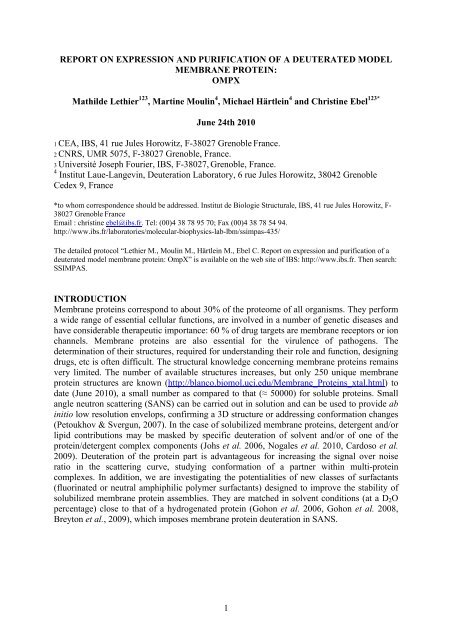
REPORT ON EXPRESSION AND PURIFICATION OF A DEUTERATED MODEL<br />
MEMBRANE PROTEIN:<br />
<strong>OMPX</strong><br />
<strong>Mathil<strong>de</strong></strong> <strong>Lethier</strong> 123 , <strong>Martine</strong> <strong>Moulin</strong> 4 , <strong>Michae</strong>l Härtlein 4 and Christine Ebel 123*<br />
June 24th 2010<br />
1 CEA, IBS, 41 rue Jules Horowitz, F-38027 Grenoble France.<br />
2 CNRS, UMR 5075, F-38027 Grenoble, France.<br />
3 Université Joseph Fourier, IBS, F-38027, Grenoble, France.<br />
4 <strong>Institut</strong> Laue-Langevin, Deuteration Laboratory, 6 rue Jules Horowitz, 38042 Grenoble<br />
Ce<strong>de</strong>x 9, France<br />
*to whom correspon<strong>de</strong>nce should be addressed. <strong>Institut</strong> <strong>de</strong> Biologie Structurale, IBS, 41 rue Jules Horowitz, F-<br />
38027 Grenoble France<br />
Email : christine ebel@ibs.fr. Tel: (00)4 38 78 95 70; Fax (00)4 38 78 54 94.<br />
http://www.ibs.fr/laboratories/molecular-biophysics-lab-lbm/ssimpas-435/<br />
The <strong>de</strong>tailed protocol “<strong>Lethier</strong> M., <strong>Moulin</strong> M., Härtlein M., Ebel C. Report on expression and purification of a<br />
<strong>de</strong>uterated mo<strong>de</strong>l membrane protein: OmpX” is available on the web site of IBS: http://www.ibs.fr. Then search:<br />
SSIMPAS.<br />
INTRODUCTION<br />
Membrane proteins correspond to about 30% of the proteome of all organisms. They perform<br />
a wi<strong>de</strong> range of essential cellular functions, are involved in a number of genetic diseases and<br />
have consi<strong>de</strong>rable therapeutic importance: 60 % of drug targets are membrane receptors or ion<br />
channels. Membrane proteins are also essential for the virulence of pathogens. The<br />
<strong>de</strong>termination of their structures, required for un<strong>de</strong>rstanding their role and function, <strong>de</strong>signing<br />
drugs, etc is often difficult. The structural knowledge concerning membrane proteins remains<br />
very limited. The number of available structures increases, but only 250 unique membrane<br />
protein structures are known (http://blanco.biomol.uci.edu/Membrane_Proteins_xtal.html) to<br />
date (June 2010), a small number as compared to that (≈ 50000) for soluble proteins. Small<br />
angle neutron scattering (SANS) can be carried out in solution and can be used to provi<strong>de</strong> ab<br />
initio low resolution envelops, confirming a 3D structure or addressing conformation changes<br />
(Petoukhov & Svergun, 2007). In the case of solubilized membrane proteins, <strong>de</strong>tergent and/or<br />
lipid contributions may be masked by specific <strong>de</strong>uteration of solvent and/or of one of the<br />
protein/<strong>de</strong>tergent complex components (Johs et al. 2006, Nogales et al. 2010, Cardoso et al.<br />
2009). Deuteration of the protein part is advantageous for increasing the signal over noise<br />
ratio in the scattering curve, studying conformation of a partner within multi-protein<br />
complexes. In addition, we are investigating the potentialities of new classes of surfactants<br />
(fluorinated or neutral amphiphilic polymer surfactants) <strong>de</strong>signed to improve the stability of<br />
solubilized membrane protein assemblies. They are matched in solvent conditions (at a D 2 O<br />
percentage) close to that of a hydrogenated protein (Gohon et al. 2006, Gohon et al. 2008,<br />
Breyton et al., 2009), which imposes membrane protein <strong>de</strong>uteration in SANS.<br />
1
RESULTS<br />
We <strong>de</strong>scribe here a protocol for obtaining <strong>de</strong>uterated at OmpX in large amounts. OmpX is<br />
known from X-Ray and NMR studies (Pautsch et al. 1999; Vogt and Shultz 1999; Fernan<strong>de</strong>z<br />
et al., 2004, Catoire et al. 2010). OmpX relatively easily obtained in hydrogenated and<br />
<strong>de</strong>uterated forms (see in the references above). The protocol presented here reports the<br />
modifications to known protocols,nee<strong>de</strong>d to carry out high cell <strong>de</strong>nsity fermention at the ILL-<br />
EMBL Deuteration Facility (Grenoble, France). The aim is to obtain large amounts. Full<br />
<strong>de</strong>uteration is not required: the required level of <strong>de</strong>uteration is ≈ 75% to match-out OmpX in<br />
100% D 2 O. We have also introduced a His-Tag on the protein for facilitating surfactant<br />
exchange procedure.<br />
Our results show:<br />
1: Cell lysis and solubilisation of the inclusion body is tricky with the high <strong>de</strong>nsity<br />
culture in <strong>de</strong>uterated minimum media.<br />
2 : Introducing a His Tag confers new folding properties: refolding of the protein from<br />
inclusion bodies has to be optimised for new construct.<br />
3: Refolding rate is the same for hydrogenated and <strong>de</strong>uterated forms.<br />
4 : An aggregation propensity is noted for the construct bearing a His-Tag after elution<br />
from NiNTA affinity columns.<br />
Step 1: Modification of the plasmid of OmpX for high <strong>de</strong>nsity cell culture.<br />
• The plasmid encoding OmpX was a gift from Laurent Catoire (IBPC), who obtained it from<br />
G.E. Schultz (Germany). The gene lacks the sequence coding for the signal pepti<strong>de</strong>. The<br />
truncated gene was inserted into the ampicillin resistant vector pET3b, between the N<strong>de</strong>I<br />
and BamHI restriction sites.<br />
• Since the requirement for over-expression in D 2 O in the Dlab fermentors is a kanamycinresistant<br />
vector, we cloned OmpX into pETM11, between the NcoI and BamHI restriction<br />
sites. The vector provi<strong>de</strong>s also a poly-histidine-tag at the N-terminal position of OmpX. The<br />
His-tag can be eventually removed with the TEV protease. After E. coli BL21 (DE3) strain<br />
transformation, the expression and purification of OmpX in hydrogenated LB medium was<br />
tested, but unfortunately, the protein could not be refol<strong>de</strong>d efficiently (data not shown),<br />
most probably because of the presence of the His-tag at the N-ter position.<br />
• We thus cloned OmpX into pET24a+ plasmid, between the XhoI and N<strong>de</strong>I restriction sites,<br />
this second construction providing a poly-histidine-tag at the C-terminal position of OmpX.<br />
A single mutation H100N, which was <strong>de</strong>scribed to favour protein crystallisation (Pautsch et<br />
al., 1999), was ad<strong>de</strong>d by site-directed mutagenesis. In the following, OmpX and, for the<br />
<strong>de</strong>uterated form, D-OmpX, correspond to proteins from this construct.<br />
Step 2: Purification of hydrogenated OmpX.<br />
E. coli BL21 DE3 strain was transformed, and OmpX was over-expressed in inclusion bodies.<br />
After cell breakage, they were recovered and washed by centrifugation as <strong>de</strong>scribed in<br />
(Pautch et al., 1999). As minor modifications, 1: the buffer for re-suspension of the bacterial<br />
cells (20 mM Tris pH 8.5) contained DNase I (250 U) and a cocktail tablet of anti-protease<br />
(one tablet /50mL solution) from Roche whereas EDTA was removed. 2: a microfluidizer<br />
apparatus (Microfluidics M-110P) was used to break the cell instead of sonication. Inclusion<br />
bodies solubilisation and refolding steps were performed similarly to (Pautch et al., 1999).<br />
However, the His-tagged OmpX refolding in 5% C8POE, was much lower when compared to<br />
the protein without His-tag. The yield was ~50% instead of 90-100%.<br />
2
Screening the <strong>de</strong>tergent for refolding:<br />
Optimization of refolding yield was done following Zhang et al. (2008): 33 µL of unfol<strong>de</strong>d<br />
OmpX (3.5 mg/mL, 20 mM Tris-HCl pH 8.5, 6 M urea) was diluted 30-fold with 1 mL of 1%<br />
(w/v) <strong>de</strong>tergent solution at 25°C. The refolding buffer was Tris-HCl, 20 mM (pH of 7.5, 8.5<br />
and 9.5), 1 mM EDTA plus 1% (w/v) of the candidate <strong>de</strong>tergent. The solution was incubated<br />
at room temperature for 3 hours un<strong>de</strong>r gentle shaking and loa<strong>de</strong>d on a 15 % SDS<br />
polyacrylami<strong>de</strong> gel. The folding efficiency can be analyzed because the migration on SDS<br />
PAGE differs for the native and non-fol<strong>de</strong>d form (Pautch et al., 1999). As an example, Figure<br />
1 shows clearly that Foscholine-12 and LDAO are good <strong>de</strong>tergent to refold OmpX. Whereas<br />
C8POE, which is efficient for the non-His tagged protein, does not allow significant<br />
refolding. LDAO was selected for refolding OmpX in large amounts, since less expensive<br />
than Foscholine-12.<br />
A protocol using LDAO was established to refold OmpX at a yield of ~ 90%. After<br />
solubilisation of inclusion bodies with 20 mM Tris-HCl at pH 8.5 and 6 M urea, the solution<br />
is centrifuged (48000g 20 min) to remove urea impurities and non-solubilised proteins. Then,<br />
at 4°C, 37 mg of unfol<strong>de</strong>d OmpX at a concentration of 3.5 mg/mL (10.5 mL) is ad<strong>de</strong>d to 6X<br />
Vol. (63 mL) of refolding buffer (20 mM Tris-HCl at pH 8.5, 0.6 M arginine and 2 %(w/v)<br />
LDAO) at a very low flow rate of ~1 mL/h with vigorous stirring. The resulting OmpX<br />
solution remains vigorously stirred at 4°C for ~24 h and is then dialyzed three times, over a<br />
total of 24 hours, against three times 400 mL (≈ 5 X Vol.) of 20 mM Tris-HCl pH 8.5, 0.05%<br />
LDAO (2 x cmc (critical micelle concentration)). SDS PAGE is used to control the refolding<br />
yield. However, because of the large amount of <strong>de</strong>tergent in the solution, protein migration is<br />
irregular at those stages.<br />
The solution (≈ 80 mL) is filtered on a 22 µm pore size filter. The protein solution is<br />
then loa<strong>de</strong>d on a 5 mL Ni-NTA affinity column (GE healthcare) equilibrated with 25 mL (5<br />
column volumes) of 20 mM Tris-HCl pH 8.5, 0.1 % (w/v) LDAO, 250 mM NaCl. The<br />
column is then washed with 25 mL of the same buffer containing 20 mM imidazole, and<br />
OmpX is eluted with the same buffer containing 200 mM imidazole (25 mL). Fractions of 1<br />
mL are collected and 50 µL of 100 mM EDTA is immediately ad<strong>de</strong>d (final concentration 5<br />
mM) to each of the fractions. SDS-PAGE allows <strong>de</strong>termining in a couple of hours the<br />
fractions to be pooled (typically 10 mL). The pool is then concentrated to 3 mL by<br />
ultrafiltration (10 kDa amicon from Millipore) and the solvent is exchanged to 20 mM Tris<br />
NaCl pH 8.5, 100 mM NaCl, 0.1 % LDAO, using a <strong>de</strong>salting column (10DG-from Biorad).<br />
The protein is recovered in a volume of 4 mL. OmpX at this stage is pure and > 90% refol<strong>de</strong>d<br />
according to SDS PAGE (Figure 2).<br />
3
Comment:<br />
Adding EDTA is necessary to avoid the aggregation of the His-tagged OmpX. It is<br />
actually known that small amounts of Nickel ion can be eluted from the Ni-NTA column and<br />
interact with the histidine residues favouring aggregates of some His-tagged proteins (see e.g.<br />
Zoonens, 2004). We observed that the solvent after Ni-NTA column has to be quickly<br />
exchanged to avoid OmpX aggregation. Aggregation in the absence of EDTA was visually<br />
probed (the solution may become cloudy!). The signature of large (>20 nm) aggregates in<br />
these solutions could also be found by light scattering measured as a drift of the absorbance in<br />
the 300-400 nm range of an UV spectrum.<br />
We tested that the pure and 90 % refol<strong>de</strong>d His-tagged protein may be crystallised. We<br />
used the facilities of the High Throughput Crystallization Laboratory at EMBL Grenoble<br />
(https://embl.fr/htxlab/) (see Dimasi et al 2007). The pure protein was concentrated at two<br />
different concentrations of 10 and 20 mg/mL by ultrafiltration (10 kDa amicon from<br />
Millipore). Hampton and Biorad screening conditions were tested for the two concentrations.<br />
Crystals were obtained in one solvent condition: 0.1M citric acid pH 4, 20 % (v/v) 2-Methyl-<br />
2.4-pentanediol (MPD) (Figure 3).<br />
Similar solvent conditions, with 10% instead of 20% MDP were found to provi<strong>de</strong><br />
crystal for the non-His-tagged pure and 100% fol<strong>de</strong>d protein (Saab 2009). His tagged OmpX<br />
crystals obtained were rapidly soaked in a cryoprotective solution (crystallisation solvent with<br />
60 % MPD) before analysing. They diffracted at 13 A° on the synchrotron ID23-2 line. This<br />
result shows that OmpX can be successfully crystallized as a c-ter His-tagged protein, <strong>de</strong>spite<br />
the presence of 5-10% miss-fol<strong>de</strong>d protein. Improvement of crystal quality is un<strong>de</strong>r way.<br />
Consi<strong>de</strong>ring these favourable results, the construction is adapted for the expression in<br />
D 2 O as well as the protein purification protocol.<br />
4
Step 3: Express and purify OmpX-His <strong>de</strong>uterated at 75% for neutron scattering<br />
experiment.<br />
The high cell <strong>de</strong>nsity culture and expression of <strong>de</strong>uterated OmpX-His was performed<br />
at the Deuteration Laboratory. The web site of the laboratory provi<strong>de</strong>s accessible protocols:<br />
http://www.ill.eu/sites/<strong>de</strong>uteration/in<strong>de</strong>x.htm.<br />
A freshly transformed BL21-DE3-pET24-OmpX clone was used. In or<strong>de</strong>r to adapt the<br />
strain, the clone was first transferred to hydrogenated filtered “Enfors” minimal medium<br />
(6.86 g (NH 4 ) 2 SO 4 + 1.56 g KH 2 PO 4 + 6.48 g Na 2 HPO 4 .2H 2 O + 0.49 g (NH 4 ) 2 -H-citrate +<br />
5 g glycerol + 1mL (1M Mg SO 4 sterilized by filtration) + 1 mL “trace” metal salt solution<br />
(for 1 liter: 0.5 g CaCl 2 .2H 2 O; 16.7 g FeCl 3 .6H 2 O; 0.18 g ZnSO 4 .7H 2 O; 0.16 g CuSO 4 .5H 2 O;<br />
0.15 g MnSO 4 .4H 2 O ; 0.18 g CoCl 2 .6H 2 O; 20.1 g Na-EDTA) sterilised by filtration), as<br />
<strong>de</strong>scribed in the section: Protocols for growth media (ILL/EMBL), minimal medium on the<br />
web site. Successive cultures were grown at 37°C (~5 successive inoculations in fresh<br />
medium). This step takes about one week. Then the pre-adapted strain was inoculated into 85<br />
% <strong>de</strong>uterated “Enfors” minimal medium. (The component of the “Enfors” minimal medium<br />
were diluted into 850mL 100% D 2 O. The trace solution was ad<strong>de</strong>d and the medium was<br />
completed with ddH 2 O). The strain was adapted as above as successive cultures at 37°C.<br />
The expression was performed in a Labfors System fermenter during three days.<br />
Fermentation parameters (e.g. stirrer speed, temperature, pH, pO 2 and gas flow rate) are<br />
simultaneously controlled. Nutrient feeding (solution of glycerol at 12 %) is then ad<strong>de</strong>d, with<br />
an increasing feed rate, in the fed-batch stage. Induction with 1 mM IPTG was ma<strong>de</strong> at<br />
D0 600nm = 16.2 and the culture was finally stopped at D0 600nm = 25 (Figure 4). Cells are<br />
harvested by centrifugation at 6000 rpm for 30 min, 4 °C. The recovered cells (67.5 gram)<br />
were frozen at -80°C.<br />
Figure 4: Profile of D-OmpX expression:<br />
The horizontal scale gives the duration in days and hours (end at 2 days and ~10hours). The vertical scales<br />
give the following parameters: stirrer speed (rpm),0 2 pressure, base pump (controlled the pH); feed pump,<br />
and OD.<br />
5
Protein purification was done with ~1/10 of the whole available cell paste, i.e. ≈ 6<br />
grams. The protocol very similar to that <strong>de</strong>fined for the hydrogenated protein can be used to<br />
purify the protein. However, the cell lysis is less easy. The solution of the resuspen<strong>de</strong>d cells is<br />
more viscous. Thus, a spatula tip of lysozyme (Eurome<strong>de</strong>x) is ad<strong>de</strong>d to the lysis buffer and<br />
the solution is incubated during 20min at room temperature un<strong>de</strong>r vigorous stirring before<br />
lysis using the microfluidizer apparatus (Microfluidics M-110P). The second indicative<br />
difference when compared to the purification of the hydrogenated protein is the solubilisation<br />
step of the inclusion bodies in urea. About 25 % of the proteins are lost. Fortunatly, a major<br />
part of the lost proteins concerns contaminant proteins. Figure 5 shows that D-OmpX at 17<br />
kDa is mainly solubilised whereas the pellet contains contaminants (e.g. at 36 kDa).<br />
We tested the refolding condition with different <strong>de</strong>tergents (Zhang et al. 2008) in the<br />
same way as we did for the H-protein. Deuterated OmpX presented the same refolding<br />
properties (Figure 6, to be compared with Figure 1).<br />
Next steps: refolding, purification on Ni-NTA column and <strong>de</strong>salting were the same as<br />
in hydrogenated conditions. The pure D-OmpX obtained was refol<strong>de</strong>d at 90% (Figure 7).<br />
From Table 1, the purification yield from the inclusion body step is not significantly different<br />
for the hydrogenated and <strong>de</strong>uterated proteins. MALDI TOF was used to evaluate the<br />
<strong>de</strong>uteration level. OmpX and D-OmpX shows some heterogeneity with three closed peaks<br />
separated by 186 Da, with two main peaks being 17411 and 17597 for OmpX, and 18006 and<br />
18196 Da for D-OmpX, to be compared to the theoretical values of 17556 and 18407 (100%<br />
6
D-OmpX is hydrogenated solvent), respectively. From the difference between these values,<br />
the <strong>de</strong>uteration level is estimated at 70.4%.<br />
Steps<br />
Constructions<br />
Inclusion<br />
bodies<br />
Solubilisation in 6M Urea<br />
(yield calculated from 40<br />
mg inclusion bodies)<br />
Refolding<br />
*<br />
Purification (Hi-<br />
Trap Q/Ni-NTA)<br />
Final<br />
yield<br />
pET 3b-OmpX 200 mg<br />
100% 90-100% 40% 40%<br />
(a)<br />
pET 24a-OmpX 150 mg<br />
100% 90% 32% 32%<br />
Hydrogenated (a)<br />
pET 24a-OmpX 200 mg<br />
80% 90% 42% 33%<br />
Deuterated (b)<br />
Table 1: Comparison of OmpX purified yield between the different constructs and hydrogenated / <strong>de</strong>uterated<br />
forms. Values were scaled for 1L of culture * Refolding yield was analysed on 15% Acrylami<strong>de</strong> SDS-PAGE<br />
from the comparison of protein heated and non-heated protein. (a) Inclusion bodies purified from 1L of LB<br />
culture. (b) Inclusion bodies purified from 6g (1/10 cell paste) of high cell <strong>de</strong>nsity culture in fermenter.<br />
Crystallisation of <strong>de</strong>uterated OmpX:<br />
Manual screening was attempted in a variety of conditions which were successful for the<br />
hydrogenated proteins (see above and Saab 2009). Three crystals were obtained without<br />
particular optimisation, e.g. in 0.1M citric acid pH 4, 20% v/v 2-Methyl, 2, 4-pentanediol.<br />
Their characterisation and optimisation is un<strong>de</strong>r way.<br />
Acknowledgments:<br />
We are in<strong>de</strong>bted to Laurent Catoire and Jean-Luc Popot (IBPC Paris) for the gift of the<br />
plasmid, the High Throughput Crystallization Laboratory at EMBL Grenoble for use of their<br />
facilities. We would like to thank Delphine Blot for involvement in crystallization, Jacques<br />
Philippe Colletier for crystal characterisation and crystallography, and Cécile Breyton for her<br />
advices in membrane protein biochemistry. This work was supported by the European Union<br />
(FP7 Grant agreement N°2265507 - NMI3).<br />
References:<br />
Breyton, C., F. Gabel, M. Abla, Y. Pierre, F. Lebaupain, G. Durand, J. L. Popot, C. Ebel and B. Pucci (2009).<br />
"Micellar and biochemical properties of (hemi)fluorinated surfactants are controlled by the size of the<br />
polar head." Biophys J 97(4): 1077-86.<br />
7
Catoire, L. J., M. Zoonens, C. van Heijenoort, F. Giusti, E. Guittet and J. L. Popot (2010). "Solution NMR<br />
mapping of water-accessible residues in the transmembrane beta-barrel of OmpX." Eur Biophys J<br />
39(4): 623-30.<br />
Dimasi, N., D. Flot, F. Dupeux and J. A. Marquez (2007). "Expression, crystallization and X-ray data collection<br />
from microcrystals of the extracellular domain of the human inhibitory receptor expressed on myeloid<br />
cells IREM-1." Acta Crystallogr Sect F Struct Biol Cryst Commun 63(Pt 3): 204-8.<br />
Fernan<strong>de</strong>z, C., C. Hilty, G. Wi<strong>de</strong>r, P. Guntert and K. Wuthrich (2004). "NMR structure of the integral membrane<br />
protein OmpX." J Mol Biol 336(5): 1211-21.<br />
Gohon, Y., T. Dahmane, R. W. Ruigrok, P. Schuck, D. Charvolin, F. Rappaport, P. Timmins, D. M. Engelman,<br />
C. Tribet, J.-L. Popot and C. Ebel (2008). "Bacteriorhodopsin/amphipol complexes: structural and<br />
functional properties." Biophys. J. 94(9): 3523-37.<br />
Gohon, Y., F. Giusti, C. Prata, D. Charvolin, P. Timmins, C. Ebel, C. Tribet and J. L. Popot (2006). "Well<strong>de</strong>fined<br />
nanoparticles formed by hydrophobic assembly of a short and polydisperse random terpolymer,<br />
amphipol A8-35." Langmuir 22(3): 1281-90.<br />
Johs, A., M. Hammel, I. Waldner, R. P. May, I. Laggner and R. Prass (2006). "Modular structure of solubilized<br />
human apolipoprotein B-100 Low resolution mo<strong>de</strong>l revealed by small angle neutron scattering." J.<br />
Biol. Chem. 281(28): 19732-39.<br />
Nogales, A., C. Garcia, J. Perez, P. Callow, T. A. Ezquerra and J. Gonzalez-Rodriguez (2010). "Threedimensional<br />
mo<strong>de</strong>l of human platelet integrin alphaIIb beta3 in solution obtained by small angle<br />
neutron scattering." J Biol Chem 285(2): 1023-31.<br />
Nogales, A., C. Garcia, J. Perez, P. Callow, T. A. Ezquerra and J. Gonzalez-Rodriguez (2010). "Threedimensional<br />
mo<strong>de</strong>l of human platelet integrin alphaIIb beta3 in solution obtained by small angle<br />
neutron scattering." J Biol Chem 285(2): 1023-31.<br />
Pautsch, A., J. Vogt, K. Mo<strong>de</strong>l, C. Siebold and G. E. Schulz (1999). "Strategy for membrane protein<br />
crystallization exemplified with OmpA and OmpX." Proteins 34(2): 167-72.<br />
Petoukhov, M. V. and D. I. Svergun (2007). "Analysis of X-ray and neutron scattering from biomacromolecular<br />
solutions." Curr Opin Struct Biol 17(5): 562-71.<br />
Saab, S. (2009). Rapport <strong>de</strong> maitrise: Purification et cristallisation d'OmpX et échange <strong>de</strong> détergent contre un<br />
tensio-actif fluoré, le F6-TAC., Université Joseph Fourrier, Grenoble, France.<br />
Vogt, J. and G. E. Schulz (1999). "The structure of the outer membrane protein OmpX from Escherichia coli<br />
reveals possible mechanisms of virulence." Structure 7(10): 1301-9.<br />
Zhang, Q., R. Horst, M. Geralt, X. Ma, W. X. Hong, M. G. Finn, R. C. Stevens and K. Wuthrich (2008).<br />
"Microscale NMR screening of new <strong>de</strong>tergents for membrane protein structural biology." J Am Chem<br />
Soc 130(23): 7357-63.<br />
Zoonens, M. (2004). Thèse: Caractérisation <strong>de</strong>s complexes formés entre le domaine transmembraniare <strong>de</strong> la<br />
protéine OmpA et <strong>de</strong>s polymères amphiphiles, les amphipols. Applications à l'étu<strong>de</strong> structurale <strong>de</strong>s<br />
protéines membranaires par RMN à haute résolution Paris, Université Pierre et Marie Curie, Paris,<br />
France.<br />
8