presentation here - ICS
presentation here - ICS
presentation here - ICS

You also want an ePaper? Increase the reach of your titles
YUMPU automatically turns print PDFs into web optimized ePapers that Google loves.
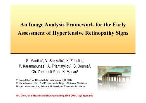
An Image Analysis Framework for the Early<br />
Assessment of Hypertensive Retinopathy Signs<br />
G. Manikis 1 , V. Sakkalis 1 , X. Zabulis 1 ,<br />
P. Karamaounas 1 , A. Triantafyllou 2 , S. Douma 2 ,<br />
Ch. Zampoulis 2 and K. Marias 1<br />
(1)<br />
Foundation for Research & Technology (FORTH)<br />
(2)<br />
Hypertension Unit, 2nd Propaedeutic Dept. of Internal Medicine,<br />
Hippokration Hospital, Aristotle University of Thessaloniki, Hellas<br />
Int. Conf. on e-Health and Bioengineering, EHB 2011, Iaşi, Romania
Introduction<br />
• This study presents a framework for the detection and measurement<br />
of retinal vessels in fundoscopy images.<br />
• The development of advanced fundus cameras along with image processing<br />
techniques offer an accurate, objective, and repeatable re<strong>presentation</strong> of retinal<br />
blood vessels.<br />
• Retinal vessels can be easily visualized with non-invasive techniques providing<br />
valuable information in the diagnosis, classification and surveillance of<br />
retinopathy signs.<br />
• In this study, a method was implemented to segment retinal vessels,<br />
enabling the actual measurement of the vessel diameter, along with a<br />
• Graphical User Interface (GUI) to support automatic and interactive measurement of<br />
vessel diameters at any selected point or region of interest. Editing the vessel<br />
re<strong>presentation</strong> in order to recover from possible segmentation misclassifications.<br />
• The proposed methodology may support vascular risk stratification in<br />
persons with hypertension.
Experimental Data<br />
• Our main aim was to provide a tool which assists<br />
the clinician to handle High Resolution (HR) raw<br />
retinal images. T<strong>here</strong>fore, 10 images of size<br />
2912X2912 were acquired.<br />
• In order to verify the applicability of the proposed<br />
scheme in lower resolutions we evaluated our<br />
methodology on two publicly available datasets;<br />
• DRIVE [1] : 40 retinal images along with manual<br />
segmentations of the vessels with size of 768X584<br />
• STARE [2] : 20 digitized slides to 700X605<br />
• Many of the post-processing steps were applied<br />
only to HR images
Flowchart of the framework<br />
The applied techniques are illustrated<br />
as a pipeline of processes consisting<br />
of two separate modules.<br />
• The first includes the detection and<br />
measurement of vessels in retinal<br />
images.<br />
• The second provides the ability to<br />
validate, edit and represent vessel<br />
information in multiple ways, by<br />
interactively selecting segments of<br />
interest and extracting their statistical<br />
information within spatial regions.<br />
A GUI encapsulates both modules and<br />
increases the automation and usability<br />
of the measurement process.
Pre-Processing 1/2<br />
The green channel of the retinal image was<br />
extracted for use as it provides the greatest<br />
contrast for blood vessels. The acquired<br />
retinal images were transformed from colored<br />
to monochromatic.<br />
HR Image processing was implemented<br />
through distinct blocking. Processing HR<br />
images was a computationally daunting task<br />
w<strong>here</strong> most algorithms fail to achieve because<br />
of memory constraints.<br />
An edge-preserving anisotropic diffusion<br />
filter [3] was applied to smooth the images<br />
within homogeneous regions. Then, the<br />
smoothed image was enhanced using a<br />
contrast limited adaptive histogram<br />
equalization [4].
Pre-Processing 2/2<br />
We then incorporated a multiple scale<br />
filtering technique for vessel enhancement,<br />
based on the eigenvalue analysis of the<br />
Hessian matrix [5] for vessel enhancement.<br />
An iterative thresholding method for<br />
segmenting the blood vessel structure was<br />
then applied for the binarization of the image.<br />
We focused on segmentation techniques that<br />
balance between accuracy and complexity<br />
and Otsu’s thresholding [6] acted fairly well<br />
under those requirements.<br />
Finally, the skeletonization of the segmented<br />
image was required for the characterization of<br />
the morphological structure of the blood<br />
vessel's network.
Evaluation of the Technique in LR Images<br />
• To facilitate the comparison with other well-known retinal vessel segmentation<br />
approaches the performance of the proposed method was evaluated on the DRIVE and<br />
STARE datasets.<br />
• LR images contribute only to the pre-processing approach in order to assess the quality<br />
of the segmented and skeletonized images before entering the post-processing phase.<br />
Performance of vessel segmentation - DRIVE<br />
Method Sensitivity Specificity Accuracy<br />
Human Observer 0.7761 0.9725 0.9473<br />
Supervised Methods Soares [7] 0.7230 0.9762 0.9446<br />
Staal [1] 0.7193 0.9773 0.9441<br />
Niemeijer [8] 0.6793 0.9801 0.9416<br />
Unsupervised Methods Mendonca [9] 0.7344 0.9764 0.9452<br />
Proposed 0.7414 0.9669 0.9371<br />
Vlachos [10] 0.7468 0.9551 0.9285<br />
Jiang [11] 0.6478 0.9625 0.9222<br />
Perez [12] 0.7086 0.9496 0.9181<br />
Chaudhuri [13] 0.2716 0.9794 0.8894<br />
Performance of vessel segmentation - STARE<br />
Method Sensitivity Specificity Accuracy<br />
Human Observer 0.8949 0.9390 0.9354<br />
Supervised Methods Staal [1] 0.6970 0.9810 0.9516<br />
Soares [7] 0.7103 0.9737 0.9480<br />
Unsupervised Methods Mendonca [9] 0.6996 0.9730 0.9440<br />
Perez [12] 0.7506 0.9569 0.9410<br />
Proposed 0.7189 0.9656 0.9318
Post-Processing 1/3<br />
Size-based filtering:<br />
The first stage of the post-processing<br />
procedure is to delete very small isolated<br />
skeleton segments as they typically<br />
correspond to noise artifacts.<br />
Pruning small vessel branches:<br />
The segmentation results obtained from HR<br />
images tend to exhibit a rich structure at<br />
vessel boundaries. In HR images such<br />
structures give rise to spurious skeleton<br />
branches. The effect of filtering the branches<br />
provided a more accurate re<strong>presentation</strong> of<br />
vessels, particularly because vessel<br />
branching points convey important information<br />
in measurements, according to the particular<br />
medical protocol.
Post-Processing 2/3<br />
Vessel width estimation:<br />
• First, a local algorithm is employed to<br />
estimate the vessel width at a skeleton point<br />
p, using both the segmented and the<br />
skeleton images.<br />
• Then, the user provides two input points for<br />
measuring the mean vessel width along a<br />
segment of a vessel.<br />
• The system finally retrieves the skeleton<br />
points of the segment in between the two<br />
points along with their estimated widths and<br />
averages them.<br />
Optical disc detection:<br />
• Optic disk is automatically detected in the acquired image and<br />
estimate of its size is provided. In this way, the application of<br />
medical protocols that are based on measurements around this<br />
disk can be automated.
Post-Processing 3/3<br />
Measuring multiple vessel segments :<br />
The measurement of average width of multiple non-branching<br />
vessels, at a range of distances from the center of the optical<br />
disk is performed. Each vessel segment initiates from the<br />
smaller to the larger circle. Depending on user selection, a<br />
number of statistics can be then estimated.<br />
Statistical Analysis in HR Images:<br />
The ratio of the quantities, CRAE and CRVE<br />
[14], which are determined by measurement<br />
on the arteries and veins detected in the<br />
region of interest respectively, were estimated.<br />
Such measures require the characterization of<br />
vessels, as to if they are veins or arteries and<br />
their mean widths. Hence, a GUI component<br />
is provided to facilitate this characterization by<br />
the medical professional.
User Interface Overview<br />
Semi-automatic pre-processing:<br />
The purpose of implementing this functionality is threefold;<br />
a) user can manually corrects remaining errors in the<br />
segmentation image<br />
b) performs targeted measurements (i.e. focuses on a<br />
particular vessel and ignore its branches)<br />
c) acquire ground truth results by superimposing a basis<br />
segmented image to the original image.<br />
Other utilities:<br />
Saving of data file with measurements, reviewing and<br />
editing of old measurement files.<br />
Image view allowing measurement and skeleton<br />
points calculation using magnification options (visible<br />
image segment in thumbnail view).<br />
Rulers display allowing calibrated grid measurements.
Conclusions<br />
• The presented application employs an image segmentation algorithm,<br />
along with pre-processing and skeletonization techniques in order to<br />
extract a re<strong>presentation</strong> of vessels.<br />
• Additionally, techniques that analyze this re<strong>presentation</strong> and measure<br />
vessel width are introduced and adapted appropriately, in order to<br />
provide measurements according to particular measurement protocols.<br />
• The above functionalities are integrated through a GUI, which assists<br />
the medical professional to perform measurements in an ergonomic<br />
fashion and apply targeted measurements.<br />
• This interface provides also functionalities that allow the clinician to edit<br />
the vessel segmentation result and update the corresponding<br />
measurements, in order to recover from segmentation errors.
Future Work<br />
• Future work will be pursued along two research avenues.<br />
• The first regards the improvement of the segmentation algorithm.<br />
• The second regards the registration of fundoscopy images of the same patient,<br />
acquired across large time intervals.<br />
• The goal is to provide medical professionals with the capability of<br />
automatically comparing vessel measurements, thus offering a<br />
valuable tool in the monitoring, diagnosis, and estimation of the<br />
condition of the cardiovascular system.
References<br />
1. Staal, J.; Abramoff, M.D.; Niemeijer, M.; Viergever, M.A.; van Ginneken, B.; , "Ridge-based vessel segmentation in color images of the<br />
retina," Medical Imaging, IEEE Transactions on , vol.23, no.4, pp.501-509, April 2004.<br />
2. A. Hoover, V. Kouznetsova, and M. Goldbaum, “Locating blood vessels in retinal images by piecewise threshold probing of a matched<br />
filter response,” IEEE Trans. Med. Imag., vol. 19, no. 3, pp. 203–211, Mar. 2000.<br />
3. P. Perona, J. Malik, “Scale-space and edge detection using anisotropic diffusion,” IEEE Trans. PAMI, V. 12, no. 7, pp. 629-639, July<br />
1990.<br />
4. K. Zuiderveld. Contrast limited adaptive histogram equalization. Graphics gems IV, pages 474-485, 1994.<br />
5. A.F. Frangi, W.J. Niessen, K.L. Vincken, M.A. Viergever (1998). Multiscale vessel enhancement filtering. In Medical Image Computing<br />
and Computer-Assisted Intervention - MICCAI'98, W.M. Wells, A. Colchester and S.L. Delp (Eds.), Lecture Notes in Computer Science,<br />
vol. 1496 - Springer Verlag, Berlin, Germany, pp. 130-137.<br />
6. Otsu, N., "A Threshold Selection Method from Gray-Level Histograms," IEEE Transactions on Systems, Man, and Cybernetics, Vol. 9,<br />
No. 1, 1979, pp. 62-66.<br />
7. João V. B. Soares, Jorge J. G. Leandro, Roberto M. Cesar Jr., Herbert F. Jelinek, and Michael J. Cree, “Retinal Vessel Segmentation<br />
Using the 2-D Gabor Wavelet and Supervised Classification,” IEEE Transactions on medical Imaging, vol. 25, no. 9, September 2006.<br />
8. Niemeijer M., Staal J., van Ginneken B, Loog M, and Abràmoff M. D., “Comparative study of retinal vessel segmentation methods on a<br />
new publicly available database,” in Proc. SPIE Med. Imag., M. Fitzpatrick and M. Sonka, Eds., 2004, vol. 5370, pp. 648–656.<br />
9. A. Mendonca and A. Campilho. Segmentation of retinal blood vessels by combining the detection of centerlines and morphological<br />
reconstruction. IEEE Transactions on Medical Imaging, 25(9):1200-1213, September 2006.<br />
10. M. Vlachos, E. Dermatas, Multi-scale retinal vessel segmentation using line tracking, Computerized Medical Imaging and Graphics,<br />
Volume 34, Issue 3, April 2010, Pages 213-227.<br />
11. Xiaoyi Jiang; Mojon, D.; , "Adaptive local thresholding by verification-based multithreshold probing with application to vessel<br />
detection in retinal images," Pattern Analysis and Machine Intelligence, IEEE Transactions on , vol.25, no.1, pp.131-137, Jan. 2003.<br />
12. M.E. Martínez-Pérez, A.D. Hughes, A.V. Stanton, S.A. Thom, A.A. Bharath, and K.H. Parker, "Retinal Blood Vessel Segmentation by<br />
Means of Scale-Space Analysis and Region Growing", in Proc. MICCAI, 1999, pp.90-97.<br />
13. S. Chaudhuri, S. Chatterjee, N. Katz, M. Nelson, and M. Goldbaum,“Detection of blood vessels in retinal images using twodimensional<br />
matched filters,” IEEE Trans. Med. Imag., pp. 263–269, 1989.<br />
14. Hubbard LD, Brothers RJ, King WN, Clegg LX, Klein R, Cooper LS, et al. Methods for evaluation of retinal microvascular abnormalities<br />
associated with hypertension/sclerosis in the Atherosclerosis Risk in Communities Study. Ophthalmology;106(12):2269-80, Dec 1999.
Acknowledgments<br />
• The authors would like to thank the Hypertension Unit of the 2 nd Propaedeutic Dept. of<br />
Internal Medicine, Hippokration Hospital in Thessaloniki for providing the High Resolution<br />
dataset.<br />
• This work was supported in part by the European Commission under the TUMOR (FP7-<br />
ICT-2009.5.4-247754) project.<br />
Vangelis Sakkalis<br />
sakkalis@ics.forth.gr