PROMUS Element⢠Plus - Boston Scientific
PROMUS Element⢠Plus - Boston Scientific
PROMUS Element⢠Plus - Boston Scientific

You also want an ePaper? Increase the reach of your titles
YUMPU automatically turns print PDFs into web optimized ePapers that Google loves.
1.5 Tesla Temperature information<br />
Non-clinical testing of RF-induced heating was performed<br />
at 64 MHz in a 1.5 Tesla Intera® Philips Medical Systems,<br />
software version Release 10.6.2.0, 2006-03-10 whole body coil<br />
MR scanner on continuously stented lengths up to 74 mm.<br />
RF power was applied for 15 minutes and the measured<br />
conductivity of the phantom material was about 0.3 S/m. The<br />
phantom average SAR was calculated using calorimetry to be<br />
2.1 W/kg. The maximal in-vitro temperature rise was calculated<br />
as 2.6°C for a measured stent length of 39 mm with the wholebody<br />
SAR scaled to 2.0 W/kg. The calculations did not include<br />
the cooling effects due to blood flow.<br />
In vivo, local SAR depends on MR Field strength and may be<br />
different than the estimated whole body averaged SAR, due to<br />
body composition, stent position within the imaging field, and<br />
scanner used, thereby affecting the actual temperature rise.<br />
image artifact information<br />
The calculated image artifact extends approximately 7 mm from<br />
the perimeter of the device diameter and 5 mm beyond each<br />
end of the length of the stent when scanned in non-clinical<br />
testing using a Spin Echo sequence. With a Gradient Echo<br />
sequence the calculated image artifact extends 5 mm beyond<br />
the perimeter of the diameter and 6 mm beyond each end of the<br />
length with both sequences partially shielding the lumen in a<br />
3.0 Tesla Intera (Achieva Upgrade), Philips Medical Solutions,<br />
software version Release 2.5.3.0 2007-09-28 MR system with a<br />
transmit/receive head coil.<br />
Medical registration<br />
It is recommended that patients register the conditions under<br />
which the implant can be scanned safely with the MedicAlert<br />
Foundation (www.medicalert.org) or equivalent organization.<br />
Magnetic Resonance<br />
MR Conditional<br />
6.13 Stent Handling (also see Section 14, Operational<br />
instructions)<br />
• For single use only. Do not resterilize or reuse this product.<br />
Note product “Use By” date. (see Section 1, Warning)<br />
• The premounted <strong>PROMUS</strong> Element stent and its delivery<br />
system are designed for use as a unit. The stent is not to be<br />
removed from its delivery balloon. The stent is not designed<br />
to be crimped onto another balloon. Removing the stent<br />
from its delivery balloon may damage the stent and coating<br />
and/or lead to stent embolization.<br />
• Special care must be taken not to handle or in any way<br />
disrupt the stent position on the delivery balloon. This is<br />
most important during catheter removal from packaging,<br />
placement over guidewire, and advancement through<br />
hemostasis valve adapter and guide catheter hub.<br />
• Excessive manipulation or handling may cause coating<br />
damage, contamination, or dislodgment of the stent from<br />
the delivery balloon.<br />
• Use only the appropriate balloon inflation media (see<br />
Section 14.3.3, Balloon Preparation). Do not use air or any<br />
gas medium to inflate the balloon.<br />
• In the event the <strong>PROMUS</strong> Element stent is not deployed,<br />
do not use the product and contact your local <strong>Boston</strong><br />
<strong>Scientific</strong> Representative for return information.<br />
6.14 Stent Placement<br />
Preparation<br />
• Do not prepare or pre-inflate balloon prior to stent<br />
deployment other than as directed. Use the balloon purging<br />
technique described in Section 14.3.3, Balloon Preparation.<br />
• If unusual resistance is felt at any time during lesion<br />
access before stent implantation, the stent delivery system<br />
and the guide catheter should be removed as a single unit<br />
(see Section 6.15, Stent Delivery System Removal).<br />
• An unexpanded stent should be introduced into the<br />
coronary arteries one time only. An unexpanded stent<br />
should not be subsequently moved in and out through the<br />
distal end of the guide catheter as stent or coating damage<br />
or stent dislodgment from the balloon may occur.<br />
Placement<br />
• The vessel should be pre-dilated with an appropriate sized<br />
balloon. Failure to do so may increase the risk of placement<br />
difficulty and procedural complications.<br />
• Do not expand the stent if it is not properly positioned in the<br />
vessel (see Section 6.15, Stent Delivery System Removal).<br />
• Balloon pressures should be monitored during inflation. Do not<br />
exceed rated burst pressure as indicated on product label (see<br />
Section 14.5, In Vitro Information, Table 14.5.1, Typical <strong>PROMUS</strong><br />
Element <strong>Plus</strong> Stent System Compliance). Use of pressures<br />
higher than specified on product label may result in a ruptured<br />
balloon and intimal damage and dissection. The stent inner<br />
diameter should approximate 1.1 times the reference diameter<br />
of the vessel.<br />
• Placement of the stent has the potential to compromise side<br />
branch patency (see Section 14.4, Post-Deployment Dilatation<br />
of Stented Segments).<br />
• Implanting a stent may lead to dissection of the vessel distal<br />
and/or proximal to the stented portion, and may cause acute<br />
closure of the vessel requiring additional intervention (e.g.,<br />
CABG, further dilation, placement of additional stents, or other).<br />
• When treating multiple lesions, the distal lesion should<br />
generally be stented first, followed by stenting of the more<br />
proximal lesion(s). Stenting in this order alleviates the need to<br />
cross the proximal stent in placement of the distal stent and<br />
reduces the chances of dislodging the proximal stent.<br />
6.15 Stent Delivery System removal<br />
• If unusual resistance is felt at any time during lesion access<br />
before stent implantation, the stent delivery system and the<br />
guide catheter should be removed as a single unit.<br />
• Do not attempt to pull an unexpanded stent back into the guide<br />
catheter, as stent or coating damage or stent dislodgment from<br />
the balloon may occur.<br />
• Stent retrieval methods (use of additional wires, snares and/<br />
or forceps) may result in additional trauma to the vascular<br />
site. Complications can include bleeding, hematoma, or<br />
pseudoaneurysm.<br />
When removing the entire stent delivery system and guide catheter<br />
as a single unit, the following steps should be executed under direct<br />
visualization using fluoroscopy:<br />
• Following stent placement, confirm complete balloon deflation<br />
(See Table 6.1, Delivery System Deflation Time Specifications).<br />
If greater than usual resistance is felt during delivery system<br />
withdrawal, pay particular attention to guide catheter position.<br />
In some cases it may be necessary to pull back slightly on the<br />
guide catheter in order to prevent deep seating (unplanned<br />
advancement) of the guide catheter and subsequent vessel<br />
damage. In cases where unplanned guide catheter movement<br />
has occurred, angiographic assessment of the coronary tree<br />
should be undertaken to ensure that there is no damage to the<br />
coronary vasculature.<br />
• Maintain guidewire placement across the lesion during the<br />
entire removal process.<br />
• Carefully pull back the stent delivery system until the proximal<br />
balloon marker of the stent delivery system is just distal to the<br />
guide catheter distal tip.<br />
• The stent delivery system and the guide catheter should be<br />
pulled back until the tip of the guide catheter is just distal to<br />
the arterial sheath, allowing the guide catheter to straighten.<br />
Carefully retract the stent delivery system into the guide<br />
catheter and remove the stent delivery system and the guide<br />
catheter from the patient as a single unit while leaving the<br />
guidewire across the lesion.<br />
Failure to follow these steps, and/or applying excessive force to<br />
the stent delivery system, can potentially result in stent or coating<br />
damage, stent dislodgment from the balloon, and/or damage to the<br />
delivery system.<br />
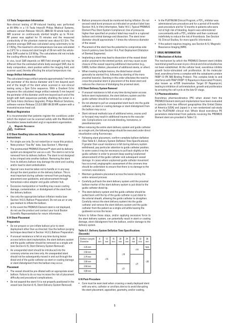
Table 6.1. Delivery System Deflation Time Specifications<br />
(Seconds)<br />
Balloon Length/<br />
Diameter<br />
2.25 mm<br />
2.50 mm<br />
2.75 mm<br />
3.00 mm<br />
3.50 mm<br />
4.00 mm<br />
8<br />
mm<br />
12<br />
mm<br />
≤16<br />
16<br />
mm<br />
5<br />
20<br />
mm<br />
≤16<br />
≤21<br />
24<br />
mm<br />
28<br />
mm<br />
≤16<br />
≤21<br />
32<br />
mm<br />
38<br />
mm<br />
6.16 Post-Procedure<br />
• Care must be exercised when crossing a newly deployed stent<br />
with any wire, catheter or ancillary device to avoid disrupting<br />
the stent placement, apposition, geometry, and/or coating.<br />
N/A<br />
≤30<br />
• In the PLATINUM Clinical Program, a P2Y12 inhibitor was<br />
administered pre-procedure and for a period of 6 months<br />
post-procedure and for 12 months in patients who were<br />
not at high risk of bleeding. Aspirin was administered<br />
concomitantly with a P2Y12 inhibitor and then continued<br />
indefinitely to reduce the risk of thrombosis. See Section<br />
10, Clinical Studies, for more specific information.<br />
• If the patient requires imaging, see Section 6.12, Magnetic<br />
Resonance Imaging (MRI).<br />
7 DrUg infOrMaTiOn<br />
7.1 Mechanism of action<br />
The mechanism by which the <strong>PROMUS</strong> Element stent inhibits<br />
neointimal growth as seen in pre-clinical and clinical studies has<br />
not been established. At the cellular level, everolimus inhibits<br />
growth factor-stimulated cell proliferation. At the molecular<br />
level, everolimus forms a complex with the cytoplasmic protein<br />
FKBP-12 (FK 506 Binding Protein). This complex binds to and<br />
interferes with FRAP (FKBP-12 Rapamycin Associated Protein),<br />
also known as mTOR (mammalian Target Of Rapamycin),<br />
leading to inhibition of cell metabolism, growth and proliferation<br />
by arresting the cell cycle at the late G1 stage.<br />
7.2 Pharmacokinetics<br />
Everolimus pharmacokinetics (PK) when eluted from the<br />
<strong>PROMUS</strong> Element stent post-implantation have been evaluated<br />
in patients from two different geographies (the United States<br />
of America [USA] and Japan) in a non-randomized sub-study<br />
of the PLATINUM clinical trial. Whole blood everolimus PK<br />
parameters determined from patients receiving the <strong>PROMUS</strong><br />
Element stent are provided in Table 7.2.<br />
<strong>Boston</strong> <strong>Scientific</strong>, (Master Brand DFU Template 8.2677in x 11.6929in A4, 90105918AL), eDFU, MB, <strong>PROMUS</strong> Element <strong>Plus</strong>, EN, 90519100-01B