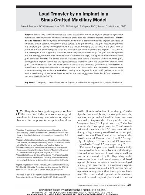
Load Transfer by an implant in a sinus-grafted Maxillary ... - Id-sc.com
Load Transfer by an implant in a sinus-grafted Maxillary ... - Id-sc.com
Load Transfer by an implant in a sinus-grafted Maxillary ... - Id-sc.com

Create successful ePaper yourself
Turn your PDF publications into a flip-book with our unique Google optimized e-Paper software.
<strong>Load</strong> <strong>Tr<strong>an</strong>sfer</strong> <strong>by</strong> <strong>an</strong> Impl<strong>an</strong>t <strong>in</strong> a<br />
S<strong>in</strong>us-Grafted <strong>Maxillary</strong> Model<br />
Mete I. F<strong>an</strong>u<strong>sc</strong>u, DDS 1 /Keisuke Iida, DDS, PhD 2 /Angelo A. Caputo, PhD 3 /Russell D. Nishimura, DDS 4<br />
Purpose: This <strong>in</strong> vitro study determ<strong>in</strong>ed the stress distribution around <strong>an</strong> impl<strong>an</strong>t placed <strong>in</strong> a posterior<br />
edentulous maxillary model with simulated s<strong>in</strong>us grafts that had different degrees of stiffness. Materials<br />
<strong>an</strong>d Methods: The <strong>com</strong>posite photoelastic model with a st<strong>an</strong>dard threaded impl<strong>an</strong>t consisted of<br />
simulated crestal cortical, c<strong>an</strong>cellous, s<strong>in</strong>us cortical, <strong>an</strong>d <strong>grafted</strong> bone. The graft maturation process<br />
<strong>an</strong>d <strong>in</strong>herent graft quality were represented <strong>in</strong> the model <strong>by</strong> vary<strong>in</strong>g the stiffness of the graft. Prior to<br />
placement of the simulated graft, axial <strong>an</strong>d <strong>in</strong>cl<strong>in</strong>ed loads were applied to the impl<strong>an</strong>t. The stresses<br />
that developed <strong>in</strong> the support<strong>in</strong>g structures were <strong>an</strong>alyzed photoelastically. The graft was then placed<br />
<strong>an</strong>d the test<strong>in</strong>g procedure was repeated over 4 consecutive days, dur<strong>in</strong>g which time the simulated<br />
graft stiffened. Results: The stress <strong>an</strong>alysis <strong>in</strong>dicated that before placement of the simulated graft,<br />
load<strong>in</strong>g on the impl<strong>an</strong>t tr<strong>an</strong>sferred the highest stresses to cortical bone. The presence of the simulated<br />
graft tr<strong>an</strong>sferred stress from the native bone simul<strong>an</strong>ts to the simulated <strong>grafted</strong> bone. Di<strong>sc</strong>ussion: As<br />
the stiffness of the graft <strong>in</strong>creased, a more equitable stress distribution was observed <strong>in</strong> the multilayer<br />
bone surround<strong>in</strong>g the impl<strong>an</strong>t. Conclusion: <strong>Load</strong><strong>in</strong>g of <strong>an</strong> impl<strong>an</strong>t <strong>in</strong> a less stiff <strong>grafted</strong> s<strong>in</strong>us could<br />
lead to overload<strong>in</strong>g of the native bone as well as the matur<strong>in</strong>g <strong>grafted</strong> bone. INT J ORAL MAXILLOFAC<br />
IMPLANTS 2003;18:667–674<br />
Key words: bone graft, bone stiffness, dental impl<strong>an</strong>t, maxillary s<strong>in</strong>us augmentation, stress distribution<br />
<strong>Maxillary</strong> s<strong>in</strong>us bone graft augmentation has<br />
be<strong>com</strong>e one of the most <strong>com</strong>mon surgical<br />
procedures for <strong>in</strong>creas<strong>in</strong>g bone volume for impl<strong>an</strong>t<br />
placement <strong>in</strong> the posterior atrophic edentulous<br />
1 Assist<strong>an</strong>t Professor <strong>an</strong>d Director, Adv<strong>an</strong>ced Education <strong>in</strong> General<br />
Dentistry, Division of Restorative Dentistry, School of Dentistry,<br />
University of California at Los Angeles, Los Angeles, California.<br />
2 Research Fellow, Division of Adv<strong>an</strong>ced Prosthodontics, Biomaterials<br />
Science, <strong>an</strong>d Hospital Dentistry, School of Dentistry, University<br />
of California at Los Angeles, Los Angeles, California.<br />
3 Professor, Division of Adv<strong>an</strong>ced Prosthodontics, Biomaterials<br />
Science, <strong>an</strong>d Hospital Dentistry, School of Dentistry, University<br />
of California at Los Angeles, Los Angeles, California.<br />
4 Associate Professor, Division of Adv<strong>an</strong>ced Prosthodontics, Biomaterials<br />
Science, <strong>an</strong>d Hospital Dentistry, School of Dentistry,<br />
University of California at Los Angeles, Los Angeles, California.<br />
Repr<strong>in</strong>t requests: Dr Mete I. F<strong>an</strong>u<strong>sc</strong>u, UCLA School of Dentistry,<br />
CHS 20-114, 10833 Le Conte Avenue, Los Angeles, CA 90095-<br />
1668. Fax: +310-794-7964. E-mail: mf<strong>an</strong>u<strong>sc</strong>u@ucla.edu<br />
Presented <strong>in</strong> part at the 78th General Session of the International<br />
Association for Dental Research, April 2000, Wash<strong>in</strong>gton, DC.<br />
maxilla. S<strong>in</strong>ce <strong>in</strong>troduction of the s<strong>in</strong>us graft technique<br />
<strong>by</strong> Boyne <strong>an</strong>d James, 1 various graft materials,<br />
impl<strong>an</strong>ts, <strong>an</strong>d procedural modifications have been<br />
proposed to improve the efficacy of the therapy.<br />
Autogenous bone, 2–5 allogenic materials, 5,6 alloplastic<br />
materials, 5,7 xenogenic materials, 5,6 <strong>an</strong>d <strong>com</strong>b<strong>in</strong>ations<br />
of these materials 4–6,8 have been utilized.<br />
S<strong>in</strong>us graft<strong>in</strong>g is usually considered for <strong>an</strong> atrophic<br />
maxilla, such as Class V <strong>an</strong>d VI accord<strong>in</strong>g to the<br />
classification of Cawood <strong>an</strong>d Howell. 9 Average<br />
ridge heights of Classes V <strong>an</strong>d VI jaws were<br />
reported to be 7.4 <strong>an</strong>d 3.2 mm, respectively. 10<br />
The edentulous posterior maxilla is <strong>an</strong>atomically<br />
characterized <strong>by</strong> th<strong>in</strong> cortical bone at both the ridge<br />
crest <strong>an</strong>d s<strong>in</strong>us floor <strong>an</strong>d low-density c<strong>an</strong>cellous<br />
bone <strong>in</strong> the rema<strong>in</strong>der (Fig 1). 11 Depend<strong>in</strong>g on the<br />
preoperative bone level, simult<strong>an</strong>eous or delayed<br />
impl<strong>an</strong>t placement techniques have been employed<br />
<strong>in</strong> s<strong>in</strong>us graft procedures. In a consensus report,<br />
Jensen <strong>an</strong>d coworkers stated a 90% success rate for<br />
impl<strong>an</strong>ts <strong>in</strong> s<strong>in</strong>us grafts with at least 3 years of function.<br />
5 The report <strong>in</strong>cluded patients with simult<strong>an</strong>eous<br />
<strong>an</strong>d delayed impl<strong>an</strong>t placement <strong>in</strong> various<br />
The International Journal of Oral & Maxillofacial Impl<strong>an</strong>ts 667
FANUSCU ET AL<br />
Fig 1 Cross-section of alveolar bone with s<strong>in</strong>us cavity <strong>an</strong>d walls<br />
<strong>in</strong> the posterior maxilla from hum<strong>an</strong> cadaver. SW = s<strong>in</strong>us walls;<br />
SB = s<strong>in</strong>us bone; AB = alveolar bone.<br />
SW<br />
SB<br />
AB<br />
the edentulous posterior maxilla, with m<strong>in</strong>eralized<br />
tissue volume of the latter r<strong>an</strong>g<strong>in</strong>g from 17.1% to<br />
26.7%. 19<br />
Premature load<strong>in</strong>g <strong>an</strong>d/or overload<strong>in</strong>g might be<br />
signific<strong>an</strong>t concerns <strong>in</strong> s<strong>in</strong>us graft cases, s<strong>in</strong>ce different<br />
graft materials <strong>an</strong>d their maturation patterns<br />
c<strong>an</strong> have variable load-bear<strong>in</strong>g capacities. Biomech<strong>an</strong>ically,<br />
control of the load tr<strong>an</strong>sfer to bone surround<strong>in</strong>g<br />
the impl<strong>an</strong>ts plays <strong>an</strong> import<strong>an</strong>t role <strong>in</strong><br />
the long-term success of impl<strong>an</strong>t therapy. 20 Several<br />
studies have reported that appropriately controlled<br />
loads c<strong>an</strong> stimulate bone remodel<strong>in</strong>g around<br />
impl<strong>an</strong>ts, 21,22 whereas excessive stresses cause marg<strong>in</strong>al<br />
bone resorption. 23–25<br />
The purpose of this study was to <strong>in</strong>vestigate the<br />
stress distribution <strong>in</strong> bone around <strong>an</strong> impl<strong>an</strong>t placed<br />
<strong>in</strong> a posterior atrophic maxilla with a simulated<br />
s<strong>in</strong>us graft <strong>by</strong> use of a <strong>com</strong>posite photoelastic<br />
model.<br />
MATERIALS AND METHODS<br />
grafts. There was no <strong>in</strong>dication of the superiority of<br />
a particular protocol or material. However, there<br />
was a statistically signific<strong>an</strong>t difference <strong>in</strong> impl<strong>an</strong>t<br />
loss when available residual or native bone was 4<br />
mm or less as opposed to 5 mm or greater.<br />
A <strong>com</strong>plex structure consist<strong>in</strong>g of bone with<br />
vary<strong>in</strong>g stiffness c<strong>an</strong> be found around impl<strong>an</strong>ts<br />
placed <strong>in</strong> the posterior maxilla with <strong>grafted</strong><br />
s<strong>in</strong>us(es). A crucial process that leads to the stability<br />
of osseo<strong>in</strong>tegrated impl<strong>an</strong>ts is m<strong>in</strong>eralization of the<br />
bone adjacent to the impl<strong>an</strong>t surface. Native bone,<br />
which is a critical factor <strong>in</strong> establish<strong>in</strong>g <strong>an</strong>d ma<strong>in</strong>ta<strong>in</strong><strong>in</strong>g<br />
osseo<strong>in</strong>tegration, consists of crestal cortical<br />
bone, c<strong>an</strong>cellous bone, <strong>an</strong>d s<strong>in</strong>us floor cortical bone.<br />
The contribution of the <strong>grafted</strong> bone to the establishment<br />
<strong>an</strong>d ma<strong>in</strong>ten<strong>an</strong>ce of impl<strong>an</strong>t stability is not<br />
yet understood. <strong>Load</strong>-bear<strong>in</strong>g characteristics of<br />
<strong>grafted</strong> bone depend on the graft material <strong>an</strong>d its<br />
maturation process. Several studies have <strong>in</strong>vestigated<br />
the total volume of hard tissue with<strong>in</strong> various<br />
<strong>grafted</strong> s<strong>in</strong>uses to predict long-term impl<strong>an</strong>t stability.<br />
Accord<strong>in</strong>g to histologic <strong>an</strong>alysis, the vital m<strong>in</strong>eralized<br />
tissue volume of the <strong>grafted</strong> s<strong>in</strong>us r<strong>an</strong>ged<br />
from 26% to 69% when only autogenous bone was<br />
used. 12–15 The r<strong>an</strong>ge of vital m<strong>in</strong>eralized tissue volume<br />
was 5% to 45% when <strong>an</strong>y other graft material<br />
or <strong>com</strong>b<strong>in</strong>ations of materials were used. 8,12,16–18 It<br />
also should be noted that the m<strong>in</strong>eralized tissue volume<br />
of <strong>grafted</strong> s<strong>in</strong>uses has been reported as relatively<br />
greater th<strong>an</strong> that of native c<strong>an</strong>cellous bone <strong>in</strong><br />
A <strong>com</strong>posite photoelastic model simulat<strong>in</strong>g a unilateral<br />
atrophic edentulous posterior maxilla was fabricated<br />
for quasi–3-dimensional test<strong>in</strong>g <strong>an</strong>d <strong>an</strong>alysis<br />
(Fig 2a). Individual photoelastic simul<strong>an</strong>ts with different<br />
stiffnesses were used for cortical bone (PLM-<br />
1, Measurements Group, Raleigh, NC) <strong>an</strong>d c<strong>an</strong>cellous<br />
bone (PL-2, Measurements Group). Simulated<br />
crestal cortical bone, c<strong>an</strong>cellous bone, <strong>an</strong>d s<strong>in</strong>us<br />
floor cortical bone were fabricated <strong>in</strong> 1-mm, 3.5-<br />
mm, <strong>an</strong>d 0.5-mm heights, respectively (Fig 2b).<br />
Therefore, the total height of simulated native bone<br />
was 5.0 mm.<br />
A st<strong>an</strong>dard threaded impl<strong>an</strong>t, 3.75 mm <strong>in</strong> diameter<br />
<strong>an</strong>d 13 mm <strong>in</strong> length (Impl<strong>an</strong>t Innovations,<br />
Palm Beach Gardens, FL), was <strong>in</strong>corporated <strong>in</strong>to<br />
the model. Photoelastic res<strong>in</strong>s represent<strong>in</strong>g the<br />
native bone <strong>com</strong>ponents were poured directly<br />
around the impl<strong>an</strong>t <strong>an</strong>d allowed to cure <strong>com</strong>pletely.<br />
An experimental photoelastic res<strong>in</strong> (modified PLM-<br />
1, Measurement Group) represent<strong>in</strong>g the graft was<br />
then placed <strong>in</strong>to the simulated maxillary s<strong>in</strong>us cavity<br />
(Fig 2b). Several res<strong>in</strong> hardener ratios of PLM-1<br />
were tested previously to establish a r<strong>an</strong>ge of stiffness<br />
values. Beams of modified PLM-1, 4.5 2.8 <br />
30 mm, were tested <strong>in</strong> flexure on <strong>an</strong> Instron test<br />
mach<strong>in</strong>e (Instron, C<strong>an</strong>ton, MA). Three beams of<br />
each formulation were tested at 3, 4, 5, 6, 7, <strong>an</strong>d 10<br />
days. From the results of these tests, a formulation<br />
with a ch<strong>an</strong>ge <strong>in</strong> stiffness over time was selected to<br />
simulate the mech<strong>an</strong>ical response of either a bone<br />
graft matur<strong>in</strong>g over time or of different qualities of<br />
graft materials (Fig 3).<br />
668 Volume 18, Number 5, 2003
FANUSCU ET AL<br />
Fig 2a (Left) Photoelastic model of the <strong>grafted</strong> posterior maxilla<br />
with <strong>an</strong> impl<strong>an</strong>t.<br />
Fig 2b (Below) Schematic cross-section of the model show<strong>in</strong>g<br />
the height of each simul<strong>an</strong>t.<br />
6<br />
Fig 3 (Left) Stiffness values of the bone simul<strong>an</strong>ts. The <strong>grafted</strong><br />
bone simul<strong>an</strong>t became stiffer with ag<strong>in</strong>g.<br />
PLM-1 <strong>com</strong>pletely cured (cortical bone simul<strong>an</strong>t)<br />
5<br />
5 2<br />
Flexural modulus (10 lb/<strong>in</strong> )<br />
4<br />
3<br />
2<br />
Modified PLM-1<br />
(graft bone simul<strong>an</strong>t)<br />
Fig 4<br />
(Below) Model under load <strong>in</strong> stra<strong>in</strong><strong>in</strong>g frame.<br />
<strong>Load</strong> cell<br />
1<br />
PLM-2 <strong>com</strong>pletely cured (trabecular bone simul<strong>an</strong>t)<br />
Stra<strong>in</strong><strong>in</strong>g frame<br />
0<br />
0 1 2 3 4 5 6 7 8 9 10 11 12<br />
Days of cur<strong>in</strong>g at room temperature<br />
Axial loads were applied to the impl<strong>an</strong>t center<br />
through <strong>an</strong> impl<strong>an</strong>t mount (Impl<strong>an</strong>t Innovations) <strong>in</strong><br />
a load<strong>in</strong>g frame <strong>by</strong> me<strong>an</strong>s of a calibrated load cell<br />
mounted on the movable head of the frame. <strong>Load</strong>s<br />
were monitored <strong>an</strong>d controlled <strong>by</strong> a digital readout<br />
(models 2130 <strong>an</strong>d 2120A, Measurement Group)<br />
(Fig 4). <strong>Load</strong>s of 13.6 kg (30 lb) were applied<br />
because they are realistic functional levels <strong>an</strong>d provide<br />
a satisfactory optical response with<strong>in</strong> the<br />
model. To represent c<strong>an</strong>tilever<strong>in</strong>g forces, <strong>in</strong>cl<strong>in</strong>ed<br />
loads of 13.6 kg were also applied while the model<br />
was tilted 15 degrees to the impl<strong>an</strong>t axis. The<br />
stresses that developed <strong>in</strong> the support<strong>in</strong>g structures<br />
were observed <strong>an</strong>d recorded photographically <strong>in</strong> the<br />
field of a circular polari<strong>sc</strong>ope. To m<strong>in</strong>imize surface<br />
refraction <strong>an</strong>d facilitate photoelastic observation,<br />
the model was submerged <strong>in</strong> a t<strong>an</strong>k of m<strong>in</strong>eral oil<br />
dur<strong>in</strong>g the <strong>an</strong>alysis. The load<strong>in</strong>g <strong>an</strong>d record<strong>in</strong>g<br />
sequences were performed before placement of the<br />
<strong>grafted</strong> bone simul<strong>an</strong>t. After the graft was placed,<br />
the load<strong>in</strong>g procedure was repeated at the predeterm<strong>in</strong>ed<br />
days (3, 4, 5, <strong>an</strong>d 6, which corresponded to<br />
the ch<strong>an</strong>ge <strong>in</strong> flexural modulus <strong>an</strong>d represented the<br />
maturity <strong>an</strong>d quality of <strong>grafted</strong> bone), to determ<strong>in</strong>e<br />
the effects of <strong>in</strong>creas<strong>in</strong>g stiffness on load tr<strong>an</strong>sfer.<br />
Test<strong>in</strong>g <strong>an</strong>d record<strong>in</strong>g procedures were repeated at<br />
least 2 times so that reproducibility of the technique<br />
could be verified. Photoelastic stress fr<strong>in</strong>ges that<br />
The International Journal of Oral & Maxillofacial Impl<strong>an</strong>ts 669
FANUSCU ET AL<br />
Fr<strong>in</strong>ge orders<br />
3<br />
2<br />
1<br />
0<br />
Fig 5 Relationship between stress level <strong>an</strong>d fr<strong>in</strong>ge order used<br />
to de<strong>sc</strong>ribe results.<br />
developed <strong>in</strong> the support<strong>in</strong>g structure were <strong>an</strong>alyzed<br />
on the <strong>sc</strong><strong>an</strong>ned data photographs, which were subsequently<br />
viewed with a <strong>com</strong>puter graphic program<br />
(Photoshop 4.0; Adobe Systems, S<strong>an</strong> Jose, CA) The<br />
stress <strong>in</strong>tensity (number of fr<strong>in</strong>ges) <strong>an</strong>d their locations<br />
were subjectively <strong>com</strong>pared. In the <strong>in</strong>terpretation<br />
of stress data, the follow<strong>in</strong>g term<strong>in</strong>ology was<br />
adopted (Fig 5): Low stress = 1 fr<strong>in</strong>ge or less; moderate<br />
stress = between 1 <strong>an</strong>d 3 fr<strong>in</strong>ges; high stress =<br />
more th<strong>an</strong> 3 fr<strong>in</strong>ges.<br />
RESULTS<br />
Medium<br />
Zero<br />
High<br />
Low<br />
Photoelastic exam<strong>in</strong>ation of the model before <strong>an</strong>d<br />
after placement of the graft simul<strong>an</strong>t revealed no<br />
signific<strong>an</strong>t <strong>in</strong>itial stresses. This observation established<br />
that the isochromatic fr<strong>in</strong>ges produced upon<br />
load<strong>in</strong>g were direct results of the applied loads. Further,<br />
no signific<strong>an</strong>t residual stresses were evident<br />
after <strong>an</strong>y of the load<strong>in</strong>g sequences.<br />
<strong>Load</strong><strong>in</strong>g Before Graft Placement<br />
Under axial load<strong>in</strong>g, high-level stresses were<br />
observed <strong>in</strong> the crestal cortical bone simul<strong>an</strong>t. Moderate<br />
stress was noted <strong>in</strong> c<strong>an</strong>cellous <strong>an</strong>d s<strong>in</strong>us floor<br />
cortical bone simul<strong>an</strong>ts (Figs 6a <strong>an</strong>d 6b). With<br />
<strong>in</strong>cl<strong>in</strong>ed load<strong>in</strong>g, higher stresses occurred <strong>in</strong> all layers<br />
of the native bone simul<strong>an</strong>ts along the impl<strong>an</strong>t. The<br />
stress concentration <strong>in</strong> the c<strong>an</strong>cellous bone was on<br />
the side away from the applied load (Figs 7a <strong>an</strong>d 7b).<br />
In both load<strong>in</strong>g conditions, low stress was observed<br />
<strong>in</strong> the cortical bone simul<strong>an</strong>t at the s<strong>in</strong>us walls.<br />
<strong>Load</strong><strong>in</strong>g After Graft Placement<br />
With the presence of the graft <strong>an</strong>d its <strong>in</strong>creas<strong>in</strong>g<br />
stiffness over time, stress distribution patterns<br />
ch<strong>an</strong>ged <strong>in</strong> bone simul<strong>an</strong>ts surround<strong>in</strong>g the impl<strong>an</strong>t.<br />
With the impl<strong>an</strong>t subjected to axial load<strong>in</strong>g when<br />
the simulated graft was at its most flexible stage, the<br />
distribution of stress with<strong>in</strong> the native bone simul<strong>an</strong>ts<br />
was similar to that observed before placement<br />
of the graft, but of a lower <strong>in</strong>tensity (Figs 8a <strong>an</strong>d<br />
8b). Further, mild stress developed around the apical<br />
portion of the impl<strong>an</strong>t <strong>in</strong> the graft. Under<br />
<strong>in</strong>cl<strong>in</strong>ed loads, the <strong>in</strong>tensity of stress <strong>in</strong> the native<br />
bone simul<strong>an</strong>ts was notably higher th<strong>an</strong> that <strong>in</strong> the<br />
graft simul<strong>an</strong>t (Figs 9a <strong>an</strong>d 9b). As the stiffness of<br />
the simulated graft gradually <strong>in</strong>creased, the graft<br />
assumed a greater proportion of the load, with con<strong>com</strong>it<strong>an</strong>t<br />
stress reduction <strong>in</strong> the native bone simul<strong>an</strong>ts.<br />
When the graft achieved its highest stiffness,<br />
<strong>an</strong>d when it was loaded axially, moderate level<br />
stresses were observed <strong>in</strong> all bone simul<strong>an</strong>ts (Figs<br />
10a <strong>an</strong>d 10b). When the graft was at its stiffest stage<br />
<strong>an</strong>d <strong>in</strong>cl<strong>in</strong>ed load was applied, moderate stresses<br />
were concentrated around the crestal <strong>an</strong>d s<strong>in</strong>us cortical<br />
bone, as well as the apical portion of the<br />
impl<strong>an</strong>t (Figs 11a <strong>an</strong>d 11b).<br />
DISCUSSION<br />
Short-term cl<strong>in</strong>ical data suggest promis<strong>in</strong>g out<strong>com</strong>es<br />
<strong>in</strong> s<strong>in</strong>us-<strong>grafted</strong> impl<strong>an</strong>t cases. However, a<br />
better underst<strong>an</strong>d<strong>in</strong>g of impl<strong>an</strong>t biomech<strong>an</strong>ics <strong>in</strong><br />
this <strong>an</strong>atomy would perhaps result <strong>in</strong> better treatment<br />
pl<strong>an</strong>n<strong>in</strong>g <strong>an</strong>d out<strong>com</strong>es. A multilayer bone<br />
structure surrounds impl<strong>an</strong>ts <strong>in</strong> the posterior maxilla<br />
with <strong>an</strong> augmented s<strong>in</strong>us. In <strong>an</strong> attempt to<br />
mimic this <strong>com</strong>plex structure, a dimensionally similar<br />
model was fabricated with photoelastic res<strong>in</strong>s<br />
with different stiffnesses. The elastic modulus of<br />
oral bone <strong>an</strong>d grafts is not well known; however,<br />
subst<strong>an</strong>tial differences <strong>in</strong> stiffness values have been<br />
<strong>in</strong>dicated among them (Table 1). 26,27<br />
Two photoelastic res<strong>in</strong>s, with <strong>an</strong> over tenfold difference<br />
<strong>in</strong> elastic modulus, <strong>an</strong>d a modified res<strong>in</strong><br />
whose elastic modulus r<strong>an</strong>ged with<strong>in</strong> that difference<br />
were employed <strong>in</strong> this study. It should be noted that<br />
stiffness of the <strong>grafted</strong> s<strong>in</strong>us could be lower or<br />
higher th<strong>an</strong> that of the c<strong>an</strong>cellous bone, depend<strong>in</strong>g<br />
on the graft material <strong>an</strong>d maturation process. The<br />
present study assumed the stiffness of the graft to be<br />
greater <strong>in</strong> light of the available literature. Simion<br />
<strong>an</strong>d coworkers demonstrated a direct correlation<br />
between the density of pre-exist<strong>in</strong>g bone <strong>an</strong>d the<br />
density of regenerated bone. 28 A graft <strong>in</strong> the s<strong>in</strong>us<br />
might assume the mech<strong>an</strong>ical characteristics of<br />
s<strong>in</strong>us cortical bone. In addition, the volume of vital<br />
m<strong>in</strong>eralized tissue <strong>in</strong> the <strong>grafted</strong> s<strong>in</strong>us has been<br />
shown to be consistently greater th<strong>an</strong> that of c<strong>an</strong>cellous<br />
bone, especially when autogenous bone is<br />
used. 19 This larger volume of m<strong>in</strong>eralized tissue <strong>in</strong><br />
670 Volume 18, Number 5, 2003
FANUSCU ET AL<br />
Fig 6a <strong>an</strong>d 6b Stresses produced <strong>in</strong> the<br />
model under 13.6-kg axial load without simulated<br />
graft. (Left) Isochromatic fr<strong>in</strong>ge patterns;<br />
(right) diagrammatic representations<br />
of stress <strong>in</strong>tensities. Stress distribution<br />
areas use blue for low-level stress, yellow<br />
for moderate stress, <strong>an</strong>d cross hatch<strong>in</strong>g for<br />
high-level stresses.<br />
Figs 7a <strong>an</strong>d 7b Stresses produced <strong>in</strong><br />
model under 13.6-kg <strong>in</strong>cl<strong>in</strong>ed load without<br />
simulated graft. (Left) Isochromatic fr<strong>in</strong>ge<br />
patterns; (right) diagrammatic representations<br />
of stress <strong>in</strong>tensities.<br />
Figs 8a <strong>an</strong>d 8b Stresses produced <strong>in</strong><br />
model under 13.6-kg axial load with simulated<br />
graft <strong>in</strong> its least stiff stage. (Left)<br />
Isochromatic fr<strong>in</strong>ge patterns; (right) diagrammatic<br />
representations of stress <strong>in</strong>tensities.<br />
Figs 9a <strong>an</strong>d 9b Stresses produced <strong>in</strong><br />
model under 13.6-kg <strong>in</strong>cl<strong>in</strong>ed load with simulated<br />
graft <strong>in</strong> its least stiff stage. (Left)<br />
Isochromatic fr<strong>in</strong>ge patterns; (right) diagrammatic<br />
representations of stress <strong>in</strong>tensities.<br />
The International Journal of Oral & Maxillofacial Impl<strong>an</strong>ts 671
FANUSCU ET AL<br />
Figs 10a <strong>an</strong>d 10b Stresses produced <strong>in</strong><br />
model under 13.6-kg axial load when the<br />
stiffness of the simulated graft <strong>in</strong>creased to<br />
its highest level. (Left) Isochromatic fr<strong>in</strong>ge<br />
patterns; (right) diagrammatic representations<br />
of stress <strong>in</strong>tensities.<br />
Figs 11a <strong>an</strong>d 11b Stresses produced <strong>in</strong><br />
model under 13.6-kg <strong>in</strong>cl<strong>in</strong>ed load when<br />
the stiffness of the simulated graft<br />
<strong>in</strong>creased to its highest level. (Left) Isochromatic<br />
fr<strong>in</strong>ge patterns; (right) diagrammatic<br />
representations of stress <strong>in</strong>tensities.<br />
Table 1 Elastic Modulus of Tissues 26,27 <strong>an</strong>d<br />
Simul<strong>an</strong>ts<br />
Elastic<br />
Elastic<br />
modulus<br />
modulus<br />
Bone (GPa) Simul<strong>an</strong>t (GPa)<br />
Cortical 14 PLM-1 2.93<br />
C<strong>an</strong>cellous 1.4 PL-2 0.21<br />
Grafted up to 11 Modified PLM-1 Variable*<br />
*Value varies with age.<br />
<strong>grafted</strong> material is potentially <strong>in</strong>dicative of higher<br />
stiffness. Even though conclusive evidence concern<strong>in</strong>g<br />
the relative mech<strong>an</strong>ical characteristics of graft<br />
<strong>an</strong>d c<strong>an</strong>cellous bone is lack<strong>in</strong>g, this study provides<br />
<strong>an</strong> <strong>in</strong>itial attempt at demonstrat<strong>in</strong>g the <strong>in</strong>teractions<br />
of different quality bones with the loaded impl<strong>an</strong>t.<br />
Further studies utiliz<strong>in</strong>g a more flexible graft simul<strong>an</strong>t<br />
are currently <strong>in</strong> progress.<br />
Photoelastic model<strong>in</strong>g, like all <strong>in</strong> vitro studies<br />
that use model systems, has adv<strong>an</strong>tages <strong>an</strong>d limitations.<br />
The most adv<strong>an</strong>tageous po<strong>in</strong>t of this technique<br />
<strong>in</strong> <strong>com</strong>parison with other methods, such as<br />
f<strong>in</strong>ite element <strong>an</strong>alysis, is the use of actual materials<br />
such as impl<strong>an</strong>ts. On the other h<strong>an</strong>d, as a disadv<strong>an</strong>tage<br />
of this technique, it is currently difficult to<br />
adjust the degree of osseo<strong>in</strong>tegration. A few studies<br />
have been carried out on bone-impl<strong>an</strong>t contact us<strong>in</strong>g<br />
retrieved tit<strong>an</strong>ium microimpl<strong>an</strong>ts from the hum<strong>an</strong><br />
posterior maxilla with <strong>grafted</strong> s<strong>in</strong>uses. 19,29 They<br />
revealed less bone-impl<strong>an</strong>t contact area <strong>in</strong> the<br />
<strong>grafted</strong> bone portion, particularly for impl<strong>an</strong>ts<br />
placed simult<strong>an</strong>eously with a bone graft, th<strong>an</strong> <strong>in</strong> the<br />
native bone portion. Although there is no evidence<br />
of a relationship between the degree of boneimpl<strong>an</strong>t<br />
contact <strong>an</strong>d stress distribution, it c<strong>an</strong> be<br />
hypothesized that potentially much higher magnitudes<br />
of stress might concentrate <strong>in</strong> the native<br />
bone/impl<strong>an</strong>t <strong>in</strong>terface when the <strong>in</strong>terface of the<br />
impl<strong>an</strong>t with <strong>grafted</strong> bone is dim<strong>in</strong>ished. In the photoelastic<br />
model, the impl<strong>an</strong>t was <strong>com</strong>pletely osseo<strong>in</strong>tegrated<br />
<strong>in</strong> both native <strong>an</strong>d <strong>grafted</strong> bone simul<strong>an</strong>ts;<br />
the magnitude of stresses might be different <strong>in</strong> the<br />
cl<strong>in</strong>ical situation. However, the trends of stress distribution<br />
would not be subst<strong>an</strong>tially ch<strong>an</strong>ged. These<br />
trends were effectively demonstrated <strong>in</strong> the <strong>com</strong>plex<br />
model under axial <strong>an</strong>d <strong>in</strong>cl<strong>in</strong>ed loads.<br />
The results of this study <strong>in</strong>dicated that higher<br />
stresses occurred <strong>in</strong> the stiffer bone simul<strong>an</strong>ts<br />
672 Volume 18, Number 5, 2003
FANUSCU ET AL<br />
around the impl<strong>an</strong>t, <strong>an</strong>d the direction of the<br />
<strong>in</strong>cl<strong>in</strong>ed load affected the localization of stresses.<br />
The stress was concentrated at the cortical bone<br />
when the <strong>grafted</strong> bone had a low stiffness value.<br />
However, some portions of the high-level stresses<br />
were tr<strong>an</strong>sferred to the graft material around the<br />
apical portion of the impl<strong>an</strong>t when the stiffness<br />
value of the <strong>grafted</strong> bone approximated that of cortical<br />
bone. Several <strong>in</strong> vivo studies have demonstrated<br />
that excessive marg<strong>in</strong>al bone loss <strong>in</strong> the cortical<br />
bone around impl<strong>an</strong>ts was associated with<br />
occlusal overloads. 20–22 These studies <strong>in</strong>dicated that<br />
the crestal cortical bone around impl<strong>an</strong>ts was most<br />
likely subjected to <strong>an</strong> excessive concentration of<br />
stress. Therefore, safe load<strong>in</strong>g of <strong>an</strong> impl<strong>an</strong>t is a<br />
critical matter, especially when the impl<strong>an</strong>t is partially<br />
supported <strong>by</strong> a graft. The graft simul<strong>an</strong>t <strong>in</strong><br />
this study had stiffened over time, <strong>an</strong>d the impl<strong>an</strong>t<br />
was loaded on 4 consecutive days to demonstrate<br />
ch<strong>an</strong>ges tak<strong>in</strong>g place <strong>in</strong> the support<strong>in</strong>g structure.<br />
Accord<strong>in</strong>g to the f<strong>in</strong>d<strong>in</strong>gs of the present study, it is<br />
suggested that load<strong>in</strong>g <strong>an</strong> impl<strong>an</strong>t <strong>in</strong> poor quality<br />
bone <strong>an</strong>d/or a premature graft <strong>in</strong> the s<strong>in</strong>us might<br />
cause <strong>in</strong>creased stress concentration <strong>in</strong> the native<br />
bone, especially around the coronal portion of the<br />
impl<strong>an</strong>t. Subsequently, marg<strong>in</strong>al bone resorption<br />
might be provoked, if the load-bear<strong>in</strong>g capacity of<br />
bone is surpassed. This may expla<strong>in</strong> the decreased<br />
success rates that result when impl<strong>an</strong>ts are placed <strong>in</strong><br />
posterior sites with less th<strong>an</strong> 5 mm of bone <strong>in</strong> <strong>com</strong>b<strong>in</strong>ation<br />
with s<strong>in</strong>us grafts. 5<br />
M<strong>in</strong>eralized tissue quality of the <strong>grafted</strong> s<strong>in</strong>us is<br />
dependent on the type <strong>an</strong>d maturity of the graft<br />
material. 8,10–13,19 R<strong>an</strong>gert <strong>an</strong>d associates reported<br />
that the load<strong>in</strong>g capacity of <strong>grafted</strong> bone <strong>in</strong> the<br />
maxillary s<strong>in</strong>us was lower th<strong>an</strong> that of native bone <strong>in</strong><br />
the posterior edentulous maxilla dur<strong>in</strong>g the heal<strong>in</strong>g<br />
period. 30 Ellegaard <strong>an</strong>d colleagues placed impl<strong>an</strong>ts<br />
<strong>in</strong>to a m<strong>in</strong>imum 3 mm of alveolar bone <strong>an</strong>d protruded<br />
the impl<strong>an</strong>ts more th<strong>an</strong> 5 mm <strong>in</strong>to the maxillary<br />
s<strong>in</strong>us, without bone grafts. 31 The results of<br />
that study showed that the success rate of s<strong>in</strong>uspenetrat<strong>in</strong>g<br />
impl<strong>an</strong>ts was reduced <strong>by</strong> half when<br />
grafts were not used, <strong>an</strong>d marg<strong>in</strong>al bone loss was<br />
greater th<strong>an</strong> 1.5 mm. However, accord<strong>in</strong>g to the<br />
f<strong>in</strong>d<strong>in</strong>gs of the current study, with better quality<br />
<strong>an</strong>d/or mature <strong>grafted</strong> bone, some of the high-level<br />
stresses might be tr<strong>an</strong>sferred to the graft, <strong>an</strong>d, as a<br />
result, con<strong>com</strong>it<strong>an</strong>t stress reduction <strong>in</strong> the native<br />
bone could take place. This process could lead to a<br />
more equitable stress distribution <strong>in</strong> the multilayer<br />
bone surround<strong>in</strong>g the impl<strong>an</strong>t.<br />
CONCLUSIONS<br />
This <strong>in</strong> vitro study determ<strong>in</strong>ed the stress distribution<br />
around <strong>an</strong> impl<strong>an</strong>t placed <strong>in</strong> a <strong>com</strong>posite photoelastic<br />
model of a posterior edentulous maxilla<br />
with a simulated s<strong>in</strong>us graft.<br />
1. In general, higher stresses were concentrated <strong>in</strong><br />
the stiffer portions of the bone simul<strong>an</strong>ts under<br />
both axial <strong>an</strong>d <strong>in</strong>cl<strong>in</strong>ed loads before <strong>an</strong>d after<br />
placement of the graft simul<strong>an</strong>t.<br />
2. Axial <strong>an</strong>d <strong>in</strong>cl<strong>in</strong>ed loads tr<strong>an</strong>sferred low-level<br />
stresses to the c<strong>an</strong>cellous bone <strong>an</strong>d graft bone<br />
simul<strong>an</strong>ts when the <strong>grafted</strong> bone simul<strong>an</strong>t was <strong>in</strong><br />
its least stiff stage, represent<strong>in</strong>g a poor quality<br />
<strong>an</strong>d/or premature graft.<br />
3. As the stiffness of the graft <strong>in</strong>creased, the graft<br />
assumed a greater proportion of the load, with a<br />
con<strong>com</strong>it<strong>an</strong>t stress reduction <strong>in</strong> the native bone<br />
simul<strong>an</strong>ts.<br />
These results suggest that the quality of a s<strong>in</strong>us<br />
graft c<strong>an</strong> be critical to avoid overload<strong>in</strong>g of native<br />
bone dur<strong>in</strong>g function.<br />
REFERENCES<br />
1. Boyne PJ, James RA. Graft<strong>in</strong>g of maxillary s<strong>in</strong>us floor with<br />
autogenous marrow <strong>an</strong>d bone. J Oral Surg 1980;38:613–618.<br />
2. Kent JN, Block MS. Simult<strong>an</strong>eous maxillary s<strong>in</strong>us floor bone<br />
graft<strong>in</strong>g <strong>an</strong>d placement of hydroxyapatite-coated impl<strong>an</strong>ts. J<br />
Oral Maxillofac Surg 1989;47:238–242.<br />
3. Blomqvist JE, Alberius P, Isaksson S. Two-stage s<strong>in</strong>us reconstruction<br />
with endosseous impl<strong>an</strong>ts: A prospective study. Int<br />
J Oral Maxillofac Impl<strong>an</strong>ts 1998;13:758–766.<br />
4. Watzek G, Weber R, Bernhart T, Ulm C, Haas R. Treatment<br />
of patients with extreme maxillary atrophy us<strong>in</strong>g s<strong>in</strong>us<br />
floor augmentation <strong>an</strong>d impl<strong>an</strong>ts: Prelim<strong>in</strong>ary results. Int J<br />
Oral Maxillofac Surg 1998;27:428–434.<br />
5. Jensen OT, Shulm<strong>an</strong> LB, Block MS, Iacono VJ. Report of<br />
the S<strong>in</strong>us Consensus Conference of 1996. Int J Oral Maxillofac<br />
Impl<strong>an</strong>ts 1998;13(suppl):758–766.<br />
6. Fugazzotto PA, Vlassis J. Long-term success of s<strong>in</strong>us augmentation<br />
us<strong>in</strong>g various surgical approaches <strong>an</strong>d graft<strong>in</strong>g<br />
materials. Int J Oral Maxillofac Impl<strong>an</strong>ts 1998;13:52–58.<br />
7. Wheeler SL, Holmes RE, Calhoun CJ. Six-year cl<strong>in</strong>ical <strong>an</strong>d<br />
histologic study of s<strong>in</strong>us-lift grafts. Int J Oral Maxillofac<br />
Impl<strong>an</strong>ts 1996;11:26–34.<br />
8. Valent<strong>in</strong>i P, Abensur D. <strong>Maxillary</strong> s<strong>in</strong>us floor elevation for<br />
impl<strong>an</strong>t placement with dem<strong>in</strong>eralized freeze-dried bone<br />
<strong>an</strong>d bov<strong>in</strong>e bone (Bio-Oss): A cl<strong>in</strong>ical study of 20 patients.<br />
Int J Periodontics Restorative Dent 1997;17:233–241.<br />
9. Cawood JI, Howell RA. A classification of the edentulous<br />
jaws. Int J Oral Maxillofac Surg 1988;17:232–236.<br />
10. Ulm CW, Solar P, Gsellm<strong>an</strong>n B, Matejka M, Watzek G. The<br />
edentulous maxillary alveolar process <strong>in</strong> the region of the<br />
maxillary s<strong>in</strong>us: A study of physiological dimensions. Int J<br />
Oral Maxillofac Surg 1995;24:279–282.<br />
The International Journal of Oral & Maxillofacial Impl<strong>an</strong>ts 673
FANUSCU ET AL<br />
11. Friberg B, Senner<strong>by</strong> L, Roos J, Lekholm U. <strong>Id</strong>entification of<br />
bone quality <strong>in</strong> conjunction with <strong>in</strong>sertion of tit<strong>an</strong>ium<br />
impl<strong>an</strong>ts. Cl<strong>in</strong> Oral Impl<strong>an</strong>ts Res 1995;6:213–219.<br />
12. Moy PK, Lundgren S, Holmes R. <strong>Maxillary</strong> s<strong>in</strong>us augmentation:<br />
Histomorphometric <strong>an</strong>alysis of graft materials for maxillary<br />
s<strong>in</strong>us floor augmentation. J Oral Maxillofac Surg 1993;<br />
51:857–862.<br />
13. Lundgren S, Moy PK, Joh<strong>an</strong>sson C, Nilsson H. Augmentation<br />
of the maxillary s<strong>in</strong>us floor with particulated m<strong>an</strong>dible:<br />
A histologic <strong>an</strong>d histomorphometric study. Int J Oral Maxillofac<br />
Impl<strong>an</strong>ts 1996;11:760–766.<br />
14. Blomqvist JE, Alberius P, Isaksson S, L<strong>in</strong>de A, Obr<strong>an</strong>t K.<br />
Import<strong>an</strong>ce of bone graft quality for impl<strong>an</strong>t <strong>in</strong>tegration<br />
after maxillary s<strong>in</strong>us reconstruction. Oral Surg Oral Med<br />
Oral Pathol Oral Radiol Endod 1998;86:268–274.<br />
15. Lorenzetti M, Mozzati M, Camp<strong>an</strong><strong>in</strong>o PP, Valente G. Bone<br />
augmentation of the <strong>in</strong>ferior floor of the maxillary s<strong>in</strong>us with<br />
autogenous bone or <strong>com</strong>posite bone grafts: A histologic-histomorphometric<br />
prelim<strong>in</strong>ary report. Int J Oral Maxillofac<br />
Impl<strong>an</strong>ts 1998;13:69–76.<br />
16. Wheeler SL. S<strong>in</strong>us augmentation for dental impl<strong>an</strong>ts: The<br />
use of alloplastic materials. J Oral Maxillofac Surg 1997;55:<br />
1287–1293.<br />
17. H<strong>an</strong>i<strong>sc</strong>h O, Lozada JL, Holmes RE, Calhoun CJ, K<strong>an</strong> JYK,<br />
Spiekerm<strong>an</strong>n H. <strong>Maxillary</strong> s<strong>in</strong>us augmentation prior to<br />
placement of endosseous impl<strong>an</strong>ts: A histomorphometric<br />
<strong>an</strong>alysis. Int J Oral Maxillofac Impl<strong>an</strong>ts 1999;14:329–336.<br />
18. Froum SJ, Tarnow DP, Wallace SS, Rohrer MD, Cho SC.<br />
S<strong>in</strong>us floor elevation us<strong>in</strong>g <strong>an</strong>org<strong>an</strong>ic bov<strong>in</strong>e bone matrix<br />
(osteograf) with <strong>an</strong>d without autogenous bone: A cl<strong>in</strong>ical<br />
histologic, radiographic, <strong>an</strong>d histomorphometric <strong>an</strong>alysis—<br />
Part 2 of <strong>an</strong> ongo<strong>in</strong>g prospective study. Int J Periodontics<br />
Restorative Dent 1998;18:529–543.<br />
19. Watzek G, Ulm CW, Haas R. Anatomic <strong>an</strong>d physiologic<br />
fundamentals of s<strong>in</strong>us floor augmentation. In: Jensen OT<br />
(ed). The S<strong>in</strong>us Bone Graft. Chicago: Qu<strong>in</strong>tessence, 1999:<br />
31–47.<br />
20. Esposito M, Hir<strong>sc</strong>h JM, Lekholm U, Thomsen P. Biological<br />
factors contribut<strong>in</strong>g to failures of osseo<strong>in</strong>tegrated oral<br />
impl<strong>an</strong>ts (II). Etiopathogenesis. Eur J Oral Sci 1998;106:<br />
721–764.<br />
21. Adell R, Lekholm U, Rockler B, et al. Marg<strong>in</strong>al tissue reactions<br />
at osseo<strong>in</strong>tegrated tit<strong>an</strong>ium fixtures (I). A 3-year longitud<strong>in</strong>al<br />
prospective study. Int J Oral Maxillofac Surg 1986;<br />
15:39–52.<br />
22. Senner<strong>by</strong> L, Lundgren S. Histologic aspects of simult<strong>an</strong>eous<br />
impl<strong>an</strong>t <strong>an</strong>d graft placement. In: Jensen OT (ed). The S<strong>in</strong>us<br />
Bone Graft. Chicago: Qu<strong>in</strong>tessence, 1999:95–105.<br />
23. L<strong>in</strong>dquist LW, Rockler B, Carlsson GE. Bone resorption<br />
around fixtures <strong>in</strong> edentulous patients treated with m<strong>an</strong>dibular<br />
fixed tissue-<strong>in</strong>tegrated prostheses. J Prosthet Dent 1988;<br />
59:59–63.<br />
24. Quirynen M, Naert I, v<strong>an</strong> Steenberghe D. Fixture design<br />
<strong>an</strong>d overload <strong>in</strong>fluence marg<strong>in</strong>al bone loss <strong>an</strong>d fixture success<br />
<strong>in</strong> the Brånemark System. Cl<strong>in</strong> Oral Impl<strong>an</strong>ts Res 1992;<br />
3:104–111.<br />
25. Isidor F. Loss of osseo<strong>in</strong>tegration caused <strong>by</strong> occlusal load of<br />
oral impl<strong>an</strong>ts. Cl<strong>in</strong> Oral Impl<strong>an</strong>ts Res 1996;7:143–152.<br />
26. Spector M. Basic pr<strong>in</strong>ciples of tissue eng<strong>in</strong>eer<strong>in</strong>g. In: Lynch<br />
SE, Genco RJ, Marx RE (eds). Tissue Eng<strong>in</strong>eer<strong>in</strong>g: Applications<br />
<strong>in</strong> Maxillofacial Surgery <strong>an</strong>d Periodontics. Chicago:<br />
Qu<strong>in</strong>tessence, 1999:3–16.<br />
27. Moroi HH, Okimoto K, Moroi R, Terada Y. Numeric<br />
approach to the biomech<strong>an</strong>ical <strong>an</strong>alysis of thermal effects <strong>in</strong><br />
coated impl<strong>an</strong>ts. Int J Prosthodont 1993;6(6):564–572.<br />
28. Simion M, Jov<strong>an</strong>ovic SA, Trisi P, Scar<strong>an</strong>o A, Piatelli A. Vertical<br />
ridge augmentation around dental impl<strong>an</strong>ts us<strong>in</strong>g a membr<strong>an</strong>e<br />
technique <strong>an</strong>d autogenous bone or allografts <strong>in</strong><br />
hum<strong>an</strong>s. Int J Periodontics Restorative Dent 1998;18(1):9–23.<br />
29. Jensen OT, Senner<strong>by</strong> L. Histologic <strong>an</strong>alysis of cl<strong>in</strong>ically<br />
retrieved tit<strong>an</strong>ium microimpl<strong>an</strong>ts placed <strong>in</strong> conjunction with<br />
maxillary s<strong>in</strong>us floor augmentation. Int J Oral Maxillofac<br />
Impl<strong>an</strong>ts 1998;13:513–521.<br />
30. R<strong>an</strong>gert B, Senner<strong>by</strong> L, Nilson H. <strong>Load</strong> factor <strong>an</strong>alysis for<br />
impl<strong>an</strong>ts <strong>in</strong> the resorbed posterior maxilla. In: Jensen OT<br />
(ed). The S<strong>in</strong>us Bone Graft. Chicago: Qu<strong>in</strong>tessence, 1999:<br />
167–176.<br />
31. Ellegaard B, Petersen JK, Baelum V. Impl<strong>an</strong>t therapy <strong>in</strong>volv<strong>in</strong>g<br />
maxillary s<strong>in</strong>us lift <strong>in</strong> periodontally <strong>com</strong>promised<br />
patients. Cl<strong>in</strong> Oral Impl<strong>an</strong>ts Res 1997;8:305–315.<br />
674 Volume 18, Number 5, 2003