Mammary Gland Neoplasia in the Cow - Veterinary Pathology
Mammary Gland Neoplasia in the Cow - Veterinary Pathology
Mammary Gland Neoplasia in the Cow - Veterinary Pathology

Create successful ePaper yourself
Turn your PDF publications into a flip-book with our unique Google optimized e-Paper software.
504 POVEY/OSBORNE<br />
swollen to <strong>the</strong> size of that of a newly-calved heavy milker, contrasted markedly<br />
with <strong>the</strong> general emaciation. Palpation <strong>in</strong>dicated a firmer than usual consistency of<br />
all quarters, but <strong>the</strong>re was a softer area <strong>in</strong> <strong>the</strong> region of <strong>the</strong> supramammary lymph<br />
nodes. The subcutaneous abdom<strong>in</strong>al (‘milk’) ve<strong>in</strong>s were engorged. No abnormality<br />
was detected on rectal exam<strong>in</strong>ation.<br />
Among cl<strong>in</strong>ical pathological f<strong>in</strong>d<strong>in</strong>gs were a few fluke eggs <strong>in</strong> <strong>the</strong> faeces,<br />
a mild anaemia, and serum glutamic oxalacetic transam<strong>in</strong>ase (SGOT) activity of<br />
124 pg pyruvate/ml (normal 56 5 148). S. progenes was isolated from <strong>the</strong> milk of all<br />
quarters but <strong>in</strong> low numbers except for <strong>the</strong> left h<strong>in</strong>d from which a moderate<br />
growth was obta<strong>in</strong>ed. Cultures for yeasts were negative, as were smears for acidfast<br />
organisms. Treatment with 1 g of oxytetracycl<strong>in</strong>e ijv was <strong>in</strong>stituted for 4 days<br />
with a lower<strong>in</strong>g of temperature to 101.5’F and of pulse, and improvement of<br />
appetite. On <strong>the</strong> morn<strong>in</strong>g of <strong>the</strong> 5th day, however, <strong>the</strong> cow was found ‘down’ and<br />
<strong>in</strong> view of its state euthanasia was performed.<br />
Postmortal F<strong>in</strong>d<strong>in</strong>gs<br />
The peritoneal cavity conta<strong>in</strong>ed some clear transudate. The liver had typical<br />
lesions of chronic fascioliasis and numerous young and adult Fasciola hepatica. The<br />
mammary gland weighed approximately 20 kg. It was firm to hard but somewhat<br />
softer dorso-posteriorly. A large tumour (approx. 45 x 40 x 25 cm dorso-ventrally)<br />
occupied <strong>the</strong> major part of <strong>the</strong> udder. Peripherally <strong>the</strong>re were large lobules of<br />
yellow-white, firm, relatively homogeneous tissue with necrosis which grew progressively<br />
more severe towards <strong>the</strong> centre, and with massive haemorrhage <strong>in</strong> <strong>the</strong><br />
actual centre, yield<strong>in</strong>g not less than 2 litres of blood and clots. There was a sharp<br />
demarcation between <strong>the</strong> tumour and normal mammary tissue. The forequarters<br />
were compressed ra<strong>the</strong>r more than <strong>the</strong> h<strong>in</strong>d-quarters, <strong>the</strong> left of which had some<br />
haemorrhage and oedema. The supramammary lymph nodes were oedematous.<br />
No metastasis of <strong>the</strong> tumour was found.<br />
Histopathology<br />
Specimens for histology were taken from <strong>the</strong> apparently more normal tissue<br />
of each of <strong>the</strong> mammary quarters and from <strong>the</strong> tumour, at its periphery adjacent to<br />
<strong>the</strong> sub-pelvic fascia, <strong>in</strong> 1 area where it abutted onto <strong>the</strong> more normal mammary<br />
tissue, and from its more central friable part. Blocks of tissue were fixed for 24 h<br />
<strong>in</strong> 5% formol-sal<strong>in</strong>e solution and paraff<strong>in</strong>-embedded sections werc cut and sta<strong>in</strong>ed<br />
by haematoxyl<strong>in</strong> and eos<strong>in</strong>, Masson’s green trichrome, Gomori’s reticul<strong>in</strong>, Ziehl-<br />
Neelsen’s, Gram’s, periodic acid-Schiff, and Grocott’s methods. Some material<br />
was fixed <strong>in</strong> cold acetone and sta<strong>in</strong>ed for alkal<strong>in</strong>e phosphatase by GOMORI’S<br />
method20.<br />
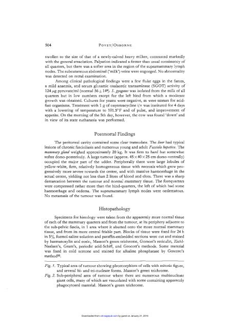
Fig. I. Typical area of tumour show<strong>in</strong>g pleomorphism of cells with mitotic figure,<br />
and several bi- and tri-nucleate forms. Masson’s green trichrome.<br />
Fig. 2. Sub-peripheral area of tumour where <strong>the</strong>re are numerous mult<strong>in</strong>ucleate<br />
giant cells, many of which are vacuolated with some conta<strong>in</strong><strong>in</strong>g apparently<br />
phagocytosed material. Masson’s green trichrome.<br />
Downloaded from vet.sagepub.com by guest on January 21, 2014