Mammary Gland Neoplasia in the Cow - Veterinary Pathology
Mammary Gland Neoplasia in the Cow - Veterinary Pathology
Mammary Gland Neoplasia in the Cow - Veterinary Pathology

You also want an ePaper? Increase the reach of your titles
YUMPU automatically turns print PDFs into web optimized ePapers that Google loves.
506 POVEY/~SBORNE<br />
The periphery of <strong>the</strong> tumour was well demarcated dorsally by dense collagenous<br />
tissue of <strong>the</strong> deep abdom<strong>in</strong>al fascia <strong>in</strong> which was <strong>in</strong>corporated a th<strong>in</strong> strip<br />
of striated muscle and ventro-laterally by th<strong>in</strong> but fairly dense collagenous septa<br />
from normal glandular tissue. The neoplastic tissue was densely cellular, with <strong>the</strong><br />
typical cell ra<strong>the</strong>r ellipsoid, averag<strong>in</strong>g 20 by 15 ,LA, with a central round or oval<br />
nucleus with sharp nuclear membrane, stippl<strong>in</strong>g of chromat<strong>in</strong>, and a variable<br />
number of small nucleoli (Fig. 1). The cytoplasm was eos<strong>in</strong>ophilic and tended to<br />
be drawn out <strong>in</strong>to stellate strands, l<strong>in</strong>k<strong>in</strong>g up with <strong>the</strong> cytoplasmic processes of<br />
neighbour<strong>in</strong>g cells. Mitotic figures were few <strong>in</strong> number (averag<strong>in</strong>g 1 per 10 fields<br />
at 320 x magnification). A strik<strong>in</strong>g feature was <strong>the</strong> large number of mult<strong>in</strong>ucleate<br />
giant cells with up to 30 nuclei, sometimes very hyperchromatic, which tended to<br />
be conglomerated towards <strong>the</strong> centre of <strong>the</strong> cell ra<strong>the</strong>r than around its periphery.<br />
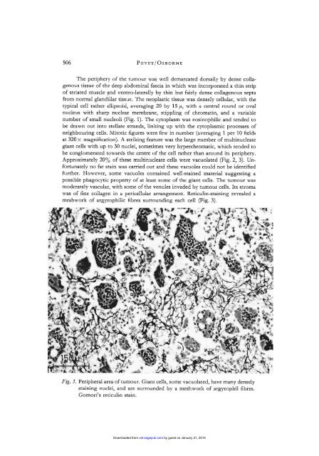
Approximately 20% of <strong>the</strong>se mult<strong>in</strong>ucleate cells were vacuolated (Fig. 2, 3). Unfortunately<br />
no fat sta<strong>in</strong> was carried out and <strong>the</strong>se vacuoles could not be identified<br />
fur<strong>the</strong>r. However, some vacuoles conta<strong>in</strong>ed well-sta<strong>in</strong>ed material suggest<strong>in</strong>g a<br />
possible phagocytic property of at least some of <strong>the</strong> giant cells. The tumour was<br />
moderately vascular, with some of <strong>the</strong> venules <strong>in</strong>vaded by tumour cells. Its stroma<br />
was of f<strong>in</strong>e collagen <strong>in</strong> a pericellular arrangement. Reticul<strong>in</strong>-sta<strong>in</strong><strong>in</strong>g revealed a<br />
meshwork of argyrophilic fibres surround<strong>in</strong>g each cell (Fig. 3).<br />
Fig. 3. Peripheral area of tumour. Giant cells, some vacuolated, have many densely<br />
sta<strong>in</strong><strong>in</strong>g nuclei, and are surrounded by a meshwork of argyrophil fibres.<br />
Gomori’s reticul<strong>in</strong> sta<strong>in</strong>.<br />
Downloaded from vet.sagepub.com by guest on January 21, 2014