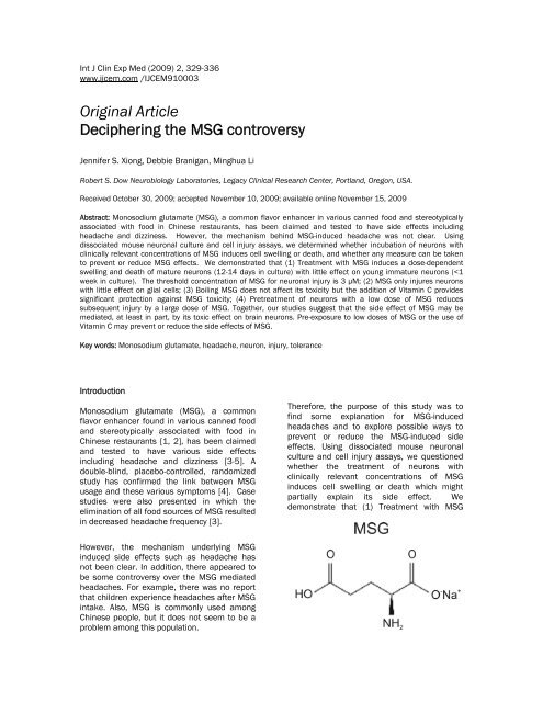
Deciphering the MSG controversy
Deciphering the MSG controversy
Deciphering the MSG controversy
Create successful ePaper yourself
Turn your PDF publications into a flip-book with our unique Google optimized e-Paper software.
Int J Clin Exp Med (2009) 2, 329-336<br />
www.ijcem.com /IJCEM910003<br />
Original Article<br />
<strong>Deciphering</strong> <strong>the</strong> <strong>MSG</strong> <strong>controversy</strong><br />
Jennifer S. Xiong, Debbie Branigan, Minghua Li<br />
Robert S. Dow Neurobiology Laboratories, Legacy Clinical Research Center, Portland, Oregon, USA.<br />
Received October 30, 2009; accepted November 10, 2009; available online November 15, 2009<br />
Abstract: Monosodium glutamate (<strong>MSG</strong>), a common flavor enhancer in various canned food and stereotypically<br />
associated with food in Chinese restaurants, has been claimed and tested to have side effects including<br />
headache and dizziness. However, <strong>the</strong> mechanism behind <strong>MSG</strong>-induced headache was not clear. Using<br />
dissociated mouse neuronal culture and cell injury assays, we determined whe<strong>the</strong>r incubation of neurons with<br />
clinically relevant concentrations of <strong>MSG</strong> induces cell swelling or death, and whe<strong>the</strong>r any measure can be taken<br />
to prevent or reduce <strong>MSG</strong> effects. We demonstrated that (1) Treatment with <strong>MSG</strong> induces a dose-dependent<br />
swelling and death of mature neurons (12-14 days in culture) with little effect on young immature neurons (
<strong>MSG</strong> toxicity in neurons activation<br />
induces a dose-dependent swelling and death<br />
of mature neurons (12-14 days in culture) with<br />
little effect on young immature neurons (
<strong>MSG</strong> toxicity in neurons activation<br />
with <strong>MSG</strong>. As shown in Figure 3, 30 minutes<br />
after <strong>the</strong> <strong>MSG</strong> treatment, a clear swelling of<br />
neurons was observed. After 2h of <strong>MSG</strong><br />
treatment, severe swelling of neurons was<br />
seen. 12h after <strong>MSG</strong> treatment, neurons were<br />
completely lysed. Thus, neuronal damage by<br />
<strong>MSG</strong> is time dependent.<br />
<strong>MSG</strong> has less effect on immature neurons<br />
Figure 1. Dose-dependent injury of mature<br />
neurons by <strong>MSG</strong> (3 - 300 µM). At day 14,<br />
cultured mouse cortical neurons in 24 wells<br />
were treated with different concentrations of<br />
<strong>MSG</strong> for 12h. 50 µl sample of culture medium<br />
was withdrawn from each well for LDH<br />
measurement. Cells in each well were <strong>the</strong>n<br />
lysed with 0.5% Triton X-100. This treatment<br />
gives total LDH. Percentage of cell death is<br />
calculated by dividing <strong>the</strong> LDH value by <strong>the</strong> total<br />
value in each well. n = 8, *p
<strong>MSG</strong> toxicity in neurons activation<br />
Figure 3. Time-dependent injury of neuronal cells by 30 µM <strong>MSG</strong>. Cultured mouse cortical neurons in 35<br />
mm dish were continuously monitored after treatment with 30 µM <strong>MSG</strong>. Neurons started to swell even after<br />
30 minutes of <strong>MSG</strong> treatment. The swelling got worse at 2h. 12h after <strong>MSG</strong> treatment, neurons were lysed.<br />
Scale bar: 15 µm.<br />
µM of <strong>MSG</strong> for 24h. As shown in Figure 5,<br />
treatment of glial cells with <strong>MSG</strong>, at<br />
concentrations as high as 300 µM, did not<br />
cause clear damage. Thus, <strong>MSG</strong>-induced cell<br />
injury is neuron specific.<br />
Boiling does not reduce damaging effect of<br />
<strong>MSG</strong><br />
Next, we explored possible ways of preventing<br />
or reducing <strong>the</strong> <strong>MSG</strong> damage of neurons. We<br />
first determined whe<strong>the</strong>r boiling <strong>MSG</strong> for 10<br />
min reduces its toxicity. 14 days old cultured<br />
neurons were treated with boiled <strong>MSG</strong> (30 µM)<br />
for 12h. As shown in Figure 6, treatment of<br />
neurons with boiled <strong>MSG</strong> produced a similar<br />
injury of neurons as non-boiled <strong>MSG</strong> (Figure<br />
6). Therefore, boiling <strong>MSG</strong> is not sufficient to<br />
reduce its damaging effect.<br />
Vitamin C reduces <strong>MSG</strong> damage of neurons<br />
Ascorbic acid or Vitamin C is an important<br />
endogenous neuroprotectant in <strong>the</strong> brain<br />
[10;11]. It has been show to be<br />
neuroprotective in various conditions.<br />
Therefore, we tested whe<strong>the</strong>r <strong>the</strong> addition of<br />
Vitamin C has any effect on <strong>MSG</strong>-induced<br />
damage of neurons. 1 mM vitamin C was<br />
added to <strong>the</strong> culture medium during <strong>MSG</strong><br />
Figure 4. Younger neurons (6-7 days old) were<br />
not sensitive to <strong>MSG</strong> damage. At day 6-7,<br />
cultured mouse cortical neurons in 24 wells<br />
were treated with different concentrations of<br />
<strong>MSG</strong> for 12h. 50 µl sample of medium was<br />
withdrawn from each well for LDH<br />
measurement. Cells were <strong>the</strong>n lysed with 0.5%<br />
Triton X-100. This treatment gives total LDH.<br />
Percentage of cell death is calculated by<br />
dividing <strong>the</strong> LDH value by <strong>the</strong> total value in<br />
each well. n = 8.<br />
treatment. As shown in Figure 7A and B,<br />
treating neurons with vitamin C provided a<br />
clear protection against <strong>MSG</strong> damage (n=3<br />
experiments).<br />
Pretreatment of neurons with a low dose of<br />
<strong>MSG</strong> reduces <strong>the</strong> injury of neurons by<br />
subsequent high dose of <strong>MSG</strong><br />
332<br />
Int J Clin Exp Med (2009) 2, 329-336
<strong>MSG</strong> toxicity in neurons activation<br />
Figure 5. <strong>MSG</strong> does not damage glial cells. Individual dishes containing cultured glial cells were treated with<br />
0, 3, 30, or 300 µM <strong>MSG</strong> for 24h. Cells were <strong>the</strong>n labeled with fluorescein diacetate. Same experiments<br />
were repeated 3 times. <strong>MSG</strong> did not cause significant damage to glial cells even at 300 µM.<br />
Figure 6. Boiling <strong>MSG</strong> does not reduce its toxic effect. At day 14, cultured mouse cortical neurons in 35 mm<br />
culture dishes were treated with 30 µM non-boiled <strong>MSG</strong> or boiled <strong>MSG</strong> for 12h. Treatment of neurons with<br />
boiled <strong>MSG</strong> produced <strong>the</strong> same damage as non-boiled <strong>MSG</strong>. The same experiments were repeated 3 times.<br />
Scale bar: 20 µm.<br />
It has been widely accepted that a stimulus or<br />
stressor capable of causing injury to a tissue<br />
or organ may, when applied close to <strong>the</strong><br />
threshold of damage, activate endogenous<br />
protective mechanisms which can potentially<br />
reduce <strong>the</strong> damaging effect of subsequent,<br />
more severe stimuli [12]. For example, a subthreshold<br />
ischemic insult applied to <strong>the</strong> brain<br />
has been shown to activate certain cellular<br />
pathways that can help to reduce damage<br />
333<br />
Int J Clin Exp Med (2009) 2, 329-336
<strong>MSG</strong> toxicity in neurons activation<br />
<strong>MSG</strong>. We selected 3 µM <strong>MSG</strong> since it did not<br />
produce much injury. Neurons were first<br />
treated with 3 µM <strong>MSG</strong> for 12h, followed by<br />
treatment with 30 µM <strong>MSG</strong> for 24h. As shown<br />
in Figure 7A and C, pre-treating neurons with 3<br />
µM <strong>MSG</strong> for 12h reduced <strong>the</strong> subsequent<br />
neuronal injury by 30 µM <strong>MSG</strong> (n=3<br />
experiments). This data suggests that taking<br />
a low and non-damaging dose of <strong>MSG</strong> may<br />
prevent injury of neurons by subsequent high<br />
doses of <strong>MSG</strong>.<br />
Discussion<br />
Figure 7. Treatment with Vitamin C or<br />
pretreatment with low dose of <strong>MSG</strong> reduced<br />
<strong>MSG</strong> damage of mature neurons. A. Neurons<br />
treated with 30 µM <strong>MSG</strong> alone for 24h. B.<br />
Neurons treated with 30 µM <strong>MSG</strong> + 1 mM<br />
Vitamin C for 24h. C. Neurons were treated<br />
with 3 µM <strong>MSG</strong> for 12h followed by 30 µM <strong>MSG</strong><br />
for 24h. Scale bar: 20 µm.<br />
caused by subsequent long ischemic episodes,<br />
a phenomenon known as ischemic<br />
preconditioning or ischemic tolerance [13].<br />
For this reason, we determined whe<strong>the</strong>r a<br />
pretreatment of mature neurons with a low<br />
dose of <strong>MSG</strong> can establish tolerance against<br />
injury of neurons by subsequent high doses of<br />
Since <strong>the</strong> report of “Chinese Restaurant<br />
Syndrome” in 1968, various studies have<br />
confirmed <strong>the</strong> link between <strong>MSG</strong> intake and its<br />
side effects including headaches [4, 5]. In<br />
those studies, an oral dose of <strong>MSG</strong> at 3g or<br />
higher reproduced restaurant syndrome within<br />
30 min [5]. A direct intravenous dose of 50<br />
mg also produced similar symptoms.<br />
Although <strong>the</strong> <strong>MSG</strong> concentration in <strong>the</strong> blood<br />
is difficult to predict with oral doses of <strong>MSG</strong>, a<br />
direct intravenous injection of 50 mg <strong>MSG</strong> is<br />
expected to yield a blood concentration of 53<br />
µM (based on <strong>the</strong> total blood volume of 5<br />
litters). Although <strong>the</strong> blood brain barrier (BBB)<br />
has low permeability to <strong>MSG</strong>, <strong>the</strong> presence of<br />
high affinity glutamate transporters located at<br />
<strong>the</strong> BBB capillary luminal membrane could<br />
facilitate <strong>the</strong> uptake of <strong>MSG</strong> into <strong>the</strong> brain.<br />
Assuming 10% of <strong>the</strong> blood concentration can<br />
be reached in <strong>the</strong> cerebral spinal fluid, it would<br />
yield ~5 µM <strong>MSG</strong> in <strong>the</strong> brain which is above<br />
<strong>the</strong> threshold concentration of <strong>MSG</strong> for<br />
causing neuronal injury in our studies. Thus,<br />
<strong>the</strong> concentrations of <strong>MSG</strong> used in our studies<br />
were clinically relevant. Our finding that<br />
neuronal swelling takes place at ~30 min after<br />
<strong>MSG</strong> treatment is also consistent with <strong>the</strong> time<br />
course of <strong>MSG</strong>-induced headache in clinical<br />
studies.<br />
Using neuronal culture technique and cell<br />
injury assay, we studied <strong>the</strong> effect of <strong>MSG</strong> on<br />
mouse cortical neurons, a commonly used in<br />
vitro preparation for cell injury studies. We<br />
demonstrated that incubation with <strong>MSG</strong>, at<br />
clinical relevant concentrations, induced<br />
swelling and injury of mature neurons. This<br />
finding may partially explain <strong>the</strong> headache<br />
induced by <strong>MSG</strong> intake.<br />
In contrast to mature neurons, immature<br />
neurons are very resistant to <strong>MSG</strong> damage.<br />
For example, incubation of immature neurons<br />
334<br />
Int J Clin Exp Med (2009) 2, 329-336
<strong>MSG</strong> toxicity in neurons activation<br />
with 30 µM <strong>MSG</strong>, a concentration sufficient to<br />
induce substantial injury of mature neurons,<br />
did not induce clear injury of immature<br />
neurons. This finding might explain why young<br />
children, whose brain neurons are not fully<br />
mature, do not experience headaches after<br />
<strong>MSG</strong> intake.<br />
We showed that boiling <strong>MSG</strong> is not sufficient<br />
to reduce its toxicity. However, taking vitamin<br />
C is effective in reducing <strong>MSG</strong>-induced<br />
neuronal injury. More interestingly, we<br />
demonstrated that pretreatment of neurons<br />
with a low dose of <strong>MSG</strong> can make neurons<br />
tolerant to subsequent high doses of <strong>MSG</strong>.<br />
Although <strong>the</strong> exact mechanism underlying this<br />
tolerance remains to be determined, such<br />
knowledge may give us an idea of how to take<br />
preventative measures for <strong>MSG</strong>-induced<br />
headaches.<br />
The reason why young neurons are not<br />
damaged by <strong>MSG</strong> is not clear. Since young<br />
neurons are not fully developed, <strong>the</strong>y may be<br />
missing some receptors that are required for<br />
<strong>MSG</strong> binding or downstream signaling<br />
pathway(s) required for <strong>MSG</strong> toxicity. The<br />
finding that pre-exposure of a low dose of <strong>MSG</strong><br />
can generate tolerance may explain why<br />
Chinese populations do not experience<br />
headaches after <strong>MSG</strong> intake. It is likely that<br />
<strong>the</strong>y have already established tolerance to<br />
<strong>MSG</strong> due to previous exposures to low doses<br />
of <strong>MSG</strong> when <strong>the</strong>y were young. In <strong>the</strong> present<br />
study, we only examined <strong>the</strong> tolerance 12h<br />
after pre-exposure to <strong>MSG</strong>. In future studies, it<br />
would be interesting to determine whe<strong>the</strong>r <strong>the</strong><br />
tolerance can last for days or longer.<br />
The results of <strong>the</strong> “Boiled <strong>MSG</strong>” revealed that<br />
no preventative measures can be made by<br />
over-cooking of <strong>MSG</strong>-containing food. We had<br />
hoped that boiling might be able to destroy <strong>the</strong><br />
structure of <strong>MSG</strong> thus reducing or eliminate its<br />
damaging effects. However, boiling <strong>MSG</strong> for<br />
10 minutes did not reduce any damaging<br />
effects at all. Future studies may consider<br />
boiling <strong>MSG</strong> for a much longer time (e.g. 1h).<br />
Glutamate is an endogenous neurotransmitter<br />
required for a variety of physiological functions<br />
of neurons [14]. Increased release of<br />
endogenous glutamate has been suggested to<br />
play an important role in neuronal injury<br />
associated with a number of neurological<br />
disorders [14, 15]. Previous studies have also<br />
shown that incubation of neurons with high<br />
concentrations of exogenously applied<br />
glutamate (e.g. 500 µM) rapidly induces<br />
neuronal cell death [16]. We show here that<br />
with a threshold concentration of as low as 3<br />
µM, <strong>MSG</strong> can induce detectable neuronal<br />
injury if incubated for a prolonged period of<br />
time. Fur<strong>the</strong>r studies may determine whe<strong>the</strong>r<br />
cell injury induced by low doses of <strong>MSG</strong> share<br />
o<strong>the</strong>r characteristics with previously reported<br />
glutamate toxicity, e.g. reduced by removal of<br />
<strong>the</strong> extracellular Ca 2+ [16] and blockade by<br />
glutamate antagonists.<br />
In conclusion, our studies suggest that <strong>the</strong><br />
headaches caused by <strong>MSG</strong> intake may be<br />
related to its injurious effect on brain neurons.<br />
Although <strong>the</strong> brain does not register pain<br />
because of <strong>the</strong> lack of nociceptors, <strong>the</strong><br />
increase of intracranial pressure due to cell<br />
swelling is well known to cause headaches.<br />
There are some possible ways, e.g. taking<br />
Vitamin C or pre-exposure to a low dose of<br />
<strong>MSG</strong>, that may ei<strong>the</strong>r prevent or reduce <strong>the</strong><br />
side effects of <strong>MSG</strong>.<br />
Address correspondence to: Minghua Li, PhD,<br />
Robert S. Dow Neurobiology Laboratories, Legacy<br />
Clinical Research Center, 1225 NE 2 nd Ave.,<br />
Portland, Oregon, USA. Tel: (503) 4135326, Fax:<br />
(503) 4135465, E-mail: mli@downeurobiology.org<br />
References<br />
[1] Morselli PL, Garattini S. Monosodium<br />
glutamate and <strong>the</strong> Chinese restaurant<br />
syndrome. Nature 1970; 227(5258):611-612.<br />
[2] Schaumburg H. Chinese-restaurant syndrome.<br />
N Engl J Med 1968; 278(20):1122.<br />
[3] Scopp AL. <strong>MSG</strong> and hydrolyzed vegetable<br />
protein induced headache: review and case<br />
studies. Headache 1991; 31(2):107-110.<br />
[4] Yang WH, Drouin MA, Herbert M, Mao Y, Karsh<br />
J. The monosodium glutamate symptom<br />
complex: assessment in a double-blind,<br />
placebo-controlled, randomized study. J Allergy<br />
Clin Immunol 1997; 99(6 Pt 1):757-762.<br />
[5] Schaumburg HH, Byck R, Gerstl R, Mashman<br />
JH. Monosodium L-glutamate: its pharmacology<br />
and role in <strong>the</strong> Chinese restaurant syndrome.<br />
Science 1969; 163(869):826-828.<br />
[6] Xiong ZG, Zhu XM, Chu XP, Minami M, Hey J,<br />
Wei WL, MacDonald JF, Wemmie JA, Price MP,<br />
Welsh MJ, Simon RP. Neuroprotection in<br />
ischemia: blocking calcium-permeable Acidsensing<br />
ion channels. Cell 2004; 118(6):687-<br />
698.<br />
[7] Chu XP, Wemmie JA, Wang WZ, Zhu XM,<br />
Saugstad JA, Price MP, Simon RP, Xiong ZG.<br />
Subunit-dependent high-affinity zinc inhibition<br />
of acid-sensing ion channels. J Neurosci 2004;<br />
335<br />
Int J Clin Exp Med (2009) 2, 329-336
<strong>MSG</strong> toxicity in neurons activation<br />
24(40):8678-8689.<br />
[8] Mukhin AG, Ivanova SA, Allen JW, Faden AI.<br />
Mechanical injury to neuronal/glial cultures in<br />
microplates: role of NMDA receptors and pH in<br />
secondary neuronal cell death. J Neurosci Res<br />
1998; 51(6):748-758.<br />
[9] Koh JY, Choi DW. Quantitative determination of<br />
glutamate mediated cortical neuronal injury in<br />
cell culture by lactate dehydrogenase efflux<br />
assay. J Neurosci Methods 1987; 20(1):83-90.<br />
[10] Rice ME. Ascorbate regulation and its<br />
neuroprotective role in <strong>the</strong> brain. Trends<br />
Neurosci 2000; 23(5):209-216.<br />
[11] Grunewald RA. Ascorbic acid in <strong>the</strong> brain. Brain<br />
Res Brain Res Rev 1993; 18(1):123-133.<br />
[12] Dirnagl U, Simon RP, Hallenbeck JM. Ischemic<br />
tolerance and endogenous neuroprotection.<br />
Trends Neurosci 2003; 26(5):248-254.<br />
[13] Stenzel-Poore MP, Stevens SL, Xiong Z, Lessov<br />
NS, Harrington CA, Mori M, Meller R,<br />
Rosenzweig HL, Tobar E, Shaw TE, Chu X,<br />
Simon RP. Effect of ischaemic preconditioning<br />
on genomic response to cerebral ischaemia:<br />
similarity to neuroprotective strategies in<br />
hibernation and hypoxia-tolerant states. Lancet<br />
2003; 362(9389):1028-1037.<br />
[14] Greenamyre JT. The role of glutamate in<br />
neurotransmission and in neurologic disease.<br />
Arch Neurol 1986; 43(10):1058-1063.<br />
[15] Rothman SM, Olney JW. Glutamate and <strong>the</strong><br />
pathophysiology of hypoxic--ischemic brain<br />
damage. Ann Neurol 1986; 19(2):105-111.<br />
[16] Choi DW. Glutamate neurotoxicity in cortical<br />
cell culture is calcium dependent. Neurosci<br />
Lett 1985; 58(3):293-297.<br />
336<br />
Int J Clin Exp Med (2009) 2, 329-336


