Hypocalcitonemia in Handigodu Disease: a spondylo epi (meta ...
Hypocalcitonemia in Handigodu Disease: a spondylo epi (meta ...
Hypocalcitonemia in Handigodu Disease: a spondylo epi (meta ...

You also want an ePaper? Increase the reach of your titles
YUMPU automatically turns print PDFs into web optimized ePapers that Google loves.
Int J Cl<strong>in</strong> Exp Med 2010;3(2):115-121<br />
www.ijcem.com /IJCEM1001003<br />
Orig<strong>in</strong>al Article<br />
<strong>Hypocalcitonemia</strong> <strong>in</strong> <strong>Handigodu</strong> <strong>Disease</strong>: a <strong>spondylo</strong><br />
<strong>epi</strong> (<strong>meta</strong>) physeal dysplasia<br />
Mallikarjun Badadani 1*, K. Taranath Shetty 1, S.S.Agarwal 2<br />
1Department of Neurochemistry, National Institute of Mental Health and Neuro Sciences, Bangalore-560029, India.<br />
2Advanced Center for Treatment Research and Education <strong>in</strong> Cancer (ACTREC), Navi Mumbai.<br />
Received January 17, 2010, accepted April 15, 2010, available onl<strong>in</strong>e April 25, 2010<br />
Abstract: <strong>Handigodu</strong> <strong>Disease</strong> (HD) is a disorder of the osteoarticular system which is highly prevalent <strong>in</strong> several villages<br />
of two districts viz, Shimoga and Chikmaglur of the state of Karnataka, southern India. The scientific name of<br />
the disease is Spondylo-<strong>epi</strong>-(<strong>meta</strong>) physeal Dysplasia, Autosomal Dom<strong>in</strong>ant variety, <strong>Handigodu</strong> syndrome. The same<br />
has been listed <strong>in</strong> the International Classification of Skeletal Dysplasias. The calcium homeostasis study was lack <strong>in</strong><br />
HD. The serum calcium, phosphorus, parathyroid hormone and calciton<strong>in</strong> levels after overnight fast state, and 24<br />
hour ur<strong>in</strong>ary excretion of calcium and phosphorus were quantified. The decreased level of calciton<strong>in</strong> associated with<br />
decreased serum total calcium and ur<strong>in</strong>ary calcium <strong>in</strong> HD were observed. The levels of parathyroid hormone, serum<br />
phosphorus and ur<strong>in</strong>ary phosphorus rema<strong>in</strong> unchanged among HD affected. The Vitam<strong>in</strong> D3 levels also noticed unchanged<br />
<strong>in</strong> HD affected. S<strong>in</strong>ce calciton<strong>in</strong> has antiresorption effect on bone, the observed low calciton<strong>in</strong> <strong>in</strong> HD may<br />
imply reosrption of bone lead<strong>in</strong>g to deformity and causes hypocalcaemia and hypocalciuria. The hypocalcitonemia<br />
without change <strong>in</strong> iPTH associated with hypocalcaemia may be a mutation <strong>in</strong> Vit D receptor (VDR) or may be an<br />
<strong>epi</strong>phenomenon.<br />
Keywords: Calciton<strong>in</strong>, hypocalciuria, parathyroid hormone<br />
Introduction<br />
<strong>Handigodu</strong> <strong>Disease</strong> (HD) is a disorder of the<br />
osteoarticular system prevalent <strong>in</strong> several villages<br />
of two districts viz, Shimoga and Chickmaglur<br />
<strong>in</strong> the state of Karnataka, southern India.<br />
The disease was first identified <strong>in</strong> a patient<br />
from <strong>Handigodu</strong> village <strong>in</strong> 1975, hence its name<br />
[1]. The data obta<strong>in</strong>ed dur<strong>in</strong>g subsequent multidiscipl<strong>in</strong>ary<br />
study by Indian Council of Medical<br />
Research (ICMR) showed that HD is a Spondylo<br />
<strong>epi</strong> (<strong>meta</strong>) physeal dysplasia, <strong>in</strong>herited as an<br />
autosomal dom<strong>in</strong>ant trait [2]. The cl<strong>in</strong>ical presentation<br />
of patients with HD has been identified<br />
<strong>in</strong>to three sub types such as Arthritic type (Type<br />
I), Dysplastic type (Type II) and Dwarf type (Type<br />
III). All three sub types segregate <strong>in</strong> the same<br />
families implies often seen <strong>in</strong> the same family.<br />
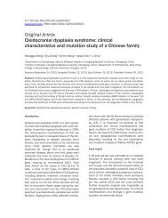
The X-ray pictures show<strong>in</strong>g the marked abnormalities<br />
are d<strong>epi</strong>cted <strong>in</strong> the Figure 1 and Figure<br />
2.<br />
The type I was characterized by late presentation<br />
of disease (45-50 years of age), characterized<br />
by pa<strong>in</strong> <strong>in</strong> hip jo<strong>in</strong>ts with difficulty <strong>in</strong> walk<strong>in</strong>g;<br />
and on exam<strong>in</strong>ation these <strong>in</strong>dividuals had<br />
near normal height and body proportions<br />
(without significant truncal shorten<strong>in</strong>g), <strong>in</strong>ability<br />
to sit cross-legged, fixed flexion deformity of<br />
hips and compensatory lumbar lordosis. Radiologically<br />
these <strong>in</strong>dividuals showed characteristic<br />
changes of osteoarthritis at the hip jo<strong>in</strong>ts<br />
bilaterally, without any abnormality of sp<strong>in</strong>e or<br />
<strong>in</strong>volvement of other bones. The type II of<br />
<strong>Handigodu</strong> disease was characterized by short<br />
stature, particularly truncal shorten<strong>in</strong>g. These<br />
patients were relatively younger (25-35 years of<br />
age) and the ma<strong>in</strong> compla<strong>in</strong>t was <strong>in</strong>ability to<br />
walk for longer distance. The X-rays of these<br />
patients showed dysplastic femoral heads bilaterally,<br />
and vary<strong>in</strong>g degrees of platyspondyly. The<br />
type III <strong>Handigodu</strong> syndrome was marked by
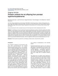
<strong>Hypocalcitonemia</strong> <strong>in</strong> <strong>Handigodu</strong> disease<br />
Figure 1. X-Ray radiograph of hip jo<strong>in</strong>ts from <strong>Handigodu</strong> <strong>Disease</strong> (HD) (A-E): The radiograph osteoarthritis <strong>in</strong> hip jo<strong>in</strong>ts<br />
of Type I subgroup are shown <strong>in</strong> A-C whereas dysplasia of hip jo<strong>in</strong>ts from Type II and Type III subgroups are presented<br />
<strong>in</strong> D and E respectively.<br />
dwarfism. They also had vary<strong>in</strong>g types of associated<br />
skeletal anomalies, particularly at the<br />
knees and hands. In these patients there was<br />
marked <strong>epi</strong>physeal dysplasia at hips, knees,<br />
hands and wrists besides significant<br />
platyspondyly [2, 3]. The similarities between<br />
the <strong>Handigodu</strong> <strong>Disease</strong> and Mseleni jo<strong>in</strong>t disease<br />
reported from South Africa [3].<br />
Unpublished f<strong>in</strong>d<strong>in</strong>gs of alterations <strong>in</strong> calcium<br />
homeostasis were observed by the group of<br />
Dr.Teotia and colleagues (as part of the ICMR<br />
multidiscipl<strong>in</strong>ary study). S<strong>in</strong>ce there was a deficit<br />
of study on m<strong>in</strong>eral <strong>meta</strong>bolism of bone with<br />
respect to hormones <strong>in</strong> HD. The calcium homeostasis<br />
was studied by quantify<strong>in</strong>g serum calcium,<br />
phosphorus, parathyroid hormone and<br />
calciton<strong>in</strong> <strong>in</strong> overnight fast state of serum. The<br />
calcium and phosphorus excretions for 24 hrs<br />
ur<strong>in</strong>e volume were also quantified to avoid <strong>in</strong>terference<br />
of diurnal rhythms and food habbits.<br />
Materials and methods<br />
Subjects<br />
The HD affected patients cl<strong>in</strong>ically and x-ray<br />
radiological confirmed (Type I; n=19, Type II; n=<br />
28 and Type III; n=8) have been selected for<br />
this study with<strong>in</strong> age group of 20-60 years old<br />
with their consent. The sixty three (Nonaffected;<br />
n=63) healthy <strong>in</strong>dividuals with age group of 20-<br />
60 years old from the non related family members<br />
of HD affected were enrolled <strong>in</strong> this study<br />
as controls. The patients were enrolled from the<br />
field. A door to door survey had been done of<br />
affected communities <strong>in</strong> all the affected villages<br />
of the Sagar taluk of District Shimoga <strong>in</strong> the<br />
state of Karnataka. X-ray exam<strong>in</strong>ation had been<br />
done on all the family members (except pregnant<br />
women), <strong>in</strong> affected families with their consent.<br />
Only cases confirmed by both physical<br />
exam<strong>in</strong>ation and evaluation of X-rays were <strong>in</strong>cluded<br />
<strong>in</strong> the study, from amongst those who<br />
were will<strong>in</strong>g to be admitted <strong>in</strong> the hospital for<br />
collection of 24 hour ur<strong>in</strong>e sample and overnight<br />
fast serum. The X-ray pictures were elucidated<br />
<strong>in</strong> Figure 1 and Figure 2. All the controls<br />
cl<strong>in</strong>ical and by x-ray diagnosis were normal.<br />
Ethics<br />
Both the affected and control subjects were<br />
expla<strong>in</strong>ed the purpose of the study and written<br />
<strong>in</strong>formed consent was obta<strong>in</strong>ed prior to the collection<br />
of specimens. The study was approved<br />
116 Int J Cl<strong>in</strong> Exp Med 2010;3(2):115-121
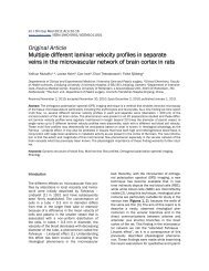
<strong>Hypocalcitonemia</strong> <strong>in</strong> <strong>Handigodu</strong> disease<br />
Figure 2. X-Ray radiograph of Lumbar sp<strong>in</strong>e from <strong>Handigodu</strong> <strong>Disease</strong> (HD) (A-C): Lateral view Lumbar sp<strong>in</strong>e show<strong>in</strong>g<br />
vary<strong>in</strong>g degrees of platyspondyly <strong>in</strong> Type I (A), Type II (B) and Type III (C) subgroups.<br />
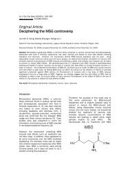
Figure 3. Calcium and Phosphorus level of sera <strong>in</strong><br />
<strong>Handigodu</strong> <strong>Disease</strong> (HD) (A-B): Serum total Calcium<br />
(A) and Phosphorus (B) levels <strong>in</strong> HD subgroups Type I,<br />
Type II, and Type III compared with nonaffected family<br />
group. Note significantly decreased level of Calcium<br />
but unchanged Phosphorus <strong>in</strong> HD subgroups compared<br />
to nonaffected family group.<br />
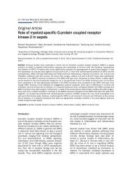
Figure 4. The excretory level of Calcium and Phosphorus<br />
<strong>in</strong> <strong>Handigodu</strong> <strong>Disease</strong> (HD) (A-B): The 24<br />
hours ur<strong>in</strong>ary excretion of Calcium (A) and Phosphorus<br />
(B) levels <strong>in</strong> HD subgroups Type I, Type II, and<br />
Type III compared with nonaffected family group.<br />
There was significant decrease <strong>in</strong> level of Calcium<br />
but unchanged Phosphorus excretion <strong>in</strong> HD subgroups<br />
compared to nonaffected family group.<br />
117 Int J Cl<strong>in</strong> Exp Med 2010;3(2):115-121
Figure 5. The Parathyroid hormone and Calciton<strong>in</strong><br />
level of sera <strong>in</strong> <strong>Handigodu</strong> <strong>Disease</strong> (HD) (A-B): Serum<br />
Parathyroid hormone (A) and Calciton<strong>in</strong> (B) levels <strong>in</strong><br />
HD subgroups Type I, Type II, and Type III compared<br />
with nonaffected family group. There was significant<br />
decrease <strong>in</strong> level of Calciton<strong>in</strong> and unchanged parathyroid<br />
hormone level <strong>in</strong> HD subgroups compared to<br />
nonaffected family group.<br />
by <strong>in</strong>stitute’s human ethics committee.<br />
Biomarkers<br />
The subjects were asked to empty their bladder<br />
prior to the collection of 24 hrs ur<strong>in</strong>e and Hydrochloric<br />
acid (HCl) was used as preservative to<br />
avoid microbial deterioration. The serum samples<br />
were collected after an overnight fast<strong>in</strong>g<br />
state. All of them (affected and control group)<br />
rema<strong>in</strong>ed on their usual home diet dur<strong>in</strong>g the<br />
24 hours ur<strong>in</strong>e collection. The serum and ur<strong>in</strong>ary<br />
levels of calcium and phosphorus were<br />
determ<strong>in</strong>ed by automated serum chemistry analyzer<br />
(Olympus). Calcium and phosphorus excretions<br />
were related to 24 hrs ur<strong>in</strong>e volume collected<br />
and creat<strong>in</strong><strong>in</strong>e levels excreted. The serum<br />
iPTH and Calciton<strong>in</strong> levels were determ<strong>in</strong>ed<br />
by Immulite 2000 reagents.<br />
Statistical analysis<br />
The Data were analyzed by us<strong>in</strong>g SPSS programme<br />
of W<strong>in</strong>dows 2000. Statistical significance<br />
was determ<strong>in</strong>ed with one- way ANOVA<br />
followed by Bonferroni T-test and represented<br />
by ‘p’ value. The ‘p’ values were determ<strong>in</strong>ed for<br />
<strong>Hypocalcitonemia</strong> <strong>in</strong> <strong>Handigodu</strong> disease<br />
different HD groups <strong>in</strong> comparison with non<br />
affected group as controls. The probability values,<br />
≤ 0.05 were considered as statistically significant.<br />
The p values were shown with respective<br />
data presented <strong>in</strong> Figures.<br />
Results<br />
The calcium homeostasis was studied by quantify<strong>in</strong>g<br />
calcium, phosphorus <strong>in</strong> ur<strong>in</strong>e and serum,<br />
and hormones from serum.<br />
The serum total calcium (mg/dl) levels<br />
(Nonaffected: 9.58 ± 0.39, Type I: 8.83 ±0.59,<br />
Type II: 8.74 ± 0.73, Type III: 8.61± 0.65) were<br />
found to be significantly less <strong>in</strong> affected group<br />
(Figure 3A). Interest<strong>in</strong>gly, serum phosphorus<br />
(mg/dl) levels (Nonaffected: 3.77 ± 0.64,Type I:<br />
3.29 ± 0.54 Type II: 3.15 ± 0.66, Type III: 3.10<br />
± 0.43) were found to be rema<strong>in</strong> unchanged<br />
(Figure 3B). The excretory levels of calcium <strong>in</strong><br />
24 hrs ur<strong>in</strong>e volume (mg/24 hrs) (Nonaffected:<br />
103.16 ± 58.65, Type I: 89.51± 44.43, Type II:<br />
83.65 ± 49.13, Type III: 76.42± 39.81) were<br />
significantly lowered <strong>in</strong> affected groups and ur<strong>in</strong>ary<br />
phosphorus levels (mg/24 hrs)<br />
(Nonaffected: 149.43 ± 39.77, Type I: 150.90 ±<br />
41.15, Type II: 182.59 ± 58.56, Type III:<br />
155.14± 73.43) found to be rema<strong>in</strong> unchanged<br />
(Figure 4A and Figure 4B).<br />
Parathyroid hormone (iPTH) (pg/ml) levels <strong>in</strong><br />
fast<strong>in</strong>g state serum between control and affected<br />
groups (Nonaffected: 36.76 ± 20.82,<br />
Type I: 40.7±14.37, Type II: 34.04± 14.91, Type<br />
III: 35.15 ± 18.08) (Figure 5A) rema<strong>in</strong> unchanged,<br />
whereas calciton<strong>in</strong> values (pg/ml)<br />
were significantly lowered <strong>in</strong> HD affected subgroups<br />
(Nonaffected: 1.97 ± 1.16, Type I: 0.522<br />
± 0.372, Type II: 1.125± 1.034, Type III: 0.996<br />
± 0.488) (Figure 5B).<br />
The serum profile of other biochemical parameters<br />
such as total prote<strong>in</strong>, album<strong>in</strong> and Alb/Glb<br />
ratio, the markers for nutritional deficiency were<br />
measured and found without significant differences.<br />
Similarly, SGOT, SGPT, Urea, Creat<strong>in</strong><strong>in</strong>e<br />
and Alkal<strong>in</strong>e Phosphatase values, the markers<br />
for liver function test and kidney function test<br />
were found to be rema<strong>in</strong> unchanged <strong>in</strong> both the<br />
HD affected and unaffected members of HD<br />
families (data not shown).<br />
Discussion<br />
Parathyroid hormone (PTH) synthesized as 115-<br />
118 Int J Cl<strong>in</strong> Exp Med 2010;3(2):115-121
am<strong>in</strong>o acid polypeptide called preproPTH <strong>in</strong><br />
parathyroid cell by polyribosomes located with<strong>in</strong><br />
cell matrix of parathyroid gland. When the grow<strong>in</strong>g<br />
polypeptide cha<strong>in</strong> ~20-30 am<strong>in</strong>o acids long,<br />
the NH2- term<strong>in</strong>us of cha<strong>in</strong> emerges first, and at<br />
this time two NH2 term<strong>in</strong>al methion<strong>in</strong>e residues<br />
clevead by methionyl am<strong>in</strong>o peptidase. As the<br />
nacent cha<strong>in</strong> cont<strong>in</strong>ues to grow the hydrophobic<br />
NH2 term<strong>in</strong>al sequence of preproPTH emerges<br />
and associates with membrane of endoplasmic<br />
reticulum. The sequence of 23 am<strong>in</strong>o acids<br />
from prepro PTH is removed by cleavage of glysyl–lysyl<br />
bond near or <strong>in</strong> reticular membrane by<br />
enzymatic activity to proPTH (90 am<strong>in</strong>o acids).The<br />
pro PTH arrives at Golgi apparatus and<br />
the tryps<strong>in</strong> like activity accomplishes conversion<br />
of proPTH to PTH (iPTH of 1-84 am<strong>in</strong>o acids)<br />
by removal of hexapeptide from NH2 term<strong>in</strong>al<br />
sequence. The transport of PTH at the site<br />
of release by plasma membrane occurs through<br />
secretory granules. PTH is the predom<strong>in</strong>ant<br />
form of hormone stored <strong>in</strong> parathyroid gland [4].<br />
Calcium is the physiologically important regulator<br />
of parathyroid glandular activity for secretion<br />
of PTH. Other secretogogues have been identified<br />
for the regulation of PTH secretion, however<br />
their physiological role not understood completely.<br />
The secretion of PTH is <strong>in</strong>versely dependent<br />
on concentration of extracellular calcium.<br />
PTH <strong>in</strong>creases the concentration calcium<br />
<strong>in</strong> blood and extracellular fluid (ECF) through its<br />
effects on bone, kidney, and gut. The negative<br />
feedback <strong>in</strong>hibition of parathyroid gland contributes<br />
to the regulation of ECF calcium concentrations.<br />
Adenylate cyclase and cAMP appears to<br />
play as an <strong>in</strong>termediate <strong>in</strong> the regulation of PTH<br />
secretion <strong>in</strong> the control of calcium, however the<br />
exact role is not known. It may be that cAMP <strong>in</strong><br />
some manner <strong>in</strong>volved <strong>in</strong> the phosphorylation of<br />
substrate phosphoprote<strong>in</strong> <strong>in</strong> parathyroid gland.<br />
This substrate is membrane prote<strong>in</strong> <strong>in</strong>volved <strong>in</strong><br />
the fusion of secretory granule with plasma<br />
membrane result<strong>in</strong>g <strong>in</strong> discharge of hormone to<br />
extracellular space. The PTH released to extracellular<br />
space by mechanism excitosis [4].<br />
PTH is responsible for regulation of calcium levels<br />
<strong>in</strong> blood and ECF through pr<strong>in</strong>cipal actions<br />
on kidney, bone and <strong>in</strong>test<strong>in</strong>e. The parathyroid<br />
hormone acts on kidney to enhance the reabsorption<br />
of calcium and dim<strong>in</strong>ish the reabsorption<br />
of phosphate. PTH stimulates the formation<br />
of 1, 25 (OH)2D (vitam<strong>in</strong> D2) through its action<br />
on renal 1- α- hydroxylase, which <strong>in</strong> turn has<br />
<strong>Hypocalcitonemia</strong> <strong>in</strong> <strong>Handigodu</strong> disease<br />
direct biological effects on <strong>in</strong>test<strong>in</strong>e. VitD2 <strong>in</strong>creases<br />
the absorption efficiency of dietry calcium<br />
through <strong>in</strong>test<strong>in</strong>e (4, 5].<br />
PTH has several actions at different sites along<br />
the nephron. PTH enhances the calcium reabsorption<br />
at cortical site with<strong>in</strong> the distal tubular<br />
portion and blocks the reabsorption of sodium,<br />
calcium, phosphate, and bicarbonate <strong>in</strong> proximal<br />
tubule. The iPTH decreases tubular reabsorption<br />
of phosphate by accelerat<strong>in</strong>g degradation<br />
of the Type-II sodium dependent phosphate<br />
transporter <strong>in</strong> the proximal tubules [5] The enzymatic<br />
activities of glucose-6-phosphate dehydrogenase<br />
and reduced nicot<strong>in</strong>e aden<strong>in</strong>e d<strong>in</strong>ucleotide<br />
phosphate (NADPH) - diaphorase <strong>in</strong>creases<br />
by exposure to PTH. The activity of latter enzyme<br />
may <strong>in</strong>volve <strong>in</strong> the activation of Vitam<strong>in</strong> D <strong>meta</strong>bolites<br />
by 1- α- hydroxylation. PTH action <strong>in</strong><br />
the kidey occurs through the formation of cAMP<br />
as second messenger cause to changes <strong>in</strong> calcium<br />
transport. PTH stimulated adenylate cyclase<br />
activity is found at basolateral renal tubular<br />
cells adjacent to the renal capillaries. The<br />
<strong>in</strong>tracellular receptor prote<strong>in</strong>s for cAMP are<br />
found <strong>in</strong> brush borders of these cells at lumial<br />
side. The cAMP generated at basolateral region<br />
of cell migrates through cytoplasm to activate<br />
cAMP dependent k<strong>in</strong>ases, <strong>in</strong> turn activate enzymes<br />
responsible for ion transport [4].<br />
In the skeleton, PTH stimulates to resorb the<br />
bone and subsequent release of calcium and<br />
phosphate <strong>in</strong>to the circulation. PTH stimulates<br />
osteocytes, osteoclasts and their precursors. In<br />
hyperparathyroidism bone destruction and concomitant<br />
release of calcium, phosphate and<br />
other salts occurs. Osteocalsts can breakdown<br />
large volume of bone when stimulated at chronical<br />
levels; osteocytes resorb areas of surround<strong>in</strong>g<br />
calcified matrix stimulated by PTH. The action<br />
of PTH on bone may largely mediated by<br />
<strong>in</strong>tracellular cAMP. The production of cAMP and<br />
elevation of <strong>in</strong>tracellular calcium levels are first<br />
events <strong>in</strong> the cascade of hormonal action. In the<br />
subsequent steps cAMP dependent prote<strong>in</strong><br />
k<strong>in</strong>ases have been activated. The <strong>in</strong>crease <strong>in</strong><br />
the <strong>in</strong>tracellular calcium level causes <strong>in</strong>creased<br />
RNA synthesis and the release of lysosomal and<br />
other enzymes associated with bone resorption<br />
[4, 6].<br />
The calciton<strong>in</strong> is a potential calciotropic hormone<br />
that has an <strong>in</strong>hibitory effect on osteoclastic<br />
activity <strong>in</strong>-vitro and can also stimulate ur<strong>in</strong>ary<br />
119 Int J Cl<strong>in</strong> Exp Med 2010;3(2):115-121
loss of calcium and phosphate [7]. The physiological<br />
role of calciton<strong>in</strong> <strong>in</strong> ma<strong>in</strong>tenance of bone<br />
mass is suggested by studies show<strong>in</strong>g that conditions<br />
associated with osteoporosis such as<br />
age and female sex show lower serum calciton<strong>in</strong><br />
level than observed <strong>in</strong> young <strong>in</strong>dividuals<br />
and <strong>in</strong> males [8,9,10]. Furthermore, the calciton<strong>in</strong><br />
adm<strong>in</strong>istration <strong>in</strong>creases bone m<strong>in</strong>eral<br />
density (BMD) <strong>in</strong> postmenopausal women. Its<br />
effectiveness <strong>in</strong> treatment of osteoporosis has<br />
been shown <strong>in</strong> a randomized trial [11, 12, 13].<br />
Calciton<strong>in</strong> <strong>in</strong>hibits extracellular Ca2+ sens<strong>in</strong>g, a<br />
potent antiresorptive signal, and by implication,<br />
calciton<strong>in</strong> withdrawal should enhance Ca2+<br />
sens<strong>in</strong>g and limit resorption [14]. Calcium Sensitive<br />
Receptor (CaSR) signal<strong>in</strong>g is required for<br />
homeostasis of iPTH levels <strong>in</strong> serum [15, 16].<br />
The lowered concentrations of calcium and<br />
phosphorus <strong>in</strong> extra-cellular fluid lead to defective<br />
m<strong>in</strong>eralization of organic bone matrix. The<br />
Defective bone matrix m<strong>in</strong>eralization of newly<br />
formed bone and growth plate cartilage leads to<br />
characteristic morphological and cl<strong>in</strong>ical signs<br />
of rickets, while at sites of bone remodel<strong>in</strong>g, it<br />
causes osteomalacia [17, 18]. However, despite<br />
negative calcium and phosphorus balance cl<strong>in</strong>ical<br />
and radiological signs of rickets and osteomalacia<br />
have not been observed <strong>in</strong> HD. It is also<br />
paradoxical that <strong>in</strong> spite oflow levels of serum<br />
and 24 hours excretion of calcium, the serum<br />
iPTH levels were normal <strong>in</strong> HD subgroups. On<br />
the basis of results observed, we can provide<br />
the follow<strong>in</strong>g hypothesis <strong>in</strong> the HD perspective<br />
to calcium homeostasis. 1) Dur<strong>in</strong>g subsequent<br />
studies of ICMR on HD observed no changes <strong>in</strong><br />
Vitam<strong>in</strong> D3 level (30.4 ± 5.8 ng/ml), values were<br />
<strong>in</strong> laboratory reference ranges. Hence these<br />
results suggest suspicious over possible mutation<br />
of Vitam<strong>in</strong> D Receptor <strong>in</strong> HD. The uptake of<br />
calcium from <strong>in</strong>test<strong>in</strong>e may disturb caus<strong>in</strong>g low<br />
level of calcium <strong>in</strong> serum, subsequently leads to<br />
low level excretion. 2) The low level of calcium<br />
without change <strong>in</strong> PTH causes dim<strong>in</strong>ished secretion<br />
of calciton<strong>in</strong>. Hence observed hypocalcitonemia<br />
may be an <strong>epi</strong>phenomenal. 3) Calciton<strong>in</strong><br />
has antiresorption effect on bone; the observed<br />
low calciton<strong>in</strong> <strong>in</strong> HD may imply<br />
reosrption of bone lead<strong>in</strong>g to deformity and<br />
causes hypocalciuria and hypocalcaemia. 4)<br />
The observed calciton<strong>in</strong> withdrawal should enhance<br />
Ca2+ sens<strong>in</strong>g, limit resorption, and <strong>in</strong>creased<br />
extracellular calcium. Surpris<strong>in</strong>gly <strong>in</strong> HD<br />
the calcium level is low and has no effect on<br />
PTH homeostasis. This result lead to suspicious<br />
over possible mutation of Calcium Sensitive<br />
<strong>Hypocalcitonemia</strong> <strong>in</strong> <strong>Handigodu</strong> disease<br />
Receptor (CaSR).<br />
The peptide bound <strong>in</strong>creased Pro / Hyp ratio<br />
suggests HD may <strong>in</strong>volve reduced bone turnover<br />
or a possible defective hydroxylation of prolyl<br />
residues dur<strong>in</strong>g posttranslational modification<br />
of collagen molecule biosynthesis [19, 20]. We<br />
have earlier reported deficiency of magnesium<br />
<strong>in</strong> HD may cause defect <strong>in</strong> bone matrix formation<br />
[21]. The varied <strong>in</strong>volvement of these factors<br />
may result <strong>in</strong> different subtypes observed <strong>in</strong><br />
HD. The scientific name of the disease is<br />
Spondylo-<strong>epi</strong>-(<strong>meta</strong>) physeal Dysplasia, Autosomal<br />
Dom<strong>in</strong>ant variety, <strong>Handigodu</strong> syndrome.<br />
The same has been listed <strong>in</strong> the International<br />
Classification of Skeletal Dysplasias. It is considered<br />
to be genetically determ<strong>in</strong>ed. The disease<br />
is highly prevalent <strong>in</strong> a localized pocket, restricted<br />
to a particular geo-ethnic community. It<br />
would be <strong>in</strong>terest<strong>in</strong>g to carry out a follow up<br />
study to evaluate it as an early biomarker of the<br />
disease.<br />
Acknowledgements<br />
Authors thank the social workers,<br />
Shri.Chandrashekhar Bhat and<br />
Smt.Rajaratnakka Bhat, <strong>in</strong> mobiliz<strong>in</strong>g the cooperation<br />
of patients and their families. Dr. B.G.<br />
Sangam, Chief Medical Officer, Sagar Taluq<br />
Hospital, was responsible for gett<strong>in</strong>g the patients<br />
admitted <strong>in</strong> the hospital for collection of<br />
24 hour ur<strong>in</strong>ary samples. Authors are also<br />
thankful to Directorate of Health Services, Govt<br />
of Karnataka for provid<strong>in</strong>g logistic support for<br />
the study. Authors are extremely thankful to the<br />
patients and family members for their cooperation.<br />
The study was supported by the ICMR<br />
Research Grant 48/1/2000.BMS received from<br />
the Indian Council of Medical Research for<br />
<strong>Handigodu</strong> <strong>Disease</strong> Phase II, and the Senior<br />
Research Fellowship (45/24/2003/Bio/BMS)<br />
received from the Indian Council of Medical Research.<br />
Please address correspondence to: Mallikarjun<br />
Badadani PhD, University of California, Irv<strong>in</strong>e, 2501,<br />
Hewitt Hall, Irv<strong>in</strong>e, CA-92697, USA. Tel: 1-903-618-<br />
9413, Fax: (949)824-6388, E-mail:<br />
badadani@gmail.com<br />
References<br />
[1] Bhat RV, Krishnamachari KVAR. Endemic familial<br />
arthritis of Malnad. Indian J Med Res.<br />
1977;66:777–786.<br />
120 Int J Cl<strong>in</strong> Exp Med 2010;3(2):115-121
[2] Agarwal SS, Phadke SR, Phadke RV, Das SK,<br />
S<strong>in</strong>gh GK, Sharma JP, Teotia SPS, Saxena BN.<br />
<strong>Handigodu</strong> disease: A radiological study. Skeletal<br />
Radiol. 1994; 23:611–619.<br />
[3] Agarwal SS, Phadke SR, Fredlund V, Viljoen D,<br />
Beighton P. Mseleni and <strong>Handigodu</strong> Familial<br />
Osteoarthropathies, Syndromic Identity?<br />
Am.J.Med.Genetics. 1997;72:435–439.<br />
[4] Hobener JF, Rosenblatt M, PottsJr JT. PTH Chemistry<br />
biosynthesis, secretion, action and <strong>meta</strong>bolism.<br />
Physiol.Review. 1984; 64:985-1053.<br />
[5] Murer H, Forster I, Hernando N, Lambert G,<br />
Traebert M, Biber J. Post transcriptional regulation<br />
of the proximal tubule Na Pi-II transporter <strong>in</strong><br />
response to PTH and dietry Pi. Am. J.Physiol.<br />
1999; 277: F.676-684.<br />
[6] Potts JT. Review. Parathyroid hormone: past and<br />
present J.Endocr<strong>in</strong>ology. 2005; 187: 311-325.<br />
[7] Chambers JJ, Chambers JC, Symonds J, Darby<br />
JA. The Effect of human calciton<strong>in</strong> on the cytoplasmic<br />
spread<strong>in</strong>g of rat osteoclasts.<br />
J.Cl<strong>in</strong>.Endo.Met, 1986; 63: 1080-1085.<br />
[8] Heath III H, Sizemore GW. Calciton<strong>in</strong> <strong>in</strong> normal<br />
man, difference between men and women.<br />
J.Cl<strong>in</strong>.Invest, 1977; 60:1135-1140.<br />
[9] Body JJ. Calciton<strong>in</strong>;from the determ<strong>in</strong>ation of<br />
circulat<strong>in</strong>g levels <strong>in</strong> various physiological and<br />
pathological conditions to the demonstration of<br />
lymphocyte receptors. Hormone research. 1993;<br />
39:166-170.<br />
[10] Body JJ, Heath IIIH. Estimates of circulat<strong>in</strong>g<br />
monomeric calciton<strong>in</strong>. physiological studies <strong>in</strong><br />
normal and thyroidectomised men.<br />
J.Cl<strong>in</strong>.Endo.Met, 1983; 57:897-903.<br />
[11] R<strong>in</strong>gster J-Y, Denis D, Albert A, Deroisy R, Hecart<br />
MP, Fonta<strong>in</strong>e MA, Lambel<strong>in</strong> P, Franch<strong>in</strong>ent P.<br />
One year controlled randomized trial of prevention<br />
of early postmenopausal bone loss by <strong>in</strong>tranasal<br />
calciton<strong>in</strong>. Lancet. 1987; 2:1481-1483.<br />
[12] McIntyre I, Tiegs RD, Wahner HW, Whitehead MI,<br />
Wimallawasna SJ, Banks LM, Healy MJ. Calciton<strong>in</strong><br />
for prevention of postmenopausal bone<br />
loss. Lancet. 1988; 1: 900-902.<br />
[13] Civitelli R, Gonnelli S, Zacchei F, Bigazzi S,<br />
Vattimo A, Avioli LV, Gennari C Bone turnover <strong>in</strong><br />
postmenopausal osteoporosis. Effect of calciton<strong>in</strong><br />
treatment. J Cl<strong>in</strong> Invest. 1988; 82(4): 1268<br />
–1274<br />
<strong>Hypocalcitonemia</strong> <strong>in</strong> <strong>Handigodu</strong> disease<br />
[14] Zaidi M et al.. Regulation of extracellular calcium<br />
sens<strong>in</strong>g <strong>in</strong> rat osteoclasts by femtomolar calciton<strong>in</strong><br />
concentrations. Am. J. Physiol. 1996;<br />
271:F637–F644.<br />
[15] Brent GA, LeBoff MS, Seely EW, Conl<strong>in</strong> PR,<br />
Brown EM. Relationship between the concentration<br />
and rate of change of calcium and serum<br />
<strong>in</strong>tact parathyroid hormone levels <strong>in</strong> normal humans.<br />
J. Cl<strong>in</strong>. Endocr<strong>in</strong>ol. Metab. 1988; 67: 944<br />
–950<br />
[16] Rogers KV, Dunn CK, Conkl<strong>in</strong> RL, Hadfield S,<br />
Petty BA, Brown EM, Hebert SC, Nemeth EF,<br />
Fox J.. Calcium receptor messenger ribonucleic<br />
acid levels <strong>in</strong> the parathyroid glands and kidney<br />
of vitam<strong>in</strong> D-deficient rats are not regulated by<br />
plasma calcium or 1,25-dihydroxyvitam<strong>in</strong> D3.<br />
Endocr<strong>in</strong>ology. 1995; 136: 499–504.<br />
[17] Reichel H, Koeffler P, Norman AW..The role of<br />
the vitam<strong>in</strong> D endocr<strong>in</strong>e system <strong>in</strong> health and<br />
disease. N. Engl. J. Med. 1989; 320: 980-991.<br />
[18] Owen TA, Aronow MS, Barone LM, Bettencourt B,<br />
Ste<strong>in</strong> GS, Lian JB. Pleiotropic effects of vitam<strong>in</strong> D<br />
on osteoblast gene expression are related to the<br />
proliferative and differentiated state of bone cell<br />
phenotype: Dependency upon basal levels of<br />
gene expression, duration of exposure and bone<br />
matrix competency <strong>in</strong> normal rat osteoblast culture.<br />
Endocr<strong>in</strong>ology. 1991; 129: 3139-3146.<br />
[19] Annunen P, Autio-Harma<strong>in</strong>en H, Kivirikko KI.. The<br />
novel type II prolyl 4-hydroxylase is the ma<strong>in</strong><br />
enzyme form <strong>in</strong> chondrocytes and capillary endothelial<br />
cells, whereas the type I enzyme predom<strong>in</strong>ates<br />
<strong>in</strong> most cells. J Biol Chem. 1998 ; 273<br />
(11): 5989-92.<br />
[20] Badadani M, SureshBabu SV, Shetty KT, Agarwal<br />
SS. Peptide bound hypohydroxyprol<strong>in</strong>uria <strong>in</strong><br />
<strong>Handigodu</strong> <strong>Disease</strong>: a familial syndrome of<br />
<strong>spondylo</strong> <strong>epi</strong>(<strong>meta</strong>)physeal dysplasia. Dis Markers.<br />
2009 ; 27(1): 7-12.<br />
[21] Badadani M, Shetty KT, Babu SV, Agarwal SS.<br />
Metabolic status of magnesium and ceruloplasm<strong>in</strong><br />
<strong>in</strong> <strong>Handigodu</strong> Jo<strong>in</strong>t <strong>Disease</strong>: a variety of<br />
<strong>spondylo</strong> <strong>epi</strong> (<strong>meta</strong>) physeal dysplasia. Cl<strong>in</strong> Chim<br />
Acta. 2008; 395(1-2):170-171.<br />
121 Int J Cl<strong>in</strong> Exp Med 2010;3(2):115-121