Structure and Cleavage Specificity of the Chymotrypsin-Like Serine ...
Structure and Cleavage Specificity of the Chymotrypsin-Like Serine ...
Structure and Cleavage Specificity of the Chymotrypsin-Like Serine ...
You also want an ePaper? Increase the reach of your titles
YUMPU automatically turns print PDFs into web optimized ePapers that Google loves.
ARTICLE IN PRESS<br />
12 <strong>Structure</strong> <strong>and</strong> Characterization <strong>of</strong> PRRSV 3CLSP<br />
mutant protein is almost identical with that <strong>of</strong> <strong>the</strong><br />
native 3CLSP, with an r.m.s.d. <strong>of</strong> only 0.19 Å. The<br />
structure contains residues 3–199 but, like <strong>the</strong> wildtype<br />
structure, lacks electron density for <strong>the</strong> loop<br />
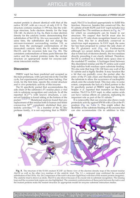
136–140. As shown in Fig. 8a, <strong>the</strong>re is clear electron<br />
density for <strong>the</strong> catalytic center, demonstrating that<br />
substitution <strong>of</strong> Ser118 by Ala was successful. At <strong>the</strong><br />
same time, <strong>the</strong> substitution did not change <strong>the</strong><br />
microenvironment surrounding residue 118, as<br />
seen from <strong>the</strong> unchanged conformations <strong>of</strong> <strong>the</strong><br />
(truncated) catalytic triad, <strong>the</strong> S1 subsite residue<br />
His133, <strong>and</strong> <strong>the</strong> oxyanion hole (see Fig. 8b). The<br />
consistent conformations <strong>of</strong> <strong>the</strong>se parts between <strong>the</strong><br />
native <strong>and</strong> <strong>the</strong> mutant proteins make <strong>the</strong> mutant<br />
structure an appropriate model for enzyme–substrate<br />
interaction studies.<br />
Discussion<br />
PRRSV nsp4 has been predicted <strong>and</strong> accepted as<br />
<strong>the</strong> main proteinase, with a pivotal role in <strong>the</strong> viral life<br />
cycle, but experimental pro<strong>of</strong> had been lacking. Our<br />
work, for <strong>the</strong> first time, reports <strong>the</strong> crystal structure<br />
<strong>and</strong> in vitro proteolytic activity <strong>of</strong> this enzyme.<br />
The S1 specificity pocket that accommodates <strong>the</strong><br />
side chain <strong>of</strong> <strong>the</strong> substrate's P1 residue plays a vital<br />
role in substrate recognition <strong>and</strong> catalysis. In all <strong>the</strong><br />
reported 3CL pro s with known structures, a conserved<br />
histidine is found sitting at <strong>the</strong> bottom <strong>of</strong> <strong>the</strong><br />
pocket. 12,22,27–33 Mutational analysis found that any<br />
replacement <strong>of</strong> this residue both in human <strong>and</strong> feline<br />
coronavirus M pro completely abolished <strong>the</strong>ir proteolytic<br />
activities. 34,35 As a member <strong>of</strong> <strong>the</strong> 3C-like<br />
protease family, it is not surprising that in PRRSV<br />
nsp4, His133 is localized appropriately to fulfill this<br />
function. However, besides this conserved His, <strong>the</strong><br />
S1 subsite <strong>of</strong> both SGPE <strong>and</strong> EAV nsp4 includes two<br />
additional Ser/Thr residues as shown in Fig. 4a, 12,20<br />
for which no counterparts can be found in our<br />
structure. We suspect that Ser136 must also be<br />
involved in P1 side-chain recognition based on two<br />
facts. First, this Ser is absolutely conserved in<br />
arterivirus nsp4 sequences. In EAV nsp4, <strong>the</strong> same<br />
Ser has been proposed to contact <strong>the</strong> side chain <strong>of</strong><br />
<strong>the</strong> P1 glutamic acid (Fig. 4a). Fur<strong>the</strong>rmore,<br />
although we cannot define <strong>the</strong> position <strong>of</strong> Ser136<br />
due to total lack <strong>of</strong> electron density, <strong>the</strong> two residues<br />
directly preceding it are clearly defined. Therefore,<br />
Ser136 is confined to a limited steric space close to<br />
<strong>the</strong> modeled P1 residue. A hydrogen bond between<br />
this disordered residue <strong>and</strong> <strong>the</strong> P1 amino acid could<br />
help stabilize both residues upon substrate binding.<br />
We also note that loop 136–140 lines one wall <strong>of</strong> <strong>the</strong><br />
S1 subsite <strong>and</strong> is highly flexible. It may function like<br />
a lid that can partially cover <strong>the</strong> pocket after <strong>the</strong><br />
entry <strong>of</strong> <strong>the</strong> P1 side chain <strong>and</strong> <strong>the</strong>refore help clinch<br />
<strong>the</strong> substrate to allow <strong>the</strong> occurrence <strong>of</strong> nucleophilic<br />
attack onto <strong>the</strong> scissile bond. This may also, to some<br />
extent, compensate <strong>the</strong> lack <strong>of</strong> a third partner in <strong>the</strong><br />
S1 specificity pocket <strong>of</strong> PRRSV nsp4 (see Results).<br />
Snijder et al. reported that mutation <strong>of</strong> this third<br />
residue, Thr115 in EAV nsp4, to o<strong>the</strong>r amino acids<br />
can have various effects on catalysis, indicating its<br />
important role as part <strong>of</strong> <strong>the</strong> S1 subsite. 11<br />
It is noteworthy that our enzyme also shows weak<br />
proteolytic activity against SP8-M with a Gln at <strong>the</strong> P1<br />
position (Fig. 6e; Table 2). This might reflect <strong>the</strong><br />
flexibility <strong>of</strong> <strong>the</strong> substrate binding <strong>of</strong> <strong>the</strong> enzyme that<br />
can also accommodate Gln in addition to Glu.<br />
Fig. 8. The structure <strong>of</strong> <strong>the</strong> S118A mutant 3CLSP. (a) Stereo view <strong>of</strong> <strong>the</strong> electron density map <strong>of</strong> <strong>the</strong> mutant residue<br />
Ala118 as well as <strong>the</strong> o<strong>the</strong>r two residues <strong>of</strong> <strong>the</strong> catalytic triad. The 1.0 σ contoured map confirmed <strong>the</strong> successful<br />
substitution <strong>of</strong> Ser118 by Ala. (b) Comparison <strong>of</strong> <strong>the</strong> native structure (light blue) with that <strong>of</strong> <strong>the</strong> mutant protein (magenta).<br />
The catalytic triad is shown as sticks. His133 is also shown because <strong>of</strong> its crucial role in substrate recognition. The missing<br />
loop <strong>and</strong> <strong>the</strong> oxyanion hole region are also highlighted. The consistent conformations <strong>of</strong> <strong>the</strong>se parts between <strong>the</strong> native <strong>and</strong><br />
<strong>the</strong> mutant proteins make <strong>the</strong> mutant structure an appropriate model for enzyme–substrate interaction studies.<br />
Please cite this article as: Tian, X., et al., <strong>Structure</strong> <strong>and</strong> <strong>Cleavage</strong> <strong>Specificity</strong> <strong>of</strong> <strong>the</strong> <strong>Chymotrypsin</strong>-<strong>Like</strong> <strong>Serine</strong> Protease (3CLSP/nsp4)<br />
<strong>of</strong> Porcine Reproductive <strong>and</strong> Respiratory Syndrome Virus (PRRSV), J. Mol. Biol. (2009), doi:10.1016/j.jmb.2009.07.062


