Structure and Cleavage Specificity of the Chymotrypsin-Like Serine ...
Structure and Cleavage Specificity of the Chymotrypsin-Like Serine ...
Structure and Cleavage Specificity of the Chymotrypsin-Like Serine ...

Create successful ePaper yourself
Turn your PDF publications into a flip-book with our unique Google optimized e-Paper software.
ARTICLE IN PRESS<br />
ASB YJMBI-61638; No. <strong>of</strong> pages: 17; 4C: 3, 5, 6, 7, 8, 11, 12<br />
doi:10.1016/j.jmb.2009.07.062<br />
J. Mol. Biol. (2009) xx, xxx–xxx<br />
Available online at www.sciencedirect.com<br />
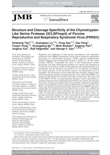
<strong>Structure</strong> <strong>and</strong> <strong>Cleavage</strong> <strong>Specificity</strong> <strong>of</strong> <strong>the</strong> <strong>Chymotrypsin</strong>-<br />
<strong>Like</strong> <strong>Serine</strong> Protease (3CLSP/nsp4) <strong>of</strong> Porcine<br />
Reproductive <strong>and</strong> Respiratory Syndrome Virus (PRRSV)<br />
Xinsheng Tian 1,2 †, Guangwen Lu 1,2 †, Feng Gao 1,3 , Hao Peng 1 ,<br />
Youjun Feng 1,2 , Guangpeng Ma 1,4 , Mark Bartlam 5 , Kegong Tian 6 ,<br />
Jinghua Yan 1 , Rolf Hilgenfeld 7 <strong>and</strong> George F. Gao 1,2,8,9 ⁎<br />
1 CAS Key Laboratory <strong>of</strong><br />
Pathogenic Microbiology <strong>and</strong><br />
Immunology (CASPMI),<br />
Institute <strong>of</strong> Microbiology,<br />
Chinese Academy <strong>of</strong> Sciences<br />
(CAS), Beijing 100101, China<br />
2 Graduate University, Chinese<br />
Academy <strong>of</strong> Sciences, Beijing<br />
100049, China<br />
3 College <strong>of</strong> Life Science, China<br />
Agricultural University, Beijing<br />
100094, China<br />
4 Department <strong>of</strong> Preventive<br />
Veterinary Medicine, College <strong>of</strong><br />
Veterinary Medicine, Nor<strong>the</strong>ast<br />
Agricultural University, Harbin<br />
150030, China<br />
5 College <strong>of</strong> Life Sciences <strong>and</strong><br />
Tianjin Key Laboratory <strong>of</strong> Protein<br />
Science, Nankai University,<br />
Tianjin 300071, China<br />
6 China Animal Disease Control<br />
Center, Beijing 100094, China<br />
7 Institute <strong>of</strong> Biochemistry, Center<br />
for Structural <strong>and</strong> Cell Biology in<br />
Medicine, University <strong>of</strong> Lübeck,<br />
Lübeck, Germany<br />
8 China–Japan Joint Laboratory <strong>of</strong><br />
Molecular Immunology <strong>and</strong><br />
Molecular Microbiology,<br />
Institute <strong>of</strong> Microbiology,<br />
Chinese Academy <strong>of</strong> Sciences,<br />
Beijing 100101, China<br />
Biogenesis <strong>and</strong> replication <strong>of</strong> <strong>the</strong> porcine reproductive <strong>and</strong> respiratory<br />
syndrome virus (PRRSV) include <strong>the</strong> crucial step <strong>of</strong> replicative polyprotein<br />
processing by self-encoded proteases. Whole genome bioinformatics<br />
analysis suggests that nonstructural protein 4 (nsp4) is a 3C-like serine<br />
protease (3CLSP), responsible for most <strong>of</strong> <strong>the</strong> nonstructural protein<br />
processing. The gene encoding this protease was cloned <strong>and</strong> expressed in<br />
Escherichia coli in order to confirm this prediction. The purified protein was<br />
crystallized, <strong>and</strong> <strong>the</strong> structure was solved at 1.9 Å resolution. In addition,<br />
<strong>the</strong> crystal structure <strong>of</strong> <strong>the</strong> Ser118Ala mutant was determined at 2.0 Å<br />
resolution. The monomeric enzyme folds into three domains, similar to that<br />
<strong>of</strong> <strong>the</strong> homologous protease <strong>of</strong> equine arteritis virus, which, like PRRSV, is a<br />
member <strong>of</strong> <strong>the</strong> family Arteriviridae in <strong>the</strong> order <strong>of</strong> Nidovirales. The active site<br />
<strong>of</strong> <strong>the</strong> PRRSV 3CLSP is located between domains I <strong>and</strong> II <strong>and</strong> harbors a<br />
canonical catalytic triad comprising Ser118, His39, <strong>and</strong> Asp64. The structure<br />
also shows an atypical oxyanion hole <strong>and</strong> a partially collapsed S1 specificity<br />
pocket. The proteolytic activity <strong>of</strong> <strong>the</strong> purified protein was assessed in vitro.<br />
Three sites joining nonstructural protein domains in <strong>the</strong> PRRSV replicative<br />
polyprotein are confirmed to be processed by <strong>the</strong> enzyme. Two <strong>of</strong> <strong>the</strong>m, <strong>the</strong><br />
nsp3/nsp4 <strong>and</strong> nsp11/nsp12 junctions, are shown to be cleaved in trans,<br />
while cis cleavage is demonstrated for <strong>the</strong> nsp4/nsp5 linker. Thus, we<br />
provide structural evidence as well as enzymatic pro<strong>of</strong> <strong>of</strong> <strong>the</strong> nsp4 protein<br />
being a functional 3CLSP. We also show that <strong>the</strong> enzyme has a strong<br />
preference for glutamic acid at <strong>the</strong> P1 position <strong>of</strong> <strong>the</strong> substrate.<br />
© 2009 Published by Elsevier Ltd.<br />
*Corresponding author. E-mail address: ga<strong>of</strong>@im.ac.cn.<br />
† X.T. <strong>and</strong> G.L. contributed equally to this work.<br />
Abbreviations used: nsp, nonstructural protein; 3CLSP, 3C-like serine protease; EAV, equine arteritis virus; SARS-CoV,<br />
severe acute respiratory syndrome coronavirus; 3CL pro , 3C-like proteinase; GST, glutathione S-transferase; MR,<br />
molecular replacement; SeMet, selenomethionine; PDB, Protein Data Bank; SGPE, Streptomyces griseus proteinase E; MS,<br />
mass spectrometry; MW, molecular weight; ESI, electrospray ionization.<br />
0022-2836/$ - see front matter © 2009 Published by Elsevier Ltd.<br />
Please cite this article as: Tian, X., et al., <strong>Structure</strong> <strong>and</strong> <strong>Cleavage</strong> <strong>Specificity</strong> <strong>of</strong> <strong>the</strong> <strong>Chymotrypsin</strong>-<strong>Like</strong> <strong>Serine</strong> Protease (3CLSP/nsp4)<br />
<strong>of</strong> Porcine Reproductive <strong>and</strong> Respiratory Syndrome Virus (PRRSV), J. Mol. Biol. (2009), doi:10.1016/j.jmb.2009.07.062
ARTICLE IN PRESS<br />
2 <strong>Structure</strong> <strong>and</strong> Characterization <strong>of</strong> PRRSV 3CLSP<br />
9 Beijing Institutes <strong>of</strong> Life Science,<br />
Chinese Academy <strong>of</strong> Sciences,<br />
Lincui East Road, Beijing<br />
100101, China<br />
Received 7 May 2009;<br />
received in revised form<br />
19 July 2009;<br />
accepted 22 July 2009<br />
Edited by R. Huber<br />
Keywords: PRRSV; 3C-like serine protease; structure; trans/cis cleavage<br />
Introduction<br />
Porcine reproductive <strong>and</strong> respiratory syndrome<br />
virus (PRRSV) was first discovered in North America<br />
in 1987 1,2 <strong>and</strong> almost simultaneously (in 1990) in<br />
Europe. 3 It manifests itself clinically with severe<br />
reproductive failure in sows <strong>and</strong> respiratory distress<br />
in neonatal pigs. 2 In recent years, <strong>the</strong> virus has<br />
become endemic throughout <strong>the</strong> world, leading to<br />
significant economic losses in pig production. 4 In <strong>the</strong><br />
autumn <strong>of</strong> 2006, unparalleled outbreaks <strong>of</strong> atypical<br />
PRRS occurred in more than 10 provinces (or<br />
autonomous cities) <strong>of</strong> China, affecting more than 2<br />
million pigs with nearly 400,000 fatal cases. Adult<br />
<strong>and</strong> growing pigs were also affected, which was<br />
different from classical PRRS. 5 In 2007, an almost<br />
identical atypical PRRSV was found to be epidemic<br />
in Vietnam <strong>and</strong> China. 6 Presently, only inactivated<br />
or attenuated vaccines are available to combat PRRS,<br />
but <strong>the</strong>ir efficacy is less than satisfactory due to<br />
adaptive evolution <strong>of</strong> PRRSV, 7,8 especially in <strong>the</strong><br />
cases <strong>of</strong> <strong>the</strong> atypical PRRS outbreaks in China <strong>and</strong><br />
Vietnam. 5,6 This urgent issue prompted us to exploit<br />
<strong>the</strong> possibility <strong>of</strong> drug discovery for treatment <strong>of</strong> <strong>the</strong><br />
highly pathogenic PRRSV infection.<br />
<strong>Like</strong> o<strong>the</strong>r RNA viruses in <strong>the</strong> order Nidovirales,<br />
such as equine arteritis virus (EAV), human coronavirus<br />
229E, <strong>and</strong> severe acute respiratory syndrome<br />
coronavirus (SARS-CoV), PRRSV has a singlestr<strong>and</strong>ed<br />
positive-sense RNA genome that is about<br />
15.3 kb in size, containing a large 5′-terminal<br />
replicase gene <strong>and</strong> a downstream set <strong>of</strong> structural<br />
protein genes. 9 The replicase gene is translated into<br />
two large polyprotein precursors, 1a <strong>and</strong> 1ab (with<br />
molecular masses <strong>of</strong> about 269.1 <strong>and</strong> 430.3 kDa for<br />
pp1a <strong>and</strong> pp1ab, respectively), with <strong>the</strong> latter being<br />
syn<strong>the</strong>sized following a ribosomal frame-shifting<br />
event. 10 After <strong>the</strong> rapid autocatalytic release <strong>of</strong><br />
nonstructural protein 1 (nsp1) <strong>and</strong> nsp2, <strong>the</strong> remainder<br />
<strong>of</strong> <strong>the</strong> polyproteins are cleaved into 10 mature<br />
nonstructural proteins (nsp3–nsp12) by <strong>the</strong> 3C-like<br />
serine protease (3CLSP), which is also <strong>of</strong>ten simply<br />
called 3C-like proteinase (3CL pro ). 9,11 The functional<br />
importance <strong>of</strong> <strong>the</strong> 3CL proteinase in <strong>the</strong> viral life<br />
cycle makes it an attractive target for <strong>the</strong> development<br />
<strong>of</strong> drugs directed against PRRSV.<br />
Here, we confirm that PRRSV nsp4, which we<br />
demonstrate to extend from residue G1780 to E1983<br />
in <strong>the</strong> viral polyprotein <strong>of</strong> <strong>the</strong> virus strain JXA1, is a<br />
3CLSP from both <strong>the</strong> structural <strong>and</strong> enzymatic<br />
points <strong>of</strong> view. The structure <strong>of</strong> <strong>the</strong> enzyme reveals<br />
two chymotrypsin-like β-barrel domains <strong>and</strong> an<br />
extra C-terminal α/β domain similar to that<br />
observed in EAV nsp4. 12 A canonical catalytic<br />
triad that is composed <strong>of</strong> Ser118, His39, <strong>and</strong><br />
Asp64 is located in <strong>the</strong> open cleft between <strong>the</strong> two<br />
β-barrel domains. An S1 subsite consisting <strong>of</strong><br />
His133 <strong>and</strong> very likely Ser136 <strong>and</strong> an oxyanion<br />
hole without <strong>the</strong> signature helix-like turn are seen in<br />
our structure. Our results also clearly show that <strong>the</strong><br />
recombinant enzyme displays proteolytic activity in<br />
vitro. Besides, based on <strong>the</strong> results <strong>of</strong> <strong>the</strong> cleavage<br />
assay, three nsp junction sites in <strong>the</strong> PRRSV replicase<br />
precursor are confirmed, <strong>the</strong>reby shedding<br />
light on <strong>the</strong> processing pathway <strong>of</strong> pp1a <strong>and</strong> pp1ab<br />
in <strong>the</strong> virus.<br />
Results<br />
Overall description <strong>of</strong> <strong>the</strong> recombinant PRRSV<br />
3CLSP used in this study<br />
As a member <strong>of</strong> <strong>the</strong> Arteriviridae, PRRSV shows<br />
substantial similarities in genome organization to<br />
EAV—<strong>the</strong> prototype virus <strong>of</strong> this family. 13–15<br />
Accordingly, whole genome bioinformatics analysis<br />
predicts that <strong>the</strong> PRRSV nsp4 domain should be <strong>the</strong><br />
main protease responsible for processing most <strong>of</strong><br />
polyproteins pp1a <strong>and</strong> pp1ab. 15 Previously, we<br />
reported on <strong>the</strong> successful expression <strong>of</strong> a gene<br />
construct encoding this putative serine protease in<br />
Escherichia coli as well as <strong>the</strong> crystallization <strong>of</strong> <strong>the</strong><br />
protein. 16 As shown in Fig. 1a, after removal <strong>of</strong> <strong>the</strong><br />
glutathione S-transferase (GST) tag, nine additional<br />
amino acid residues were still left at <strong>the</strong> N-terminus<br />
<strong>of</strong> <strong>the</strong> protein. The in vitro peptide cleavage assay in<br />
this study was performed using this recombinant<br />
enzyme with <strong>the</strong> nine extra residues present, while<br />
for <strong>the</strong> cis cleavage assay, even more additional N-<br />
terminal residues were present (Fig. 1b). In both<br />
cases, <strong>the</strong> recombinant protein displayed proteolytic<br />
activity in vitro, <strong>the</strong>reby confirming <strong>the</strong> identity <strong>of</strong><br />
nsp4 as <strong>the</strong> main protease in PRRSV replication (see<br />
results below).<br />
Please cite this article as: Tian, X., et al., <strong>Structure</strong> <strong>and</strong> <strong>Cleavage</strong> <strong>Specificity</strong> <strong>of</strong> <strong>the</strong> <strong>Chymotrypsin</strong>-<strong>Like</strong> <strong>Serine</strong> Protease (3CLSP/nsp4)<br />
<strong>of</strong> Porcine Reproductive <strong>and</strong> Respiratory Syndrome Virus (PRRSV), J. Mol. Biol. (2009), doi:10.1016/j.jmb.2009.07.062
ARTICLE IN PRESS<br />
<strong>Structure</strong> <strong>and</strong> Characterization <strong>of</strong> PRRSV 3CLSP<br />
3<br />
Fig. 1. Graphic representation <strong>of</strong> <strong>the</strong> recombinant 3CLSP used in this study. (a) Overview <strong>of</strong> <strong>the</strong> proteolytic processing<br />
<strong>of</strong> PRRSV replicase. The rapid autocatalytic release <strong>of</strong> nsp1α, nsp1β, <strong>and</strong> nsp2 after translation <strong>of</strong> <strong>the</strong> ORF1a or ORF1ab is<br />
indicated. The rest <strong>of</strong> <strong>the</strong> junction sites are processed by nsp4. All <strong>the</strong> predicted cleavage sites are listed above <strong>the</strong><br />
schematic boxes representing nsp1 to nsp12. Three <strong>of</strong> <strong>the</strong> putative cleavage sites were confirmed by our work. The rest <strong>of</strong><br />
<strong>the</strong>m were not evidenced to be processed in our study <strong>and</strong> <strong>the</strong>refore are labeled with a question mark on <strong>the</strong> top. The<br />
numerals indicated in each cleavage site are based on <strong>the</strong> genomic information <strong>of</strong> PRRSV strain JXA1 (GenBank accession<br />
no. EF112445). The recombinant protein for crystallization trials <strong>and</strong> in vitro peptide cleavage assay was produced using<br />
<strong>the</strong> GST fusion expression system (GE Healthcare). Nine extra N-terminal amino acid residues remained after removal <strong>of</strong><br />
<strong>the</strong> GST tag. (b) The translational fusion proteins used in <strong>the</strong> cis cleavage assay. The red-colored portion indicates <strong>the</strong><br />
additional residues introduced by <strong>the</strong> pET-28b vector. The eight N-terminal amino acid residues <strong>of</strong> nsp5 are colored pink<br />
<strong>and</strong> <strong>the</strong> Myc epitope is highlighted in blue.<br />
<strong>Structure</strong> determination <strong>and</strong> quality <strong>of</strong> <strong>the</strong> refined<br />
structure<br />
Although <strong>the</strong> sequence identity between PRRSV<br />
<strong>and</strong> EAV nsp4s is more than 34%, we failed to solve<br />
<strong>the</strong> structure <strong>of</strong> PRRSV 3CLSP by molecular<br />
replacement (MR) using <strong>the</strong> structure <strong>of</strong> EAV nsp4<br />
as a search model. The presence <strong>of</strong> two methionine<br />
residues in <strong>the</strong> recombinant PRRSV 3CLSP suggested<br />
that selenomethionine (SeMet)-based multiple-wavelength<br />
anomalous dispersion 17 could be<br />
used to solve <strong>the</strong> phase problem. The unit-cell<br />
dimensions <strong>of</strong> <strong>the</strong> crystals (a=112.4 Å, b=49.2 Å,<br />
c=42.9 Å, β=110.3°; space group C2) indicate that<br />
<strong>the</strong>re is only one molecule per asymmetric unit, <strong>and</strong><br />
we successfully located both <strong>of</strong> <strong>the</strong> two crystallographically<br />
independent selenium sites. Based on<br />
<strong>the</strong> obtained phases, a final model with good<br />
stereochemistry was built (for details, see Materials<br />
<strong>and</strong> Methods).<br />
As shown in Table 1, <strong>the</strong> structure was refined to<br />
1.9 Å resolution, with R cryst =19.6% <strong>and</strong><br />
R free =24.6%; 89.5% <strong>of</strong> <strong>the</strong> residues lie in <strong>the</strong> most<br />
favored regions <strong>of</strong> <strong>the</strong> Ramach<strong>and</strong>ran plot <strong>and</strong><br />
10.5% lie in <strong>the</strong> additionally allowed regions. 18 The<br />
quality <strong>of</strong> <strong>the</strong> electron density map <strong>of</strong> <strong>the</strong> recombinant<br />
protease at 1.9 Å is excellent, with an overall<br />
temperature factor <strong>of</strong> 22.2 Å 2 , <strong>and</strong> allowed us to<br />
model <strong>the</strong> positions <strong>of</strong> <strong>the</strong> main chain, most sidechain<br />
atoms, <strong>and</strong> 208 solvent molecules unambiguously.<br />
At <strong>the</strong> polypeptide chain termini, electron<br />
density is visible starting from residue 3 <strong>and</strong> ending<br />
at residue 199, but loop 136–140 is not seen in <strong>the</strong><br />
maps due to disorder.<br />
The overall structure <strong>of</strong> PRRSV 3CLSP <strong>and</strong><br />
comparison with EAV nsp4<br />
The PRRSV 3CLSP is folded into three domains,<br />
including two antiparallel β-barrels <strong>and</strong> an extra C-<br />
terminal α/β domain (Fig. 2a <strong>and</strong> b). The N-<br />
terminal β-barrel (domain I) consists <strong>of</strong> six β-str<strong>and</strong>s<br />
(aI to fI) <strong>and</strong> a short α-helix (helix A) that closes <strong>the</strong><br />
barrel like a lid, whereas <strong>the</strong> middle β-barrel<br />
(domain II) comprises seven β-str<strong>and</strong>s (aII to gII)<br />
<strong>and</strong> is connected to domain I via a very long loop<br />
(amino acids 69–89) (Fig. 2a). The catalytic site is<br />
located at <strong>the</strong> opening <strong>of</strong> <strong>the</strong> cleft between domains I<br />
<strong>and</strong> II <strong>and</strong> consists <strong>of</strong> residues Ser118, His39, <strong>and</strong><br />
Asp64 (Fig. 2a), which are totally conserved among<br />
members <strong>of</strong> <strong>the</strong> Arteriviridae (Fig. 2c). The extra C-<br />
terminal domain (domain III) comprises residues<br />
157–199 <strong>and</strong> consists <strong>of</strong> two pairs <strong>of</strong> short antiparallel<br />
β-sheets (str<strong>and</strong>s aIII–cIII <strong>and</strong> bIII–dIII) <strong>and</strong><br />
two α-helices. The crossing angle is ∼90° between<br />
helix B <strong>and</strong> helix C (Fig. 2a). A long N-terminal loop<br />
extends over domain II to <strong>the</strong> vicinity <strong>of</strong> domain III.<br />
The orientation <strong>of</strong> this loop is stabilized by <strong>the</strong> three<br />
hydrogen bonds Arg4 N…Glu100 OE1, Thr5 N…<br />
Val99 O, <strong>and</strong> Thr5 OG1…Val99 N (Fig. 2a). The<br />
phenyl ring <strong>of</strong> Phe3 is directed into <strong>the</strong> hydrophobic<br />
interface between domains II <strong>and</strong> III <strong>and</strong>, <strong>the</strong>refore,<br />
also helps to stabilize <strong>the</strong> orientation <strong>of</strong> <strong>the</strong> N-<br />
terminal loop (Fig. 5a, left image).<br />
Please cite this article as: Tian, X., et al., <strong>Structure</strong> <strong>and</strong> <strong>Cleavage</strong> <strong>Specificity</strong> <strong>of</strong> <strong>the</strong> <strong>Chymotrypsin</strong>-<strong>Like</strong> <strong>Serine</strong> Protease (3CLSP/nsp4)<br />
<strong>of</strong> Porcine Reproductive <strong>and</strong> Respiratory Syndrome Virus (PRRSV), J. Mol. Biol. (2009), doi:10.1016/j.jmb.2009.07.062
ARTICLE IN PRESS<br />
4 <strong>Structure</strong> <strong>and</strong> Characterization <strong>of</strong> PRRSV 3CLSP<br />
Table 1. Data collection <strong>and</strong> refinement statistics<br />
Native protein<br />
S118A mutant<br />
Data collection<br />
Space group C2 C2<br />
Cell dimensions<br />
a, b, c (Å) 112.4, 49.2, 42.9 113.3, 48.6, 42.9<br />
α, β, γ (°) 90.0, 110.3, 90.0 90.0, 110.4, 90.0<br />
SeMet multiple-wavelength<br />
anomalous dispersion<br />
Native Peak Remote<br />
Wavelength (Å) 1.5418 0.9793 0.9000 1.5418<br />
Resolution range (Å) 32.3–1.9 53.0–1.9 50.0–2.0 44.19–2.01<br />
a,b<br />
R merge 0.045 (0.167) 0.213 (0.459) 0.123 (0.216) 0.049 (0.169)<br />
Average I/σ(I) a 22.1 (4.3) 12.5 (1.4) 10.1 (2.4) 47.1 (11.8)<br />
Completeness (%) a 95.2 (91.6) 95.7 (77.6) 89.8 (75.7) 98.9 (96.3)<br />
Redundancy 4.5 (4.3) 4.5 (3.4) 2.4 (2.0) 5.2 (4.8)<br />
Refinement<br />
R cryst /R free (%) c 19.6/24.6 18.2/21.5<br />
Ramach<strong>and</strong>ran plot (%) d<br />
Most favored regions 89.5 90.2<br />
Additionally allowed regions 10.5 9.8<br />
Generously allowed regions 0 0<br />
Disallowed regions 0 0<br />
r.m.s.d. from ideality<br />
Bonds (Å) 0.017 0.018<br />
Angles (°) 1.572 1.529<br />
Average B-factor (Å 2 )<br />
Protein 19.933 28.120<br />
Water 36.572 37.980<br />
a Values for <strong>the</strong> outermost resolution shell are given in paren<strong>the</strong>ses.<br />
b R merge =∑ i ∑ hkl │ I i −〈I〉│/∑ i ∑ hkl I i , where I i is <strong>the</strong> observed intensity <strong>and</strong> 〈I〉 is <strong>the</strong> average intensity from multiple measurements.<br />
c R cryst =∑ ││F o │ −│ F c ││/∑│ F o │, where F o <strong>and</strong> F c are <strong>the</strong> structure-factor amplitudes from <strong>the</strong> data <strong>and</strong> <strong>the</strong> model, respectively. R free<br />
is <strong>the</strong> R-factor for a subset (5%) <strong>of</strong> reflections that was selected prior to refinement calculations <strong>and</strong> was not included in <strong>the</strong> refinement.<br />
d Ramach<strong>and</strong>ran plots were generated by using <strong>the</strong> program PROCHECK.<br />
A Dali search 19 <strong>of</strong> <strong>the</strong> Protein Data Bank (PDB)<br />
database gave a variety <strong>of</strong> proteins with significant<br />
structural similarities to our structure, among which<br />
<strong>the</strong> EAV nsp4 expectedly had <strong>the</strong> highest Z score<br />
(21.7). As <strong>the</strong> prototype structure <strong>of</strong> nsp4s in<br />
Arteriviridae, <strong>the</strong> EAV 3CLSP also folds into three<br />
domains. 12 Superposing domains I <strong>and</strong> II <strong>of</strong> PRRSV<br />
3CLSP onto those <strong>of</strong> <strong>the</strong> EAV nsp4 (copy A <strong>of</strong> <strong>the</strong> four<br />
copies in <strong>the</strong> asymmetric unit) yielded an r.m.s.d. <strong>of</strong><br />
2.04 Å for 122 equivalent (out <strong>of</strong> 134 compared) C α<br />
pairs (Fig. 3a), while domain III alone exhibited an r.<br />
m.s.d. <strong>of</strong> 0.52 Å for 30 (out <strong>of</strong> 38) C α pairs (Fig. 3d),<br />
indicating that <strong>the</strong> two enzymes share great structural<br />
similarity at <strong>the</strong> tertiary level. However, <strong>the</strong> steric<br />
arrangement <strong>of</strong> <strong>the</strong> domains in <strong>the</strong> two proteases is<br />
quite different. An ∼8 Å shift <strong>of</strong> domain III relative to<br />
domains I <strong>and</strong> II was observed in our structure, when<br />
compared to EAV nsp4. This shift causes <strong>the</strong> whole<br />
domain III to move away from <strong>the</strong> cleft where <strong>the</strong><br />
catalytic triad is located (Fig. 3a). Clear electron<br />
density for <strong>the</strong> loop Asn153-Ile157 that connects<br />
domains II <strong>and</strong> III, as shown in Fig. 3c, leaves no<br />
doubt about <strong>the</strong> chain tracing <strong>of</strong> <strong>the</strong> loop <strong>and</strong> verifies<br />
<strong>the</strong> 8-Å shift <strong>of</strong> domain III, compared to EAV nsp4.<br />
The shift may also account for our failure to solve <strong>the</strong><br />
structure by MR using EAV nsp4 as <strong>the</strong> search model.<br />
Fur<strong>the</strong>r discrepancies were also observed in two<br />
loop regions. One is <strong>the</strong> loop connecting str<strong>and</strong>s cII<br />
<strong>and</strong> dII (Cys111-Pro121 in PRRSV nsp4). In EAV<br />
nsp4, this loop is much like a tadpole with its head<br />
lying next to <strong>the</strong> catalytic nucleophile Ser120 (Fig.<br />
3b). However, <strong>the</strong> corresponding loop in our<br />
structure is approximately “⨼” shaped with a<br />
protuberance pointing to domain III (Fig. 3a <strong>and</strong> b)<br />
on which <strong>the</strong> hydrophobic side chain <strong>of</strong> Phe112<br />
resides. This residue is conserved in PRRSV <strong>and</strong><br />
lactate dehydrogenase-elevating virus 3CLSP but<br />
not in EAV nsp4, where it is substituted by Trp (Fig.<br />
2c). The phenyl ring <strong>of</strong> Phe112 protrudes into <strong>the</strong><br />
hydrophobic interface between domains II <strong>and</strong> III<br />
(Fig. 5a, left image) <strong>and</strong> <strong>the</strong>reby causes <strong>the</strong> “⨼”-<br />
shaped conformation <strong>of</strong> <strong>the</strong> cII–dII loop.<br />
Ano<strong>the</strong>r loop <strong>of</strong> great variance is <strong>the</strong> one connecting<br />
str<strong>and</strong>s eII <strong>and</strong> fII. In PRRSV 3CLSP, this loop is<br />
eight residues in length, from Gly135 to Gly142, <strong>and</strong><br />
is highly flexible as proved by <strong>the</strong> total lack <strong>of</strong><br />
electron density for residues 136–140. However, in<br />
EAV nsp4, <strong>the</strong> corresponding region is mainly a β-<br />
str<strong>and</strong> with a much shorter loop, indicating that <strong>the</strong><br />
conformation <strong>of</strong> this part is relatively rigid (Fig. 3b).<br />
Interestingly <strong>the</strong>se two regions showing significant<br />
variance are actually believed to cover, respectively,<br />
<strong>the</strong> S1 specificity pocket <strong>and</strong> <strong>the</strong> oxyanion<br />
hole in EAV nsp4, 12 two highly important features<br />
involved in substrate recognition <strong>and</strong> catalysis by<br />
3CL proteases.<br />
The S1 specificity pocket<br />
In 3CL proteases, <strong>the</strong> S1 specificity pocket<br />
accommodates <strong>the</strong> side chain <strong>of</strong> <strong>the</strong> substrate P1<br />
residue <strong>and</strong> <strong>the</strong>refore determines <strong>the</strong> enzyme's<br />
Please cite this article as: Tian, X., et al., <strong>Structure</strong> <strong>and</strong> <strong>Cleavage</strong> <strong>Specificity</strong> <strong>of</strong> <strong>the</strong> <strong>Chymotrypsin</strong>-<strong>Like</strong> <strong>Serine</strong> Protease (3CLSP/nsp4)<br />
<strong>of</strong> Porcine Reproductive <strong>and</strong> Respiratory Syndrome Virus (PRRSV), J. Mol. Biol. (2009), doi:10.1016/j.jmb.2009.07.062
ARTICLE IN PRESS<br />
<strong>Structure</strong> <strong>and</strong> Characterization <strong>of</strong> PRRSV 3CLSP<br />
5<br />
Fig. 2. Crystal structure <strong>of</strong> PRRSV nsp4. (a) Cartoon representation <strong>of</strong> <strong>the</strong> overall structure <strong>of</strong> PRRSV nsp4. The N-<br />
terminal (aI–fI) <strong>and</strong> middle (aII–gII) β-barrels are colored green <strong>and</strong> cyan, respectively, with <strong>the</strong> red broken line indicating<br />
<strong>the</strong> loop 136–140, which is totally devoid <strong>of</strong> visible electron density. The C-terminal extra domain is shown in yellow.<br />
Members <strong>of</strong> <strong>the</strong> catalytic triad <strong>and</strong> <strong>the</strong> three hydrogen bonds that are believed to stabilize <strong>the</strong> orientation <strong>of</strong> <strong>the</strong> long N-<br />
terminus are featured. The long loop connecting domains I <strong>and</strong> II is highlighted in red. (b) Topological presentation <strong>of</strong> <strong>the</strong><br />
same structure. (c) Multiple sequence alignment <strong>of</strong> <strong>the</strong> 3CLSPs <strong>of</strong> <strong>the</strong> members in <strong>the</strong> family Arteriviridae. The catalytic<br />
triad is indicated by red arrows, <strong>and</strong> <strong>the</strong> three residues that are believed to constitute <strong>the</strong> S1 specificity pocket in EAV nsp4<br />
are marked with blue stars. The Phe residue, which is thought to lead to <strong>the</strong> atypical oxyanion hole in PRRSV 3CLSP, is<br />
labeled by a dark triangle. Cys111 <strong>and</strong> Cys115, which have been referred to in <strong>the</strong> text, are marked with black squares.<br />
LDVC <strong>and</strong> LDVP, lactate dehydrogenase-elevating virus neurovirulent type C <strong>and</strong> strain Plagemann, respectively;<br />
PRRSVAS <strong>and</strong> PRRSVES, PRRSV American strain <strong>and</strong> European strain, respectively.<br />
Please cite this article as: Tian, X., et al., <strong>Structure</strong> <strong>and</strong> <strong>Cleavage</strong> <strong>Specificity</strong> <strong>of</strong> <strong>the</strong> <strong>Chymotrypsin</strong>-<strong>Like</strong> <strong>Serine</strong> Protease (3CLSP/nsp4)<br />
<strong>of</strong> Porcine Reproductive <strong>and</strong> Respiratory Syndrome Virus (PRRSV), J. Mol. Biol. (2009), doi:10.1016/j.jmb.2009.07.062
ARTICLE IN PRESS<br />
6 <strong>Structure</strong> <strong>and</strong> Characterization <strong>of</strong> PRRSV 3CLSP<br />
Fig. 3. Structural alignments between EAV (magenta) <strong>and</strong> PRRSV (light blue) nsp4s. (a) Superposition <strong>of</strong> <strong>the</strong> two β-<br />
barrel domains <strong>of</strong> PRRSV 3CLSP with those <strong>of</strong> EAV nsp4. A shift <strong>of</strong> about 8 Å observed in <strong>the</strong> C-terminal extra domain is<br />
highlighted. The well-aligned catalytic triad <strong>of</strong> <strong>the</strong> two proteases is shown in stick representation. The two loop regions<br />
that display great discrepancies between <strong>the</strong> two enzymes are also featured. Residue Phe112, which makes hydrophobic<br />
interactions with domain III in PRRSV nsp4, is highlighted by line representation. (b) Magnified view <strong>of</strong> <strong>the</strong> two loop<br />
regions referred to in <strong>the</strong> text. (c) Stereo view <strong>of</strong> <strong>the</strong> electron density map <strong>of</strong> <strong>the</strong> loop (N153-I157) connecting domains II<br />
<strong>and</strong> III in PRRSV nsp4. This 2|F o |−|F c | map is contoured at 1.0 σ above <strong>the</strong> mean. (d) Alignment <strong>of</strong> domain III alone <strong>of</strong><br />
PRRSV <strong>and</strong> EAV nsp4 to show <strong>the</strong>ir well superposition.<br />
preference for specific amino acid residues at this<br />
position. To identify <strong>the</strong> possible residues that may<br />
be involved in <strong>the</strong> formation <strong>of</strong> <strong>the</strong> S1 pocket in<br />
PRRSV 3CLSP, we used <strong>the</strong> structure <strong>of</strong> Streptomyces<br />
griseus proteinase E (SGPE) in complex with a<br />
tetrapeptide as <strong>the</strong> reference structure for superposition.<br />
SGPE is ano<strong>the</strong>r chymotrypsin-like proteinase<br />
that has a substrate preference for Glu at <strong>the</strong> P1<br />
position. 20 Both <strong>the</strong> EAV nsp4 (copy A) <strong>and</strong> our<br />
structure were aligned. As shown in Fig. 4a, <strong>the</strong> S1<br />
specificity pocket <strong>of</strong> both <strong>the</strong> SGPE <strong>and</strong> EAV nsp4 is<br />
composed <strong>of</strong> three residues, with a conserved His<br />
(His134 in EAV nsp4 <strong>and</strong> His213 in SGPE) lying at<br />
<strong>the</strong> bottom <strong>of</strong> <strong>the</strong> pocket <strong>and</strong> one Ser (EAV nsp4:<br />
Ser137; SGPE: Ser216) lining one side <strong>and</strong> ano<strong>the</strong>r<br />
Thr/Ser (EAV nsp4: Thr115; SGPE: Ser192) located<br />
on <strong>the</strong> opposite side. However, only <strong>the</strong> histidine<br />
(His133) could be found in our structure. The first<br />
serine involved in <strong>the</strong> S1 subsite is conserved in<br />
PRRSV 3CLSP (Ser136) (Fig. 2c), but we cannot<br />
define its position due to <strong>the</strong> absence <strong>of</strong> electron<br />
density for <strong>the</strong> loop 136–140 as mentioned before.<br />
Never<strong>the</strong>less, <strong>the</strong>re is no residue in PRRSV nsp4 to<br />
match <strong>the</strong> Thr/Ser (Thr115 in EAV nsp4, Ser192 in<br />
SGPE) on <strong>the</strong> o<strong>the</strong>r side <strong>of</strong> <strong>the</strong> pocket. Thr113 <strong>of</strong><br />
PRRSV nsp4, which is also highly conserved among<br />
Please cite this article as: Tian, X., et al., <strong>Structure</strong> <strong>and</strong> <strong>Cleavage</strong> <strong>Specificity</strong> <strong>of</strong> <strong>the</strong> <strong>Chymotrypsin</strong>-<strong>Like</strong> <strong>Serine</strong> Protease (3CLSP/nsp4)<br />
<strong>of</strong> Porcine Reproductive <strong>and</strong> Respiratory Syndrome Virus (PRRSV), J. Mol. Biol. (2009), doi:10.1016/j.jmb.2009.07.062
ARTICLE IN PRESS<br />
<strong>Structure</strong> <strong>and</strong> Characterization <strong>of</strong> PRRSV 3CLSP<br />
7<br />
Fig. 4. The S1 specificity pocket <strong>and</strong> <strong>the</strong> oxyanion hole <strong>of</strong> PRRSV nsp4. Black broken lines indicate hydrogen bonds.<br />
Residues referred to in <strong>the</strong> text are labeled. The position <strong>of</strong> Ser136 in PRRSV 3CLSP is extrapolated from <strong>the</strong> position <strong>of</strong> <strong>the</strong><br />
residue directly preceding it (Gly135) <strong>and</strong> is <strong>the</strong>refore encircled in black. (a) Superposition <strong>of</strong> <strong>the</strong> S1 subsites <strong>of</strong> PRRSV<br />
nsp4 (light blue), EAV nsp4 copy A (magenta) (PDB code: 1MBM), <strong>and</strong> SGPE (cyan) (PDB code: 1HPG). (b) Comparison <strong>of</strong><br />
<strong>the</strong> oxyanion holes <strong>of</strong> <strong>the</strong> three serine proteases. Gly192-Ser195 <strong>of</strong> SGPE is also shown with line representation. The<br />
signature helix-like turn is highlighted.<br />
members <strong>of</strong> Arteriviridae (Fig. 2c), is too far away to<br />
contact <strong>the</strong> substrate P1 residue (Fig. 4a).<br />
An atypical oxyanion hole without <strong>the</strong> signature<br />
helix-like turn<br />
Both EAV nsp4 <strong>and</strong> SGPE form a canonical<br />
oxyanion hole that is characterized by a helix-like<br />
turn preceding <strong>the</strong> nucleophilic serine. As shown in<br />
Fig. 4b, <strong>the</strong> backbone amides <strong>of</strong> Ser195 <strong>and</strong> Gly193 in<br />
SGPE can form hydrogen bonds with <strong>and</strong> <strong>the</strong>refore<br />
stabilize <strong>the</strong> carbonyl oxyanion in <strong>the</strong> tetrahedral<br />
intermediate during catalysis. Although, in EAV<br />
nsp4, only copy B has <strong>the</strong> normal active oxyanion<br />
hole geometry while <strong>the</strong> o<strong>the</strong>r three copies only form<br />
collapsed ones, this difference is mainly caused by<br />
flipping <strong>of</strong> <strong>the</strong> peptide bond between Ser117 <strong>and</strong><br />
Gly118 ra<strong>the</strong>r than a big change <strong>of</strong> <strong>the</strong> orientation <strong>of</strong><br />
<strong>the</strong> signature turn. 12 However, in PRRSV nsp4, this<br />
oxyanion-specific turn is missing. Instead, a much<br />
more stretched loop leaves only <strong>the</strong> amide group <strong>of</strong><br />
<strong>the</strong> catalytic nucleophile (Ser118) in contact with <strong>the</strong><br />
oxyanion. We term this rare property an atypical<br />
oxyanion hole.<br />
A strong hydrophobic interaction between<br />
domains II <strong>and</strong> III<br />
Compared to that <strong>of</strong> <strong>the</strong> EAV nsp4, an even<br />
stronger hydrophobic interaction between domains<br />
II <strong>and</strong> III is observed in our structure. With Phe3,<br />
Val103, Phe108, Phe110, Phe112, Ile157, Leu162,<br />
Phe165, Phe166, Ile182, L196, <strong>and</strong> L197, <strong>the</strong> interface<br />
harbors 12 hydrophobic residues, whereas in EAV<br />
nsp4, this interface only includes eight such residues<br />
(Fig. 5a). This hydrophobic core holds domains II<br />
<strong>and</strong> III toge<strong>the</strong>r <strong>and</strong> likely forces <strong>the</strong> loop connecting<br />
<strong>the</strong> two domains to adopt its observed orientation,<br />
leading to <strong>the</strong> 8-Å shift as described above.<br />
Ano<strong>the</strong>r unexpected feature <strong>of</strong> <strong>the</strong> extra domain is<br />
<strong>the</strong> presence <strong>of</strong> two patches <strong>of</strong> solvent-exposed<br />
hydrophobic residues, implying possible interactions<br />
<strong>of</strong> PRRSV nsp4 with o<strong>the</strong>r proteins. One patch,<br />
which is also present in EAV nsp4 (Fig. 5b, right<br />
image), 12 consists <strong>of</strong> Leu173, Ile178, <strong>and</strong> Ile183 (Fig.<br />
5b, left image). Ano<strong>the</strong>r patch, comprising Val171,<br />
Val176, Ala195, Ala198, <strong>and</strong> Ala199, is even more<br />
extended <strong>and</strong> locates on a surface that is almost<br />
perpendicular to <strong>the</strong> former one (Fig. 5c, left image,<br />
<strong>and</strong> 5b, left image). It is noteworthy that this second<br />
hydrophobic patch is totally absent in EAV nsp4<br />
(Fig. 5c, right image).<br />
Trans cleavage <strong>of</strong> domain junctions in pp1a/pp1ab<br />
Peptides corresponding to eight predicted junctions<br />
in PRRSV pp1a or pp1ab (Fig. 1a; Table 2)were<br />
syn<strong>the</strong>sized, tested for trans cleavage, <strong>and</strong> fur<strong>the</strong>r<br />
analyzed by high-performance liquid chromatography<br />
(HPLC) in order to see whe<strong>the</strong>r <strong>the</strong> recombinant<br />
Please cite this article as: Tian, X., et al., <strong>Structure</strong> <strong>and</strong> <strong>Cleavage</strong> <strong>Specificity</strong> <strong>of</strong> <strong>the</strong> <strong>Chymotrypsin</strong>-<strong>Like</strong> <strong>Serine</strong> Protease (3CLSP/nsp4)<br />
<strong>of</strong> Porcine Reproductive <strong>and</strong> Respiratory Syndrome Virus (PRRSV), J. Mol. Biol. (2009), doi:10.1016/j.jmb.2009.07.062
ARTICLE IN PRESS<br />
8 <strong>Structure</strong> <strong>and</strong> Characterization <strong>of</strong> PRRSV 3CLSP<br />
Fig. 5. Surface representation <strong>of</strong><br />
(a) <strong>the</strong> hydrophobic interface<br />
between domains II <strong>and</strong> III; (b)<br />
<strong>the</strong> exposed hydrophobic patch on<br />
one face <strong>of</strong> <strong>the</strong> C-terminal extra<br />
domain <strong>and</strong> (c) ano<strong>the</strong>r patch <strong>of</strong><br />
exposed hydrophobic residues<br />
located on a different face <strong>of</strong> <strong>the</strong><br />
same extra domain <strong>of</strong> both PRRSV<br />
(left) <strong>and</strong> EAV (right) nsp4s. Residues<br />
involved are labeled <strong>and</strong><br />
colored magenta. The surface is<br />
shown in cyan for PRRSV nsp4<br />
<strong>and</strong> in yellow for EAV nsp4.<br />
nsp4 is proteolytically active in vitro. In our assay,<br />
we showed that peptides corresponding to two<br />
junction sites can be cleaved by <strong>the</strong> enzyme. For<br />
each predicted cleavage site, <strong>the</strong> scissile peptide<br />
bond was between Glu <strong>and</strong> Gly.<br />
Just like o<strong>the</strong>r reports on peptide-based cleavage<br />
assays, 21,22 our enzyme also shows a remarkable<br />
difference in efficiency towards different peptides.<br />
Of <strong>the</strong> nine potential substrates investigated, only<br />
three can be cleaved to <strong>the</strong> extent <strong>of</strong> being detectable<br />
on an HPLC system (see Table 2). Peptide SP8-1,<br />
derived from <strong>the</strong> junction sequence between nsp11<br />
<strong>and</strong> 12, is most susceptible to this enzyme. After<br />
incubation with <strong>the</strong> recombinant enzyme, two new<br />
peaks with retention times <strong>of</strong> 16.9 <strong>and</strong> 17.4 min,<br />
respectively, were observed after analyzing a reaction<br />
volume <strong>of</strong> 40 μl on a C18 column (Fig. 6b). The<br />
uncleaved peptide gave a peak at 18.5 min, consistent<br />
with that for <strong>the</strong> peptide-alone control (Fig.<br />
6a). The two product peaks were collected <strong>and</strong><br />
analyzed by mass spectrometry (MS). The results<br />
showed that <strong>the</strong> calculated molecular masses corresponding<br />
to <strong>the</strong> peaks were 1241.6293 <strong>and</strong> 802.3875,<br />
respectively, matching <strong>the</strong>ir <strong>the</strong>oretical monoisotopic<br />
molecular weight (MW) perfectly (<strong>the</strong> <strong>the</strong>oretical<br />
MW for fragment KDKTAYFQLE is 1241.6291, <strong>and</strong><br />
for GRHFTW, it is 802.3874). A typical representation<br />
<strong>of</strong> <strong>the</strong> MS result is shown in Fig. 6f. When <strong>the</strong><br />
catalytic serine residue was replaced by alanine<br />
(Ser118Ala mutant), no detectable peaks <strong>of</strong> cleaved<br />
peptides were observed in <strong>the</strong> HPLC pr<strong>of</strong>ile (Fig.<br />
6c). This confirmed <strong>the</strong> crucial function <strong>of</strong> Ser118 as<br />
<strong>the</strong> nucleophile in catalysis.<br />
Ano<strong>the</strong>r peptide that was shown to be processed<br />
is SP1, which corresponds to <strong>the</strong> nsp3/nsp4 junction.<br />
However, <strong>the</strong> proteolytic efficiency for SP1 was<br />
much lower than that for SP8-1. Only a small portion<br />
(b20%) <strong>of</strong> <strong>the</strong> peptide was cleaved after a 15-h<br />
incubation (data not shown).<br />
The kinetic parameters <strong>of</strong> <strong>the</strong> protein were<br />
determined in vitro using a fluorescent assay system<br />
to quantitatively test <strong>the</strong> proteolytic activity <strong>of</strong> <strong>the</strong><br />
recombinant enzyme. The results showed that <strong>the</strong><br />
enzyme exhibits a K m <strong>of</strong> about 2.24 mM <strong>and</strong> a k cat <strong>of</strong><br />
about 0.435 min − 1 for <strong>the</strong> substrate Dabsyl-<br />
KTAYFQLE↓GRHFE-Edans.<br />
Please cite this article as: Tian, X., et al., <strong>Structure</strong> <strong>and</strong> <strong>Cleavage</strong> <strong>Specificity</strong> <strong>of</strong> <strong>the</strong> <strong>Chymotrypsin</strong>-<strong>Like</strong> <strong>Serine</strong> Protease (3CLSP/nsp4)<br />
<strong>of</strong> Porcine Reproductive <strong>and</strong> Respiratory Syndrome Virus (PRRSV), J. Mol. Biol. (2009), doi:10.1016/j.jmb.2009.07.062
ARTICLE IN PRESS<br />
<strong>Structure</strong> <strong>and</strong> Characterization <strong>of</strong> PRRSV 3CLSP<br />
9<br />
Table 2. Comparative cleavage <strong>of</strong> peptides corresponding to polyprotein junctions in PRRSV JXA1<br />
Peptide<br />
Polyprotein<br />
junction<br />
Sequence(P–P′)<br />
P10 P9 P8 P7 P6 P5 P4 P3 P2 P1 P1′ P2′ P3′ P4′ P5′ P6′ P7′ P8′<br />
<strong>Cleavage</strong><br />
SP1 nsp3/nsp4 S Q L G S L L E G A F R T Q K P +<br />
SP2 nsp4/nsp5 L A A K P E L E G G L S T V Q L −<br />
SP3 nsp5/nsp6 F F L R Y F A E G K L R E G V S −<br />
SP4 nsp6/nsp7 Q S C G M S H E S L T G A L A M −<br />
SP5 nsp7/nsp8 E N R R A Q W E A A K L S V E Q −<br />
SP6 nsp9/nsp10 E K L R S N H E G K K S R M C G −<br />
SP7 nsp10/nsp11 R A I C A D L E G S S S P L P K −<br />
SP8 nsp11/nsp12 K T A Y F Q L E G R H F T W Y Q +++<br />
SP8-1 nsp11/nsp12 K D K T A Y F Q L E G R H F T W +++<br />
SP8-M K D K T A Y F Q L Q G R H F T W +/−<br />
Syn<strong>the</strong>tic 16-mer peptides that represent <strong>the</strong> eight predicted junction sequences in <strong>the</strong> replicative polyprotein were incubated with 25 μM<br />
recombinant PRRSV nsp4 as described in Materials <strong>and</strong> Methods <strong>and</strong> analyzed by HPLC for <strong>the</strong> extent <strong>of</strong> cleavage. After incubation at<br />
37 °C for 15 h, if more than 50% <strong>of</strong> <strong>the</strong> peptide was found to be cleaved as is determined by <strong>the</strong> decrease in area <strong>of</strong> <strong>the</strong> peak representing<br />
<strong>the</strong> parental peptide, “+++” is given; if 10–20% <strong>of</strong> <strong>the</strong> parental peptide is cleaved, we perceive it as “+”; if less than 10% is cleaved but still<br />
detectable amounts <strong>of</strong> products are observed as marked by <strong>the</strong> appearance <strong>of</strong> new peaks in <strong>the</strong> HPLC pr<strong>of</strong>ile, we mark it “+/−”. “−”<br />
means no visible product peaks in <strong>the</strong> HPLC pr<strong>of</strong>ile could be observed at all. All <strong>the</strong> cleaved products were verified by MS <strong>and</strong>/or<br />
comparison with HPLC traces <strong>of</strong> peptide st<strong>and</strong>ards. SP8-1 <strong>and</strong> SP8 correspond to <strong>the</strong> same junction site in pp1b. SP8-1 was syn<strong>the</strong>sized<br />
<strong>and</strong> analyzed for <strong>the</strong> purpose <strong>of</strong> separating <strong>the</strong> two product peaks from each o<strong>the</strong>r.<br />
The glutamic acid at <strong>the</strong> P1 position <strong>of</strong> <strong>the</strong> peptide<br />
substrate is crucial for effective cleavage <strong>and</strong><br />
cannot be replaced by glutamine<br />
3C-like proteases <strong>of</strong> RNA viruses are specific for<br />
ei<strong>the</strong>r Gln or Glu at <strong>the</strong> P1 position <strong>of</strong> <strong>the</strong> substrate.<br />
Among <strong>the</strong> nidoviruses, coronavirus 3CL pro cleavage<br />
sites always have a Gln at <strong>the</strong> P1 position. 9,23,24<br />
However, in <strong>the</strong> same order Nidovirales, <strong>the</strong> 3CLSPs<br />
<strong>of</strong> Arteriviridae, which are represented by <strong>the</strong> prototype<br />
EAV nsp4, show a clear preference for glutamic<br />
acid over glutamine at <strong>the</strong> P1 position <strong>of</strong> <strong>the</strong> substrate.<br />
9 In EAV, <strong>of</strong> <strong>the</strong> eight cleavage sites <strong>of</strong> <strong>the</strong><br />
replicative polyprotein processed by <strong>the</strong> main<br />
protease, only one junction contains a glutamine at<br />
<strong>the</strong> P1 position (Q2837/S joining nsp10/11), while<br />
all o<strong>the</strong>r cleavage sites have glutamic acid in this<br />
position. 11,25,26 Based on <strong>the</strong> similarity in genome<br />
organization between PRRSV <strong>and</strong> EAV, <strong>the</strong> putative<br />
cleavage sites processed by 3CLSP were predicted in<br />
PRRSV, all <strong>of</strong> which harbor a Glu at <strong>the</strong> P1<br />
position. 15 Therefore, it is a reasonable assumption<br />
that <strong>the</strong> 3CLSP <strong>of</strong> PRRSV also shows preference for<br />
Glu over Gln at <strong>the</strong> P1 position <strong>of</strong> <strong>the</strong> substrate. The<br />
peptide SP8-M was syn<strong>the</strong>sized <strong>and</strong> tested for<br />
cleavage by 3CLSP to confirm this prediction. SP8-<br />
M is identical in sequence to SP8-1 except that <strong>the</strong><br />
Glu in P1 was replaced by Gln. As shown in Fig. 6d,<br />
<strong>the</strong> retention time for this peptide is 18.3 min. As<br />
expected, after incubation <strong>of</strong> <strong>the</strong> peptide with 3CLSP<br />
for 15 h at 37 °C, only tiny amounts <strong>of</strong> cleavage<br />
products can be detected (Fig. 6e), indicating that <strong>the</strong><br />
glutamic acid at <strong>the</strong> P1 position in <strong>the</strong> substrate is<br />
crucial for effective cleavage by 3CLSP <strong>and</strong> cannot<br />
be replaced by glutamine.<br />
The nsp4/nsp5 junction site is processed by<br />
3CLSP via cis cleavage<br />
Whereas <strong>the</strong> linker between nsp3 <strong>and</strong> nsp4 was<br />
shown to be processed in vitro, <strong>the</strong> SP2 peptide that<br />
harbors <strong>the</strong> junction <strong>of</strong> nsp4 <strong>and</strong> nsp5 was not<br />
observed to be cleaved in trans in our in vitro assay<br />
(Table 2). However, processing <strong>of</strong> <strong>the</strong> nsp4/nsp5<br />
junction is crucial for <strong>the</strong> release <strong>of</strong> <strong>the</strong> enzyme to<br />
free itself from <strong>the</strong> polyprotein precursor. Therefore,<br />
a cis cleavage assay was designed in order to<br />
investigate processing <strong>of</strong> this site. As shown in Fig.<br />
1b, two constructs bearing <strong>the</strong> nsp4/nsp5 junction<br />
fused to a Myc epitope at <strong>the</strong> C-terminus <strong>of</strong> wildtype<br />
3CLSP or its S118A mutant were made. Extra<br />
N-terminal <strong>and</strong> C-terminal hexa-histidine tags were<br />
introduced into <strong>the</strong> fusion proteins by <strong>the</strong> pET-28b<br />
vector to facilitate detection, resulting in <strong>the</strong> wildtype<br />
protein his-nsp4-[nsp4/nsp5]-Myc-his <strong>and</strong> <strong>the</strong><br />
mutant product his-nsp4(S118A)-[nsp4/nsp5]-Mychis.<br />
After expression <strong>of</strong> <strong>the</strong> two gene constructs in E.<br />
coli, an apparent molecular mass difference between<br />
<strong>the</strong> two protein products was observed in SDS-<br />
PAGE, where <strong>the</strong> mass <strong>of</strong> <strong>the</strong> wild-type protein is<br />
smaller than that <strong>of</strong> <strong>the</strong> mutant by about 3 kDa,<br />
indicating <strong>the</strong> occurrence <strong>of</strong> cis cleavage at <strong>the</strong> C-<br />
terminal junction site <strong>of</strong> his-nsp4-[nsp4/nsp5]-Mychis<br />
(Fig. 7a). Both <strong>the</strong> wild-type <strong>and</strong> <strong>the</strong> mutant<br />
proteins were purified <strong>and</strong> subjected to Western<br />
blot analysis for detection <strong>of</strong> <strong>the</strong> Myc epitope as well<br />
as <strong>the</strong> hexa-histidine tag to confirm this conclusion.<br />
Two different amounts (2 <strong>and</strong> 1 μg) <strong>of</strong> both proteins<br />
were loaded. At both amounts, <strong>the</strong> wild-type<br />
protein showed no signal for <strong>the</strong> Myc epitope, in<br />
contrast to a strong Myc signal given by <strong>the</strong> mutant<br />
protein (Fig. 7c). Both proteins displayed an<br />
unambiguous His-tag signal, which was much<br />
stronger in <strong>the</strong> case <strong>of</strong> <strong>the</strong> mutant product,<br />
consistent with <strong>the</strong> fact that it contains a hexa-His<br />
tag at both termini while <strong>the</strong> wild-type protein only<br />
has <strong>the</strong> N-terminal tag left (Fig. 7b). These results<br />
clearly showed that his-nsp4-[nsp4/nsp5]-Myc-his<br />
removed its C-terminal tail via cis cleavage after<br />
expression.<br />
The purified wild-type protein was sent to <strong>the</strong><br />
National Center <strong>of</strong> Biochemical Analysis, Academy<br />
Please cite this article as: Tian, X., et al., <strong>Structure</strong> <strong>and</strong> <strong>Cleavage</strong> <strong>Specificity</strong> <strong>of</strong> <strong>the</strong> <strong>Chymotrypsin</strong>-<strong>Like</strong> <strong>Serine</strong> Protease (3CLSP/nsp4)<br />
<strong>of</strong> Porcine Reproductive <strong>and</strong> Respiratory Syndrome Virus (PRRSV), J. Mol. Biol. (2009), doi:10.1016/j.jmb.2009.07.062
Please cite this article as: Tian, X., et al., <strong>Structure</strong> <strong>and</strong> <strong>Cleavage</strong> <strong>Specificity</strong> <strong>of</strong> <strong>the</strong> <strong>Chymotrypsin</strong>-<strong>Like</strong> <strong>Serine</strong> Protease (3CLSP/nsp4)<br />
<strong>of</strong> Porcine Reproductive <strong>and</strong> Respiratory Syndrome Virus (PRRSV), J. Mol. Biol. (2009), doi:10.1016/j.jmb.2009.07.062<br />
Fig. 6. Proteolytic activity <strong>of</strong> recombinant PRRSV nsp4 in an in vitro peptide cleavage assay using ei<strong>the</strong>r SP8-1 or SP8-M as substrate. Two hundred fifty micromolar <strong>of</strong> ei<strong>the</strong>r<br />
peptide was incubated ei<strong>the</strong>r with 25 μM nsp4 or 25 μM S118A mutant protein or without any enzyme added at 37 °C for 15 h <strong>and</strong> analyzed by HPLC (for details, see Materials <strong>and</strong><br />
Methods). Two additional peaks were observed for <strong>the</strong> peptides incubated with nsp4. The retention time <strong>and</strong> <strong>the</strong> identity <strong>of</strong> each peptide are given. (a) SP8-1 alone. (b) SP8-1 plus<br />
nsp4. (c) SP8-1 plus nsp4 (S118A). (d) SP8-M alone. (e) SP8-M plus nsp4. (f) A typical MS result <strong>of</strong> <strong>the</strong> product peptide with a retention time <strong>of</strong> 16.89 min in <strong>the</strong> HPLC pr<strong>of</strong>ile for<br />
SP8-1 incubated with nsp4. Its calculated MW is determined using <strong>the</strong> formula shown in <strong>the</strong> figure. 1.0074 represents <strong>the</strong> accurate MW <strong>of</strong> <strong>the</strong> hydrogen atom.<br />
10 <strong>Structure</strong> <strong>and</strong> Characterization <strong>of</strong> PRRSV 3CLSP<br />
ARTICLE IN PRESS
ARTICLE IN PRESS<br />
<strong>Structure</strong> <strong>and</strong> Characterization <strong>of</strong> PRRSV 3CLSP<br />
11<br />
Fig. 7. The cis cleavage activity <strong>of</strong> PRRSV nsp4. Both his-nsp4-[nsp4/nsp5]-Myc-his <strong>and</strong> his-nsp4(S118A)-[nsp4/<br />
nsp5]-Myc-his proteins bear <strong>the</strong> nsp4/nsp5 junction sequence fused to a Myc epitope at <strong>the</strong> C-terminus. The N- <strong>and</strong> C-<br />
terminal hexa-His tags were introduced by <strong>the</strong> pET-28b vector. After induction by IPTG at 37 °C for 5 h, <strong>the</strong> cells were<br />
harvested, lysed, <strong>and</strong> <strong>the</strong>n purified by affinity chromatography on a Ni-NTA column followed by gel filtration.<br />
Coomassie staining as well as Western blot analysis results are shown. (a) The 12% SDS-PAGE separation pr<strong>of</strong>ile <strong>of</strong> both<br />
<strong>the</strong> wild-type <strong>and</strong> <strong>the</strong> mutant fusion proteins. The uninduced cell sample, cell lysate after induction, <strong>and</strong> <strong>the</strong> purified<br />
protein are indicated. (b) Detection <strong>of</strong> <strong>the</strong> wild-type <strong>and</strong> <strong>the</strong> mutant fusion proteins by <strong>the</strong> anti-His antibody. (c) Detection<br />
<strong>of</strong> both proteins using an anti-Myc antibody. (d) C-terminal sequencing result <strong>of</strong> <strong>the</strong> purified his-nsp4-[nsp4/nsp5]-Mychis<br />
protein. After digestion <strong>of</strong> <strong>the</strong> protein by trypsin, a peptide fragment <strong>of</strong> 2117.06 Da was chosen to sequence by nano-<br />
ESI-MS/MS. The deduced sequence (read from right to left) is identical with that <strong>of</strong> <strong>the</strong> C-terminal portion <strong>of</strong> <strong>the</strong> his-nsp4-<br />
[nsp4/nsp5]-Myc-his protein after cleavage at <strong>the</strong> E1983/G junction, confirming <strong>the</strong> cleavage.<br />
<strong>of</strong> Military Medical Sciences, for C-terminal sequencing<br />
to fur<strong>the</strong>r ascertain <strong>the</strong> exact peptide bond cut<br />
by <strong>the</strong> enzyme. After digestion <strong>of</strong> <strong>the</strong> protein using<br />
trypsin, a peptide fragment with observed mass <strong>of</strong><br />
2117.06 was found <strong>and</strong> selected for fur<strong>the</strong>r sequencing.<br />
As shown in Fig. 7d, <strong>the</strong> amino acid sequence<br />
determined for this fragment is 100% identical with<br />
that <strong>of</strong> <strong>the</strong> C-terminal portion <strong>of</strong> <strong>the</strong> wild-type<br />
protein after cleavage at <strong>the</strong> E1983/G junction,<br />
which, in turn, confirms this site to be <strong>the</strong> nsp4/<br />
nsp5 junction in pp1a or pp1ab.<br />
The structure <strong>of</strong> <strong>the</strong> S118A mutant <strong>of</strong> 3CLSP<br />
The 2. 0-Å structure <strong>of</strong> <strong>the</strong> S118A mutant protein<br />
was solved by MR using <strong>the</strong> wild-type structure as<br />
<strong>the</strong> search model <strong>and</strong> refined to R cryst =18.2% <strong>and</strong><br />
R free =21.3 (see Table 1). The overall structure <strong>of</strong> <strong>the</strong><br />
Please cite this article as: Tian, X., et al., <strong>Structure</strong> <strong>and</strong> <strong>Cleavage</strong> <strong>Specificity</strong> <strong>of</strong> <strong>the</strong> <strong>Chymotrypsin</strong>-<strong>Like</strong> <strong>Serine</strong> Protease (3CLSP/nsp4)<br />
<strong>of</strong> Porcine Reproductive <strong>and</strong> Respiratory Syndrome Virus (PRRSV), J. Mol. Biol. (2009), doi:10.1016/j.jmb.2009.07.062
ARTICLE IN PRESS<br />
12 <strong>Structure</strong> <strong>and</strong> Characterization <strong>of</strong> PRRSV 3CLSP<br />
mutant protein is almost identical with that <strong>of</strong> <strong>the</strong><br />
native 3CLSP, with an r.m.s.d. <strong>of</strong> only 0.19 Å. The<br />
structure contains residues 3–199 but, like <strong>the</strong> wildtype<br />
structure, lacks electron density for <strong>the</strong> loop<br />
136–140. As shown in Fig. 8a, <strong>the</strong>re is clear electron<br />
density for <strong>the</strong> catalytic center, demonstrating that<br />
substitution <strong>of</strong> Ser118 by Ala was successful. At <strong>the</strong><br />
same time, <strong>the</strong> substitution did not change <strong>the</strong><br />
microenvironment surrounding residue 118, as<br />
seen from <strong>the</strong> unchanged conformations <strong>of</strong> <strong>the</strong><br />
(truncated) catalytic triad, <strong>the</strong> S1 subsite residue<br />
His133, <strong>and</strong> <strong>the</strong> oxyanion hole (see Fig. 8b). The<br />
consistent conformations <strong>of</strong> <strong>the</strong>se parts between <strong>the</strong><br />
native <strong>and</strong> <strong>the</strong> mutant proteins make <strong>the</strong> mutant<br />
structure an appropriate model for enzyme–substrate<br />
interaction studies.<br />
Discussion<br />
PRRSV nsp4 has been predicted <strong>and</strong> accepted as<br />
<strong>the</strong> main proteinase, with a pivotal role in <strong>the</strong> viral life<br />
cycle, but experimental pro<strong>of</strong> had been lacking. Our<br />
work, for <strong>the</strong> first time, reports <strong>the</strong> crystal structure<br />
<strong>and</strong> in vitro proteolytic activity <strong>of</strong> this enzyme.<br />
The S1 specificity pocket that accommodates <strong>the</strong><br />
side chain <strong>of</strong> <strong>the</strong> substrate's P1 residue plays a vital<br />
role in substrate recognition <strong>and</strong> catalysis. In all <strong>the</strong><br />
reported 3CL pro s with known structures, a conserved<br />
histidine is found sitting at <strong>the</strong> bottom <strong>of</strong> <strong>the</strong><br />
pocket. 12,22,27–33 Mutational analysis found that any<br />
replacement <strong>of</strong> this residue both in human <strong>and</strong> feline<br />
coronavirus M pro completely abolished <strong>the</strong>ir proteolytic<br />
activities. 34,35 As a member <strong>of</strong> <strong>the</strong> 3C-like<br />
protease family, it is not surprising that in PRRSV<br />
nsp4, His133 is localized appropriately to fulfill this<br />
function. However, besides this conserved His, <strong>the</strong><br />
S1 subsite <strong>of</strong> both SGPE <strong>and</strong> EAV nsp4 includes two<br />
additional Ser/Thr residues as shown in Fig. 4a, 12,20<br />
for which no counterparts can be found in our<br />
structure. We suspect that Ser136 must also be<br />
involved in P1 side-chain recognition based on two<br />
facts. First, this Ser is absolutely conserved in<br />
arterivirus nsp4 sequences. In EAV nsp4, <strong>the</strong> same<br />
Ser has been proposed to contact <strong>the</strong> side chain <strong>of</strong><br />
<strong>the</strong> P1 glutamic acid (Fig. 4a). Fur<strong>the</strong>rmore,<br />
although we cannot define <strong>the</strong> position <strong>of</strong> Ser136<br />
due to total lack <strong>of</strong> electron density, <strong>the</strong> two residues<br />
directly preceding it are clearly defined. Therefore,<br />
Ser136 is confined to a limited steric space close to<br />
<strong>the</strong> modeled P1 residue. A hydrogen bond between<br />
this disordered residue <strong>and</strong> <strong>the</strong> P1 amino acid could<br />
help stabilize both residues upon substrate binding.<br />
We also note that loop 136–140 lines one wall <strong>of</strong> <strong>the</strong><br />
S1 subsite <strong>and</strong> is highly flexible. It may function like<br />
a lid that can partially cover <strong>the</strong> pocket after <strong>the</strong><br />
entry <strong>of</strong> <strong>the</strong> P1 side chain <strong>and</strong> <strong>the</strong>refore help clinch<br />
<strong>the</strong> substrate to allow <strong>the</strong> occurrence <strong>of</strong> nucleophilic<br />
attack onto <strong>the</strong> scissile bond. This may also, to some<br />
extent, compensate <strong>the</strong> lack <strong>of</strong> a third partner in <strong>the</strong><br />
S1 specificity pocket <strong>of</strong> PRRSV nsp4 (see Results).<br />
Snijder et al. reported that mutation <strong>of</strong> this third<br />
residue, Thr115 in EAV nsp4, to o<strong>the</strong>r amino acids<br />
can have various effects on catalysis, indicating its<br />
important role as part <strong>of</strong> <strong>the</strong> S1 subsite. 11<br />
It is noteworthy that our enzyme also shows weak<br />
proteolytic activity against SP8-M with a Gln at <strong>the</strong> P1<br />
position (Fig. 6e; Table 2). This might reflect <strong>the</strong><br />
flexibility <strong>of</strong> <strong>the</strong> substrate binding <strong>of</strong> <strong>the</strong> enzyme that<br />
can also accommodate Gln in addition to Glu.<br />
Fig. 8. The structure <strong>of</strong> <strong>the</strong> S118A mutant 3CLSP. (a) Stereo view <strong>of</strong> <strong>the</strong> electron density map <strong>of</strong> <strong>the</strong> mutant residue<br />
Ala118 as well as <strong>the</strong> o<strong>the</strong>r two residues <strong>of</strong> <strong>the</strong> catalytic triad. The 1.0 σ contoured map confirmed <strong>the</strong> successful<br />
substitution <strong>of</strong> Ser118 by Ala. (b) Comparison <strong>of</strong> <strong>the</strong> native structure (light blue) with that <strong>of</strong> <strong>the</strong> mutant protein (magenta).<br />
The catalytic triad is shown as sticks. His133 is also shown because <strong>of</strong> its crucial role in substrate recognition. The missing<br />
loop <strong>and</strong> <strong>the</strong> oxyanion hole region are also highlighted. The consistent conformations <strong>of</strong> <strong>the</strong>se parts between <strong>the</strong> native <strong>and</strong><br />
<strong>the</strong> mutant proteins make <strong>the</strong> mutant structure an appropriate model for enzyme–substrate interaction studies.<br />
Please cite this article as: Tian, X., et al., <strong>Structure</strong> <strong>and</strong> <strong>Cleavage</strong> <strong>Specificity</strong> <strong>of</strong> <strong>the</strong> <strong>Chymotrypsin</strong>-<strong>Like</strong> <strong>Serine</strong> Protease (3CLSP/nsp4)<br />
<strong>of</strong> Porcine Reproductive <strong>and</strong> Respiratory Syndrome Virus (PRRSV), J. Mol. Biol. (2009), doi:10.1016/j.jmb.2009.07.062
ARTICLE IN PRESS<br />
<strong>Structure</strong> <strong>and</strong> Characterization <strong>of</strong> PRRSV 3CLSP<br />
13<br />
Recognition <strong>of</strong> both residues has been reported in<br />
o<strong>the</strong>r 3C proteinases, for example, 3C from members<br />
<strong>of</strong> <strong>the</strong> Picornaviridae. 22,36 Taking <strong>the</strong> similarity <strong>of</strong> <strong>the</strong><br />
side chains <strong>of</strong> Glu <strong>and</strong> Gln into account, this might not<br />
be surprising. Fur<strong>the</strong>rmore, <strong>the</strong> flexibility <strong>of</strong> Ser136<br />
might also be attributed to this partial compatibility.<br />
We also report a rare form <strong>of</strong> oxyanion hole in our<br />
structure, which is totally lacking <strong>the</strong> signature helixlike<br />
turn. In serine proteinases, <strong>the</strong> catalytic cleavage<br />
<strong>of</strong> <strong>the</strong> substrate peptide bond is assumed to undergo<br />
a transformation from <strong>the</strong> ground planar state to a<br />
transition state that is characterized by <strong>the</strong> formation<br />
<strong>of</strong> a tetrahedral intermediate. 37 The backbone NH<br />
groups (in <strong>the</strong> case <strong>of</strong> subtilisin also including <strong>the</strong><br />
side chain <strong>of</strong> Asn155) can hydrogen-bond with <strong>the</strong><br />
resultant carbonyl oxyanion <strong>of</strong> <strong>the</strong> substrate P1<br />
residue during cleavage at <strong>the</strong> scissile bond <strong>and</strong> are<br />
<strong>the</strong>refore very important in catalysis. 37–40 Both in<br />
EAV nsp4 <strong>and</strong> SARS-CoV M pro , two forms <strong>of</strong> <strong>the</strong><br />
oxyanion hole were observed, an active form <strong>and</strong> a<br />
collapsed one. 12,28 Never<strong>the</strong>less, in both forms, <strong>the</strong><br />
signature turn remains almost <strong>the</strong> same. In EAV<br />
nsp4, <strong>the</strong> different forms <strong>of</strong> <strong>the</strong> oxyanion hole are<br />
believed to arise mainly from <strong>the</strong> flipping <strong>of</strong> <strong>the</strong><br />
peptide bond between Ser117 <strong>and</strong> Gly118, which is<br />
common in pairs <strong>of</strong> homologous protein structures.<br />
12,41 However, as shown in Fig. 4b, <strong>the</strong> stretched<br />
form observed in our structure (lacking <strong>the</strong><br />
helix-like turn) can only support <strong>the</strong> formation <strong>of</strong><br />
one hydrogen bond with <strong>the</strong> oxyanion to stabilize<br />
<strong>the</strong> negative charge. We suspect that <strong>the</strong> hydrophobic<br />
strength <strong>of</strong> Phe112 might be <strong>the</strong> main cause<br />
for this atypical oxyanion hole. This is supported by<br />
our recent mutational work <strong>of</strong> Phe112Ala. The<br />
mutant enzyme shows some higher enzymatic<br />
activity, implying <strong>the</strong> role that Phe112 played in<br />
<strong>the</strong> formation <strong>of</strong> <strong>the</strong> atypical oxyanion hole, which<br />
might be attributed to <strong>the</strong> observed low enzymatic<br />
activity (data not shown). This needs to be verified<br />
by a clear crystal structure <strong>and</strong> fur<strong>the</strong>r mutational<br />
work in <strong>the</strong> future. The unusual properties <strong>of</strong> both<br />
<strong>the</strong> S1 subsite <strong>and</strong> <strong>the</strong> oxyanion hole observed in our<br />
structure, as we discussed above, might well explain<br />
<strong>the</strong> low in vitro catalytic activity <strong>of</strong> PRRSV nsp4<br />
compared to that <strong>of</strong> o<strong>the</strong>r 3C-like proteases in similar<br />
experiments. 21,22<br />
Ano<strong>the</strong>r interesting point is that in <strong>the</strong> neighborhood<br />
<strong>of</strong> <strong>the</strong> substrate-binding site, Cys111 is located<br />
in close proximity to Cys115 but has its side chain<br />
directed away from that <strong>of</strong> <strong>the</strong> latter, so that a<br />
disulfide bond cannot be formed. Disulfide bonds<br />
are rare in proteins that function in <strong>the</strong> cytosol, but<br />
for instance in coronavirus nonstructural proteins,<br />
several cases have been observed. 42–44 We presume<br />
that <strong>the</strong> side chain <strong>of</strong> Cys111 can adopt a different<br />
conformation <strong>and</strong> form a disulfide bond with<br />
Cys115, perhaps after substrate binding to facilitate<br />
<strong>the</strong> proteolysis. This should be addressed in <strong>the</strong><br />
future with a substrate–enzyme complex structure.<br />
With great efforts, we failed to obtain <strong>the</strong> complex<br />
crystals so far.<br />
As <strong>the</strong> main proteinase in Arteriviridae, nsp4 must<br />
first release itself from <strong>the</strong> replicative polyprotein to<br />
fulfill its proteolytic function <strong>of</strong> processing <strong>the</strong><br />
downstream domains into functional replicative<br />
subunits. Unlike EAV, whose nsp4/nsp5 cleavage<br />
site is reported to be cleaved in <strong>the</strong> major processing<br />
pathway with <strong>the</strong> presence <strong>of</strong> nsp2 as <strong>the</strong> c<strong>of</strong>actor, 26<br />
we have shown in our cis cleavage assay that a quick<br />
<strong>and</strong> thorough cleavage at <strong>the</strong> C-terminus <strong>of</strong> PRRSV<br />
nsp4 occurs automatically in vitro. Therefore, based<br />
on <strong>the</strong> results obtained in this study, we propose a<br />
possible nsp4 release model for PRRSV: Translation<br />
<strong>of</strong> ORF1a is followed by a rapid autocatalytic release<br />
<strong>of</strong> nsp1α, nsp1β, <strong>and</strong> nsp2; 45,46 subsequently, <strong>the</strong><br />
nsp4/nsp5 junction site is quickly processed via cis<br />
cleavage by <strong>the</strong> 3CLSP to yield nsp3–nsp4 <strong>and</strong><br />
nsp5–nsp8. Then, a trans cleavage <strong>of</strong> <strong>the</strong> nsp3/nsp4<br />
joining sequence occurs to generate nsp3 <strong>and</strong> free<br />
nsp4, which can <strong>the</strong>n fulfill its role as <strong>the</strong> main<br />
proteinase. Never<strong>the</strong>less, taking into account that<br />
<strong>the</strong> trans processing <strong>of</strong> <strong>the</strong> nsp3/nsp4 junction is<br />
very slow as shown by <strong>the</strong> peptide cleavage assay<br />
(see Results), we believe that cis cleavage at this site<br />
might also happen, especially in <strong>the</strong> early stage <strong>of</strong><br />
translation <strong>of</strong> ORF1a. In support <strong>of</strong> this, a very long<br />
N-terminal loop was observed in our structure (Fig.<br />
2a). This terminus can be brought into an appropriate<br />
position for cis cleavage by a conformational<br />
rearrangement upon breaking <strong>of</strong> <strong>the</strong> three hydrogen<br />
bonds mentioned above. Refinement <strong>of</strong> this cleavage<br />
model will require fur<strong>the</strong>r studies in <strong>the</strong> future.<br />
For coronaviral 3CL pro s such as in SARS-CoV,<br />
<strong>the</strong>re is plenty <strong>of</strong> evidence showing that <strong>the</strong> N-<br />
terminus <strong>of</strong> <strong>the</strong> protease is crucial for its proteolytic<br />
activity. Ei<strong>the</strong>r addition <strong>of</strong> extra residues to <strong>the</strong> N-<br />
terminus or deletion <strong>of</strong> <strong>the</strong> N-terminal residues is<br />
deleterious to enzyme activity. 47–49 There are also<br />
many reports showing that <strong>the</strong> homodimeric form<br />
<strong>of</strong> <strong>the</strong> SARS-CoV 3CL pro is <strong>the</strong> catalytically active<br />
form <strong>and</strong> <strong>the</strong> unique C-terminal extra domain plays<br />
a key role in controlling <strong>the</strong> dimer–monomer<br />
equilibrium. 50–53 In our previous report, we showed<br />
that <strong>the</strong> recombinant PRRSV nsp4 with nine extra<br />
amino acid residues at <strong>the</strong> N-terminus is monomeric<br />
in solution. 16 These extra residues might also affect<br />
<strong>the</strong> enzymatic activity <strong>of</strong> PRRSV nsp4 <strong>and</strong> might be<br />
attributed to <strong>the</strong> high K m observed for <strong>the</strong> enzyme.<br />
Never<strong>the</strong>less, we confirm here that <strong>the</strong> recombinant<br />
enzyme displays obvious catalytic activity in vitro.<br />
Fur<strong>the</strong>rmore, <strong>the</strong> enzyme is actually active in vitro<br />
even with 21 extra N-terminal residues added as in<br />
<strong>the</strong> cis cleavage assay. Thus, <strong>the</strong> 3C-like proteases <strong>of</strong><br />
Nidovirales likely evolved different proteolytic<br />
mechanisms for Coronaviridae such as SARS-CoV<br />
<strong>and</strong> Arteriviridae such as PRRSV. These aspects merit<br />
fur<strong>the</strong>r study in <strong>the</strong> future.<br />
Materials <strong>and</strong> Methods<br />
Construction <strong>of</strong> <strong>the</strong> recombinant plasmids<br />
The construction <strong>of</strong> <strong>the</strong> recombinant plasmid encoding<br />
wild-type 3CLSP has been described previously. 16 Briefly,<br />
<strong>the</strong> coding sequence <strong>of</strong> <strong>the</strong> PRRSV nsp4 was amplified by<br />
Please cite this article as: Tian, X., et al., <strong>Structure</strong> <strong>and</strong> <strong>Cleavage</strong> <strong>Specificity</strong> <strong>of</strong> <strong>the</strong> <strong>Chymotrypsin</strong>-<strong>Like</strong> <strong>Serine</strong> Protease (3CLSP/nsp4)<br />
<strong>of</strong> Porcine Reproductive <strong>and</strong> Respiratory Syndrome Virus (PRRSV), J. Mol. Biol. (2009), doi:10.1016/j.jmb.2009.07.062
ARTICLE IN PRESS<br />
14 <strong>Structure</strong> <strong>and</strong> Characterization <strong>of</strong> PRRSV 3CLSP<br />
Table 3. Primers used in this study<br />
Primer Restriction site Nucleic acid sequence (5′–3′)<br />
3CL-F2 SmalI cccgggGCGCTTTCAGAACTCAAAA<br />
3CL-R2 XhoI CCGctcgagTTATTCCAGTTCGGGTTTGGC<br />
3CLM-F1 None TGGCGATGCTGGATCCC<br />
3CLM-R1 None GGGATCCAGCATCGCCA<br />
3CL-EA-F1 NdeI CGCcatatgGGCGCTTTCAGAACTCAAAA<br />
3CL-EA-R1 XhoI CCGctcgagCAGATCCTCTTCTGAGATGAGTTTTTGTTC<br />
AAGTTGAACTGTGGAAAGGCCTCCTTCCAGTTCGGGTTTGGC<br />
Primers used in this study. Noncapitalized letters indicate <strong>the</strong> sequence <strong>of</strong> <strong>the</strong> restriction site inserted. Bold letters indicate nucleotide<br />
changes introduced. Italic letters represent <strong>the</strong> sequence encoding <strong>the</strong> Myc epitope. Underlined letters represent <strong>the</strong> coding sequence <strong>of</strong><br />
<strong>the</strong> eight N-terminal residues <strong>of</strong> nsp5.<br />
reverse transcription-PCR reaction from <strong>the</strong> genomic RNA<br />
extracts <strong>of</strong> PRRSV isolate (GenBank accession no.<br />
EF112445) <strong>and</strong> <strong>the</strong>n inserted into SmaI <strong>and</strong> XhoI sites <strong>of</strong><br />
pGEX-6P-1 to yield plasmid pGEX-3CL. The S118A<br />
(Ser118 substituted by Ala) mutant construct was<br />
achieved by overlapping extension PCR. Using pGEX-<br />
3CL as template, with primer combinations <strong>of</strong> 3CL-F2/<br />
3CLM-R1 or 3CLM-F1/3CL-R2, two segments were<br />
obtained first <strong>and</strong> <strong>the</strong>n <strong>the</strong>y were mixed toge<strong>the</strong>r at<br />
equal molar ratio as new templates in a second round <strong>of</strong><br />
PCR by primer pairs 3CL-F2/3CL-R2. The resultant<br />
fragment that carries <strong>the</strong> S118A substitution was inserted<br />
into pGEX-6P-1 to generate pGEX-3CLM-S118A.<br />
With primer pairs 3CL-EA-F1/3CL-EA-R1, using ei<strong>the</strong>r<br />
pGEX-3CL or pGEX-3CLM-S118A as template, DNA<br />
fragments encoding translational fusion proteins hisnsp4-[nsp4/nsp5]-Myc-his<br />
<strong>and</strong> his-nsp4(S118A)-[nsp4/<br />
nsp5]-Myc-his were amplified respectively. The fragments<br />
were introduced into <strong>the</strong> pET-28b plasmid to create pET-<br />
3CL-EA <strong>and</strong> pET-3CL-S118A-EA (Fig. 1b).<br />
All <strong>the</strong> constructs were verified by direct DNA<br />
sequencing, <strong>and</strong> <strong>the</strong> sequences <strong>of</strong> <strong>the</strong> primers used in<br />
this study are listed in Table 3.<br />
Protein purification <strong>and</strong> cystallization<br />
The detailed production <strong>and</strong> purification procedure for<br />
<strong>the</strong> wild-type 3CLSP have been described previously. 16<br />
For <strong>the</strong> mutant enzyme, <strong>the</strong> same method was adopted. In<br />
brief, 1 μl <strong>of</strong> <strong>the</strong> plasmid was transformed into E. coli BL21<br />
(DE3) competent cells for protein expression. The resultant<br />
GST fusion protein was purified by glutathione-Sepharose<br />
chromatography, cleaved with PreScission Protease, <strong>and</strong><br />
<strong>the</strong> recombinant protein was fur<strong>the</strong>r purified by sizeexclusion<br />
chromatography.<br />
SeMet-labeled 3CLSP was produced in BL21 (DE3) E.<br />
coli grown in SeMet minimal medium (0.65% YNB, 5%<br />
glucose, <strong>and</strong> 1 mM MgSO 4 in M9 medium) supplemented<br />
with L-SeMet at 60 mg L − 1 ; lysine, threonine, <strong>and</strong><br />
phenylalanine at 100 mg L − 1 ; <strong>and</strong> leucine, isoleucine,<br />
<strong>and</strong> valine at 50 mg L − 1 . 54 The expressed protein was<br />
purified as described above.<br />
The purified proteins were concentrated, <strong>and</strong> <strong>the</strong>ir<br />
concentration was determined using BCA Protein<br />
Assay Kit (Pierce) according to <strong>the</strong> manufacturer's<br />
instructions.<br />
Crystallization conditions were optimized, <strong>and</strong> <strong>the</strong> best<br />
crystals for both <strong>the</strong> wild-type <strong>and</strong> SeMet-labeled 3CLSP<br />
were obtained by vapor diffusion in hanging drops<br />
consisting <strong>of</strong> 1 μl <strong>of</strong> reservoir solution (0.1 M sodium<br />
citrate, pH 5.3, <strong>and</strong> 0.7 M monobasic ammonium<br />
phosphate) <strong>and</strong> 1 μl <strong>of</strong> concentrated protein solution<br />
(12 mg ml − 1 in 10 mM Tris–HCl <strong>and</strong> 10 mM NaCl, pH 8.0),<br />
followed by incubation at 18 °C for 7 days.<br />
For <strong>the</strong> S118A mutant protein, <strong>the</strong> crystals were<br />
obtained under conditions similar to those for <strong>the</strong> wildtype<br />
3CLSP, except that <strong>the</strong> pH value was elevated to 5.7<br />
<strong>and</strong> <strong>the</strong> concentration <strong>of</strong> <strong>the</strong> mutant protein was 18 mg/<br />
ml. Crystals appeared after 10 days at 18 °C.<br />
Data collection, phasing, <strong>and</strong> refinement<br />
Crystals were flash-cooled in liquid nitrogen in<br />
cryoprotectant solution containing 15% glycerol <strong>and</strong><br />
50% reservoir solution. Multiwavelength X-ray diffraction<br />
data to 1.9 Å were collected from crystals <strong>of</strong> SeMetlabeled<br />
protein on beamline 3W1B <strong>of</strong> <strong>the</strong> Beijing<br />
Synchrotron Radiation Facility <strong>and</strong> processed with <strong>the</strong><br />
HKL2000 suite <strong>of</strong> programs. Two selenium sites were<br />
located from data collected at peak <strong>and</strong> remote wavelengths<br />
using SOLVE. 17 An initial model was built using<br />
RESOLVE 55 followed by model refinement using<br />
REFMAC5 (CCP4 suite). 56<br />
X-ray diffraction data collected from crystals <strong>of</strong> <strong>the</strong><br />
native protein on an in-house Rigaku MicroMax007<br />
rotating-anode X-ray generator were phased using MR<br />
procedures in MOLREP as implemented in CCP4 Version<br />
6.02. The initially determined structure was used as a<br />
search model. A series <strong>of</strong> iterative cycles <strong>of</strong> manual<br />
rebuilding were carried out in Coot <strong>and</strong> refinement with<br />
REFMAC5 (CCP4 suite). 56<br />
The S118A mutant structure was solved by MR method<br />
using <strong>the</strong> wild-type structure as <strong>the</strong> search model <strong>and</strong><br />
refined to 2.0 Å.<br />
The final statistics are summarized in Table 1. During <strong>the</strong><br />
course <strong>of</strong> model building <strong>and</strong> refinement, <strong>the</strong> stereochemistry<br />
<strong>of</strong> <strong>the</strong> structures was checked by PROCHECK. 18 All<br />
figures were generated using PyMOL 57 <strong>and</strong> ESPript. 58<br />
In vitro peptide trans cleavage assay<br />
The 16-mer substrate peptides (representing eight<br />
putative cleavage sites in PRRSV pp1a or pp1ab) for <strong>the</strong><br />
in vitro 3CL protease peptide cleavage assay were<br />
purchased from Beijing Scilight Biotechnology Ltd. Co.<br />
Each peptide was dissolved in 100% dimethyl sulfoxide at<br />
a concentration <strong>of</strong> 50 mM <strong>and</strong> stored at −80 °C. The nsp4<br />
activity assay was performed in a reaction volume <strong>of</strong><br />
200 μl, using 5 μl nsp4 (or S118A mutant nsp4) (25 mg/ml)<br />
<strong>and</strong> 1 μl peptide (50 mM) in <strong>the</strong> reaction buffer (20 mM<br />
Tris, pH 7.4, 150 mM NaCl, 1 mM ethylenediaminetetraacetic<br />
acid, 1 mM DTT, <strong>and</strong> 10% glycerol) to yield a<br />
final concentration <strong>of</strong> 25 μM enzyme <strong>and</strong> 250 μM peptide<br />
in <strong>the</strong> assay. <strong>Cleavage</strong> reactions were routinely incubated<br />
at 37 °C for 15 h. The reactions were terminated by <strong>the</strong><br />
addition <strong>of</strong> an equal volume <strong>of</strong> 2% trifluoroacetic acid, <strong>and</strong><br />
samples were frozen in liquid nitrogen immediately<br />
Please cite this article as: Tian, X., et al., <strong>Structure</strong> <strong>and</strong> <strong>Cleavage</strong> <strong>Specificity</strong> <strong>of</strong> <strong>the</strong> <strong>Chymotrypsin</strong>-<strong>Like</strong> <strong>Serine</strong> Protease (3CLSP/nsp4)<br />
<strong>of</strong> Porcine Reproductive <strong>and</strong> Respiratory Syndrome Virus (PRRSV), J. Mol. Biol. (2009), doi:10.1016/j.jmb.2009.07.062
ARTICLE IN PRESS<br />
<strong>Structure</strong> <strong>and</strong> Characterization <strong>of</strong> PRRSV 3CLSP<br />
15<br />
before being stored at −70 °C. The samples were<br />
centrifuged for 10 min at 12,000 rpm before analysis by<br />
reverse-phase HPLC on a C18 column (4.6 mm×250 mm).<br />
<strong>Cleavage</strong> products were resolved by using a 30-min, 0–<br />
40% linear gradient <strong>of</strong> acetonitrile in 0.1% trifluoroacetic<br />
acid. The absorbance was determined at 215 nm, <strong>and</strong> peak<br />
areas were calculated by integration. The identities <strong>of</strong> <strong>the</strong><br />
product peptides were verified by quadrupole Fourier<br />
transform-MS analysis <strong>and</strong>/or comparison with HPLC<br />
traces <strong>of</strong> peptide st<strong>and</strong>ards.<br />
Determination <strong>of</strong> <strong>the</strong> kinetic parameters <strong>of</strong> <strong>the</strong><br />
recombinant PRRSV nsp4<br />
A fluorescence-based assay with <strong>the</strong> fluorogenic peptide<br />
substrate Dabsyl-KTAYFQLE↓GRHFE-Edans (95%<br />
purity, Biosyntan GmbH, Berlin, Germany) was used to<br />
assess <strong>the</strong> activity <strong>of</strong> <strong>the</strong> PRRSV 3CLSP. This peptide<br />
contains <strong>the</strong> nsp11/nsp12 cleavage site (indicated by <strong>the</strong><br />
arrow). The enhanced fluorescence due to <strong>the</strong> cleavage <strong>of</strong><br />
this substrate as catalyzed by <strong>the</strong> enzyme was monitored<br />
at 490 nm with excitation at 340 nm, using a Cary Eclipse<br />
fluorescence spectrophotometer. The experiments were<br />
performed in a buffer consisting <strong>of</strong> 20 mM Tris–HCl<br />
(pH 7.3), 100 mM NaCl, 1 mM ethylenediaminetetraacetic<br />
acid, <strong>and</strong> 5 mM DTT. Kinetic parameters K m <strong>and</strong> k cat were<br />
determined by initial-rate measurements at 25 °C. The<br />
reaction was initiated by adding proteinase (final concentration,<br />
0.5 μM) to a solution containing different final<br />
concentrations <strong>of</strong> <strong>the</strong> fluorogenic peptide (10–40 μM). The<br />
initial rates were converted to enzyme activities (micromole<br />
substrate cleaved per second). Kinetic constants were<br />
derived by fitting <strong>the</strong> data to <strong>the</strong> Michaelis–Menten<br />
equation with <strong>the</strong> nonlinear regression analysis program<br />
Sigma Plot.<br />
Analysis <strong>of</strong> <strong>the</strong> cis proteolytic activity <strong>of</strong> 3CLSP<br />
To investigate <strong>the</strong> cis cleavage activity <strong>of</strong> 3CLSP, we<br />
designed two constructs bearing <strong>the</strong> nsp4/nsp5 scissile<br />
bond fused to a Myc epitope at <strong>the</strong> C-terminus <strong>of</strong> 3CLSP<br />
or 3CLSP (S118A), resulting in <strong>the</strong> expression vectors pET-<br />
3CL-EA <strong>and</strong> pET-3CL-S118A-EA. The N-terminal <strong>and</strong> C-<br />
terminal hexa-His tags were introduced into <strong>the</strong> recombinant<br />
proteins via vector pET-28b (Fig. 1b). The recombinant<br />
proteins were expressed <strong>and</strong> purified by using a Ni-<br />
NTA affinity chromatography column <strong>and</strong> a Superdex 75<br />
Hiload 16/60 column (GE Healthcare). The uninduced cell<br />
sample, cell lysate after induction, <strong>and</strong> <strong>the</strong> purified protein<br />
were separated by 12% SDS-PAGE for Coomassie brilliant<br />
blue staining.<br />
For Western blot analysis, 2 or 1 μg <strong>of</strong> <strong>the</strong> purified hisnsp4-[nsp4/nsp5]-Myc-his<br />
<strong>and</strong> his-nsp4(S118A)-[nsp4/<br />
nsp5]-Myc-his were loaded onto 12% SDS-PAGE for<br />
separation <strong>and</strong> <strong>the</strong>n transferred onto Hybond nitrocellulose<br />
membranes (Amersham Biosciences). Membranes<br />
were blocked overnight in 10% milk powder dissolved<br />
in Tris-buffered saline. Blotted proteins were detected<br />
with <strong>the</strong> rabbit polyclonal anti-His primary antibody<br />
(Santa Cruz) or <strong>the</strong> mouse monoclonal anti-Myc antibody<br />
(Santa Cruz). A horseradish-peroxidase-labeled anti-rabbit<br />
or anti-mouse secondary antibody (Santa Cruz) was<br />
used. After each incubation step, membranes were<br />
washed four times with 0.05% Tween-20 in Tris-buffered<br />
saline. Detection was performed by using SuperSignal<br />
West Pico Chemiluminescent substrate solution (Pierce<br />
Biotechnology).<br />
C-terminal sequencing <strong>of</strong> his-nsp4-[nsp4/nsp5]-Myc-his<br />
protein<br />
The protein was prepared in 50 mM NH 4 HCO 3 at a<br />
concentration <strong>of</strong> 1 mg/ml followed by digestion using<br />
trypsin (Roche) at 37 °C for 16 h with <strong>the</strong> mass ratio <strong>of</strong><br />
trypsin to <strong>the</strong> protein set at 1:30. The digested products<br />
were fur<strong>the</strong>r reduced by <strong>the</strong> addition <strong>of</strong> DTT to <strong>the</strong><br />
digestion volume at 10 mM followed by incubation at<br />
56 °C for 1 h. The resultant products were analyzed by<br />
electrospray ionization (ESI)-MS/MS. Then, a peptide<br />
fragment <strong>of</strong> 2117.06 Da was chosen for sequencing by ESI-<br />
MS/MS on a Q-TOF2 hybrid quadrupole/time-<strong>of</strong>-flight<br />
mass spectrometer (Micromass, UK).<br />
PDB accession numbers<br />
The coordinates <strong>and</strong> associated structure factors have<br />
been deposited in <strong>the</strong> Research Collaboratory for Structural<br />
Bioinformatics PDB (PDB code 3FAN for <strong>the</strong> native<br />
protein; PDB code 3FAO for <strong>the</strong> S118A mutant structure).<br />
Acknowledgements<br />
We thank Stefan Anemüller for measuring <strong>the</strong><br />
enzyme kinetics <strong>of</strong> PRRSV nsp4. This work was<br />
supported by National Key Technologies R&D<br />
Program (2006BAD06A04 <strong>and</strong> 2006BAD06A01)<br />
<strong>and</strong> National Basic Research Program (Project 973)<br />
<strong>of</strong> China (2005CB523001) <strong>and</strong> International Cooperation<br />
Project <strong>of</strong> <strong>the</strong> Ministry <strong>of</strong> Science <strong>and</strong><br />
Technology (2006DFB32010 <strong>and</strong> 2007DFC30240).<br />
China–Japan Joint Laboratory <strong>of</strong> Molecular Immunology<br />
<strong>and</strong> Molecular Microbiology is, in part,<br />
supported by Japan Ministry <strong>of</strong> Education, Culture,<br />
Sports, Science <strong>and</strong> Technology. G.F.G. is a distinguished<br />
young investigator <strong>of</strong> <strong>the</strong> National Natural<br />
Science Foundation <strong>of</strong> China (Grant No. 30525010).<br />
R.H. acknowledges support by <strong>the</strong> Schleswig–<br />
Holstein Innovation Fund <strong>and</strong> <strong>the</strong> Fonds der<br />
Chemischen Industrie.<br />
References<br />
1. Nielsen, H. S., Liu, G., Nielsen, J., Oleksiewicz, M. B.,<br />
Botner, A., Storgaard, T. & Faaberg, K. S. (2003).<br />
Generation <strong>of</strong> an infectious clone <strong>of</strong> VR-2332, a highly<br />
virulent North American-type isolate <strong>of</strong> porcine<br />
reproductive <strong>and</strong> respiratory syndrome virus. J. Virol.<br />
77, 3702–3711.<br />
2. Paton, D. J., Brown, I. H., Edwards, S. & Wensvoort, G.<br />
(1991). ‘Blue ear’ disease <strong>of</strong> pigs. Vet. Rec. 128, 617.<br />
3. A BVA Trust Symposium (1992). Porcine reproductive<br />
<strong>and</strong> respiratory syndrome (PRRS or blue-eared pig<br />
disease). Vet. Rec. 130, 87–89.<br />
4. Neumann, E. J., Kliebenstein, J. B., Johnson, C. D.,<br />
Mabry, J. W., Bush, E. J., Seitzinger, A. H. et al.<br />
(2005). Assessment <strong>of</strong> <strong>the</strong> economic impact <strong>of</strong><br />
porcine reproductive <strong>and</strong> respiratory syndrome on<br />
swine production in <strong>the</strong> United States. J. Am. Vet. Med.<br />
Assoc. 227, 385–392.<br />
Please cite this article as: Tian, X., et al., <strong>Structure</strong> <strong>and</strong> <strong>Cleavage</strong> <strong>Specificity</strong> <strong>of</strong> <strong>the</strong> <strong>Chymotrypsin</strong>-<strong>Like</strong> <strong>Serine</strong> Protease (3CLSP/nsp4)<br />
<strong>of</strong> Porcine Reproductive <strong>and</strong> Respiratory Syndrome Virus (PRRSV), J. Mol. Biol. (2009), doi:10.1016/j.jmb.2009.07.062
ARTICLE IN PRESS<br />
16 <strong>Structure</strong> <strong>and</strong> Characterization <strong>of</strong> PRRSV 3CLSP<br />
5. Tian, K., Yu, X., Zhao, T., Feng, Y., Cao, Z., Wang, C.<br />
et al. (2007). Emergence <strong>of</strong> fatal PRRSV variants:<br />
unparalleled outbreaks <strong>of</strong> atypical PRRS in China <strong>and</strong><br />
molecular dissection <strong>of</strong> <strong>the</strong> unique hallmark. PLoS<br />
ONE, 2, e526.<br />
6. Feng, Y., Zhao, T., Nguyen, T., Inui, K., Ma, Y.,<br />
Nguyen, T. H. et al. (2008). Porcine respiratory <strong>and</strong><br />
reproductive syndrome virus variants, Vietnam <strong>and</strong><br />
China, 2007. Emerg. Infect. Dis. 14, 1774–1776.<br />
7. Kiss, I., Sami, L., Kecskemeti, S. & Hanada, K.<br />
(2006). Genetic variation <strong>of</strong> <strong>the</strong> prevailing porcine<br />
respiratory <strong>and</strong> reproductive syndrome viruses occurring<br />
on a pig farm upon vaccination. Arch. Virol. 151,<br />
2269–2276.<br />
8. Yuan, S., Mickelson, D., Murtaugh, M. P. & Faaberg,<br />
K. S. (2001). Complete genome comparison <strong>of</strong> porcine<br />
reproductive <strong>and</strong> respiratory syndrome virus parental<br />
<strong>and</strong> attenuated strains. Virus Res. 79, 189–200.<br />
9. Ziebuhr, J., Snijder, E. J. & Gorbalenya, A. E. (2000).<br />
Virus-encoded proteinases <strong>and</strong> proteolytic processing<br />
in <strong>the</strong> Nidovirales. J. Gen. Virol. 81, 853–879.<br />
10. den Boon, J. A., Snijder, E. J., Chirnside, E. D., de Vries,<br />
A. A., Horzinek, M. C. & Spaan, W. J. (1991). Equine<br />
arteritis virus is not a togavirus but belongs to <strong>the</strong><br />
coronaviruslike superfamily. J. Virol. 65, 2910–2920.<br />
11. Snijder, E. J., Wassenaar, A. L., van Dinten, L. C.,<br />
Spaan, W. J. & Gorbalenya, A. E. (1996). The<br />
arterivirus nsp4 protease is <strong>the</strong> prototype <strong>of</strong> a novel<br />
group <strong>of</strong> chymotrypsin-like enzymes, <strong>the</strong> 3C-like<br />
serine proteases. J. Biol. Chem. 271, 4864–4871.<br />
12. Barrette-Ng, I. H., Ng, K. K., Mark, B. L., Van Aken,<br />
D., Cherney, M. M., Garen, C. et al. (2002). <strong>Structure</strong> <strong>of</strong><br />
arterivirus nsp4. The smallest chymotrypsin-like<br />
proteinase with an alpha/beta C-terminal extension<br />
<strong>and</strong> alternate conformations <strong>of</strong> <strong>the</strong> oxyanion hole. J.<br />
Biol. Chem. 277, 39960–39966.<br />
13. Blaha, T. (2000). The “colorful” epidemiology <strong>of</strong><br />
PRRS. Vet. Res. 31, 77–83.<br />
14. Zimmerman, J. J., Yoon, K. J., Wills, R. W. & Swenson,<br />
S. L. (1997). General overview <strong>of</strong> PRRSV: a perspective<br />
from <strong>the</strong> United States. Vet. Microbiol. 55, 187–196.<br />
15. Wootton, S., Yoo, D. & Rogan, D. (2000). Full-length<br />
sequence <strong>of</strong> a Canadian porcine reproductive <strong>and</strong><br />
respiratory syndrome virus (PRRSV) isolate. Arch.<br />
Virol. 145, 2297–2323.<br />
16. Tian, X., Feng, Y., Zhao, T., Peng, H., Yan, J., Qi, J. et al.<br />
(2007). Molecular cloning, expression, purification<br />
<strong>and</strong> crystallographic analysis <strong>of</strong> PRRSV 3CL protease.<br />
Acta Crystallogr., Sect. F, 63, 720–722.<br />
17. Terwilliger, T. C. & Berendzen, J. (1999). Automated<br />
MAD <strong>and</strong> MIR structure solution. Acta Crystallogr.,<br />
Sect. D: Biol. Crystallogr. 55, 849–861.<br />
18. Laskowski, R. A., MacArthur, M. W., Moss, D. S. &<br />
Thornton, J. M. (1993). PROCHECK: a program to<br />
check <strong>the</strong> stereochemical quality <strong>of</strong> protein structures.<br />
J. Appl. Crystallogr. 26, 283–291.<br />
19. Holm, L. & S<strong>and</strong>er, C. (1996). Mapping <strong>the</strong> protein<br />
universe. Science, 273, 595–603.<br />
20. Nienaber, V. L., Breddam, K. & Birkt<strong>of</strong>t, J. J. (1993). A<br />
glutamic acid specific serine protease utilizes a novel<br />
histidine triad in substrate binding. Biochemistry, 32,<br />
11469–11475.<br />
21. van Aken, D., Benckhuijsen, W. E., Drijfhout, J. W.,<br />
Wassenaar, A. L., Gorbalenya, A. E. & Snijder, E. J.<br />
(2006). Expression, purification, <strong>and</strong> in vitro activity <strong>of</strong><br />
an arterivirus main proteinase. Virus Res. 120, 97–106.<br />
22. Birtley, J. R., Knox, S. R., Jaulent, A. M., Brick, P.,<br />
Lea<strong>the</strong>rbarrow, R. J. & Curry, S. (2005). Crystal<br />
structure <strong>of</strong> foot-<strong>and</strong>-mouth disease virus 3C protease.<br />
New insights into catalytic mechanism <strong>and</strong><br />
cleavage specificity. J. Biol. Chem. 280, 11520–11527.<br />
23. Gao, F., Ou, H. Y., Chen, L. L., Zheng, W. X. &<br />
Zhang, C. T. (2003). Prediction <strong>of</strong> proteinase cleavage<br />
sites in polyproteins <strong>of</strong> coronaviruses <strong>and</strong> its applications<br />
in analyzing SARS-CoV genomes. FEBS Lett.<br />
553, 451–456.<br />
24. Thiel, V., Ivanov, K. A., Putics, A., Hertzig, T., Schelle,<br />
B., Bayer, S. et al. (2003). Mechanisms <strong>and</strong> enzymes<br />
involved in SARS coronavirus genome expression. J.<br />
Gen. Virol. 84, 2305–2315.<br />
25. van Dinten, L. C., Rensen, S., Gorbalenya, A. E. &<br />
Snijder, E. J. (1999). Proteolytic processing <strong>of</strong> <strong>the</strong> open<br />
reading frame 1b-encoded part <strong>of</strong> arterivirus replicase<br />
is mediated by nsp4 serine protease <strong>and</strong> is essential for<br />
virus replication. J. Virol. 73, 2027–2037.<br />
26. Wassenaar, A. L., Spaan, W. J., Gorbalenya, A. E. &<br />
Snijder, E. J. (1997). Alternative proteolytic processing<br />
<strong>of</strong> <strong>the</strong> arterivirus replicase ORF1a polyprotein: evidence<br />
that NSP2 acts as a c<strong>of</strong>actor for <strong>the</strong> NSP4 serine<br />
protease. J. Virol. 71, 9313–9322.<br />
27. An<strong>and</strong>, K., Palm, G. J., Mesters, J. R., Siddell, S. G.,<br />
Ziebuhr, J. & Hilgenfeld, R. (2002). <strong>Structure</strong> <strong>of</strong><br />
coronavirus main proteinase reveals combination <strong>of</strong><br />
a chymotrypsin fold with an extra alpha-helical<br />
domain. EMBO J. 21, 3213–3224.<br />
28. Yang, H., Yang, M., Ding, Y., Liu, Y., Lou, Z., Zhou, Z.<br />
et al. (2003). The crystal structures <strong>of</strong> severe acute<br />
respiratory syndrome virus main protease <strong>and</strong> its<br />
complex with an inhibitor. Proc. Natl Acad. Sci. USA,<br />
100, 13190–13195.<br />
29. Mat<strong>the</strong>ws, D. A., Smith, W. W., Ferre, R. A.,<br />
Condon, B., Budahazi, G., Sisson, W. et al. (1994).<br />
<strong>Structure</strong> <strong>of</strong> human rhinovirus 3C protease reveals a<br />
trypsin-like polypeptide fold, RNA-binding site, <strong>and</strong><br />
means for cleaving precursor polyprotein. Cell, 77,<br />
761–771.<br />
30. Mat<strong>the</strong>ws, D. A., Dragovich, P. S., Webber, S. E.,<br />
Fuhrman, S. A., Patick, A. K., Zalman, L. S. et al.<br />
(1999). <strong>Structure</strong>-assisted design <strong>of</strong> mechanism-based<br />
irreversible inhibitors <strong>of</strong> human rhinovirus 3C protease<br />
with potent antiviral activity against multiple<br />
rhinovirus serotypes. Proc. Natl Acad. Sci. USA, 96,<br />
11000–11007.<br />
31. Mosimann, S. C., Cherney, M. M., Sia, S., Plotch, S. &<br />
James, M. N. (1997). Refined X-ray crystallographic<br />
structure <strong>of</strong> <strong>the</strong> poliovirus 3C gene product. J. Mol.<br />
Biol. 273, 1032–1047.<br />
32. Bergmann, E. M., Mosimann, S. C., Chernaia, M. M.,<br />
Malcolm, B. A. & James, M. N. (1997). The refined<br />
crystal structure <strong>of</strong> <strong>the</strong> 3C gene product from hepatitis<br />
A virus: specific proteinase activity <strong>and</strong> RNA recognition.<br />
J. Virol. 71, 2436–2448.<br />
33. An<strong>and</strong>, K., Ziebuhr, J., Wadhwani, P., Mesters, J. R. &<br />
Hilgenfeld, R. (2003). Coronavirus main proteinase<br />
(3CLpro) structure: basis for design <strong>of</strong> anti-SARS<br />
drugs. Science, 300, 1763–1767.<br />
34. Ziebuhr, J., Heusipp, G. & Siddell, S. G. (1997).<br />
Biosyn<strong>the</strong>sis, purification, <strong>and</strong> characterization <strong>of</strong> <strong>the</strong><br />
human coronavirus 229E 3C-like proteinase. J. Virol.<br />
71, 3992–3997.<br />
35. Hegyi, A. & Ziebuhr, J. (2002). Conservation <strong>of</strong><br />
substrate specificities among coronavirus main proteases.<br />
J. Gen. Virol. 83, 595–599.<br />
36. Jewell, D. A., Swietnicki, W., Dunn, B. M. & Malcolm,<br />
B. A. (1992). Hepatitis A virus 3C proteinase substrate<br />
specificity. Biochemistry, 31, 7862–7869.<br />
37. Kraut, J. (1977). <strong>Serine</strong> proteases: structure <strong>and</strong> mechanism<br />
<strong>of</strong> catalysis. Annu. Rev. Biochem. 46, 331–358.<br />
Please cite this article as: Tian, X., et al., <strong>Structure</strong> <strong>and</strong> <strong>Cleavage</strong> <strong>Specificity</strong> <strong>of</strong> <strong>the</strong> <strong>Chymotrypsin</strong>-<strong>Like</strong> <strong>Serine</strong> Protease (3CLSP/nsp4)<br />
<strong>of</strong> Porcine Reproductive <strong>and</strong> Respiratory Syndrome Virus (PRRSV), J. Mol. Biol. (2009), doi:10.1016/j.jmb.2009.07.062
ARTICLE IN PRESS<br />
<strong>Structure</strong> <strong>and</strong> Characterization <strong>of</strong> PRRSV 3CLSP<br />
17<br />
38. Robertus, J. D., Kraut, J., Alden, R. A. & Birkt<strong>of</strong>t, J. J.<br />
(1972). Subtilisin; a stereochemical mechanism involving<br />
transition-state stabilization. Biochemistry, 11,<br />
4293–4303.<br />
39. Henderson, R. (1970). <strong>Structure</strong> <strong>of</strong> crystalline alphachymotrypsin.<br />
IV. The structure <strong>of</strong> indoleacryloylalpha-chyotrypsin<br />
<strong>and</strong> its relevance to <strong>the</strong> hydrolytic<br />
mechanism <strong>of</strong> <strong>the</strong> enzyme. J. Mol. Biol. 54, 341–354.<br />
40. Huber, R. & Bode, W. (1978). Structural basis <strong>of</strong> <strong>the</strong><br />
activation <strong>and</strong> action <strong>of</strong> trypsin. Acc. Chem. Res. 11,<br />
114–122.<br />
41. Hayward, S. (2001). Peptide-plane flipping in proteins.<br />
Protein Sci. 10, 2219–2227.<br />
42. Ponnusamy, R., Moll, R., Weimar, T., Mesters, J. R. &<br />
Hilgenfeld, R. (2008). Variable oligomerization modes<br />
in coronavirus non-structural protein 9. J. Mol. Biol.<br />
383, 1081–1096.<br />
43. Cao, J., Wu, C. C. & Lin, T. L. (2008). Turkey<br />
coronavirus non-structure protein NSP15—an endoribonuclease.<br />
Intervirology, 51, 342–351.<br />
44. Tan, J., Vonrhein, C., Smart, O. S., Bricogne, G., Bollati,<br />
M., Kusov, Y. et al. (2009). The SARS-unique domain<br />
(SUD) <strong>of</strong> SARS coroanvirus contains two macrodomains<br />
that bind G-quadruplexes. PLoS Pathog.Inpress.<br />
45. Kroese, M. V., Zevenhoven-Dobbe, J. C., Bos-de<br />
Ruijter, J. N., Peeters, B. P., Meulenberg, J. J.,<br />
Cornelissen, L. A. & Snijder, E. J. (2008). The nsp1alpha<br />
<strong>and</strong> nsp1 papain-like autoproteinases are essential for<br />
porcine reproductive <strong>and</strong> respiratory syndrome virus<br />
RNA syn<strong>the</strong>sis. J. Gen. Virol. 89, 494–499.<br />
46. den Boon, J. A., Faaberg, K. S., Meulenberg, J. J.,<br />
Wassenaar, A. L., Plagemann, P. G., Gorbalenya, A. E.<br />
& Snijder, E. J. (1995). Processing <strong>and</strong> evolution <strong>of</strong> <strong>the</strong><br />
N-terminal region <strong>of</strong> <strong>the</strong> arterivirus replicase ORF1a<br />
protein: identification <strong>of</strong> two papainlike cysteine<br />
proteases. J. Virol. 69, 4500–4505.<br />
47. Xue, X., Yang, H., Shen, W., Zhao, Q., Li, J., Yang, K.<br />
et al. (2007). Production <strong>of</strong> au<strong>the</strong>ntic SARS-CoV M<br />
(pro) with enhanced activity: application as a novel<br />
tag-cleavage endopeptidase for protein overproduction.<br />
J. Mol. Biol. 366, 965–975.<br />
48. Chen, S., Chen, L., Tan, J., Chen, J., Du, L., Sun, T. et al.<br />
(2005). Severe acute respiratory syndrome coronavirus<br />
3C-like proteinase N terminus is indispensable<br />
for proteolytic activity but not for enzyme dimerization.<br />
Biochemical <strong>and</strong> <strong>the</strong>rmodynamic investigation<br />
in conjunction with molecular dynamics simulations.<br />
J. Biol. Chem. 280, 164–173.<br />
49. Verschueren, K. H., Pumpor, K., Anemuller, S., Chen,<br />
S., Mesters, J. R. & Hilgenfeld, R. (2008). A structural<br />
view <strong>of</strong> <strong>the</strong> inactivation <strong>of</strong> <strong>the</strong> SARS coronavirus main<br />
proteinase by benzotriazole esters. Chem. Biol. 15,<br />
597–606.<br />
50. Graziano, V., McGrath, W. J., Yang, L. & Mangel, W.<br />
F. (2006). SARS CoV main proteinase: <strong>the</strong> monomer–<br />
dimer equilibrium dissociation constant. Biochemistry,<br />
45, 14632–14641.<br />
51. Hsu, W. C., Chang, H. C., Chou, C. Y., Tsai, P. J., Lin,<br />
P. I. & Chang, G. G. (2005). Critical assessment <strong>of</strong><br />
important regions in <strong>the</strong> subunit association <strong>and</strong><br />
catalytic action <strong>of</strong> <strong>the</strong> severe acute respiratory<br />
syndrome coronavirus main protease. J. Biol. Chem.<br />
280, 22741–22748.<br />
52. Shi, J. & Song, J. (2006). The catalysis <strong>of</strong> <strong>the</strong> SARS 3Clike<br />
protease is under extensive regulation by its extra<br />
domain. FEBS J. 273, 1035–1045.<br />
53. Shi, J., Wei, Z. & Song, J. (2004). Dissection study on<br />
<strong>the</strong> severe acute respiratory syndrome 3C-like protease<br />
reveals <strong>the</strong> critical role <strong>of</strong> <strong>the</strong> extra domain in<br />
dimerization <strong>of</strong> <strong>the</strong> enzyme: defining <strong>the</strong> extra<br />
domain as a new target for design <strong>of</strong> highly specific<br />
protease inhibitors. J. Biol. Chem. 279, 24765–24773.<br />
54. Van Duyne, G. D., St<strong>and</strong>aert, R. F., Karplus, P. A.,<br />
Schreiber, S. L. & Clardy, J. (1993). Atomic structures<br />
<strong>of</strong> <strong>the</strong> human immunophilin FKBP-12 complexes with<br />
FK506 <strong>and</strong> rapamycin. J. Mol. Biol. 229, 105–124.<br />
55. Terwilliger, T. C. (2000). Maximum-likelihood density<br />
modification. Acta Crystallogr., Sect. D: Biol. Crystallogr.<br />
56, 965–972.<br />
56. Collaborative Computing Project Number 4. (1994).<br />
The CCP4 suite: programs for protein crystallography.<br />
Acta Crystallogr., Sect. D: Biol. Crystallogr. 50, 760–763.<br />
57. DeLano, W. L. (2002). The PyMOL Molecular Graphics<br />
System. DeLano Scientific, San Carlos, CA USA.<br />
http://www.pymol.org.<br />
58. Gouet, P., Courcelle, E., Stuart, D. I. & Metoz, F.<br />
(1999). ESPript: analysis <strong>of</strong> multiple sequence alignments<br />
in PostScript. Bioinformatics, 15, 305–308.<br />
Please cite this article as: Tian, X., et al., <strong>Structure</strong> <strong>and</strong> <strong>Cleavage</strong> <strong>Specificity</strong> <strong>of</strong> <strong>the</strong> <strong>Chymotrypsin</strong>-<strong>Like</strong> <strong>Serine</strong> Protease (3CLSP/nsp4)<br />
<strong>of</strong> Porcine Reproductive <strong>and</strong> Respiratory Syndrome Virus (PRRSV), J. Mol. Biol. (2009), doi:10.1016/j.jmb.2009.07.062