Purification and characterization of four keratinases ... - ResearchGate
Purification and characterization of four keratinases ... - ResearchGate
Purification and characterization of four keratinases ... - ResearchGate

Create successful ePaper yourself
Turn your PDF publications into a flip-book with our unique Google optimized e-Paper software.
Bioresource Technology 101 (2010) 344–350<br />
Contents lists available at ScienceDirect<br />
Bioresource Technology<br />
journal homepage: www.elsevier.com/locate/biortech<br />
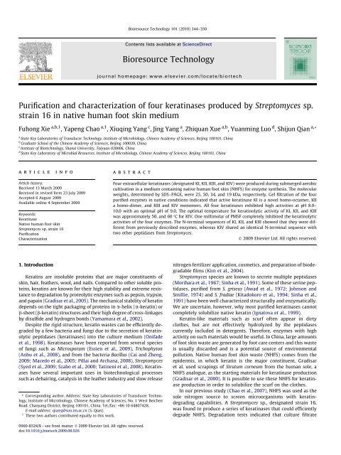
<strong>Purification</strong> <strong>and</strong> <strong>characterization</strong> <strong>of</strong> <strong>four</strong> <strong>keratinases</strong> produced by Streptomyces sp.<br />
strain 16 in native human foot skin medium<br />
Fuhong Xie a,b,1 , Yapeng Chao a,1 , Xiuqing Yang c , Jing Yang a , Zhiquan Xue a,b , Yuanming Luo d , Shijun Qian a, *<br />
a State Key Laboratories <strong>of</strong> Transducer Technology, Institute <strong>of</strong> Microbiology, Chinese Academy <strong>of</strong> Sciences, Beijing 100101, China<br />
b Graduate School <strong>of</strong> the Chinese Academy <strong>of</strong> Sciences, Beijing 100039, China<br />
c Institute <strong>of</strong> Biotechnology, Shanxi University, Taiyuan 030006, China<br />
d State Key Laboratory <strong>of</strong> Microbial Resources, Institute <strong>of</strong> Microbiology, Chinese Academy <strong>of</strong> Sciences, Beijing 100101, China<br />
article<br />
info<br />
abstract<br />
Article history:<br />
Received 13 March 2009<br />
Received in revised form 23 July 2009<br />
Accepted 6 August 2009<br />
Available online 4 September 2009<br />
Keywords:<br />
Keratinase<br />
Native human foot skin<br />
Streptomyces sp. strain 16<br />
<strong>Purification</strong><br />
Characterization<br />
Four extracellular <strong>keratinases</strong> (designated KI, KII, KIII, <strong>and</strong> KIV) were produced during submerged aerobic<br />
cultivation in a medium containing native human foot skin (NHFS) for enzyme synthesis. The molecular<br />
weights, determined by SDS–PAGE, were 25, 50, 34, <strong>and</strong> 19 kDa, respectively. Gel filtration <strong>of</strong> the <strong>four</strong><br />
purified enzymes in native conditions indicated that active keratinase KI is a novel homo-octamer, KII<br />
a homo-dimer, <strong>and</strong> KIII <strong>and</strong> KIV monomers. All <strong>four</strong> <strong>keratinases</strong> exhibited high activities at pH 8.0–<br />
10.0 with an optimal pH <strong>of</strong> 9.0. The optimal temperature for keratinolytic activity <strong>of</strong> KI, KII, <strong>and</strong> KIII<br />
was approximately 50, <strong>and</strong> 60 °C for KIV. One millimolar <strong>of</strong> PMSF completely inhibited the keratinolytic<br />
activities <strong>of</strong> the <strong>four</strong> enzymes. The N-terminal sequences <strong>of</strong> KI, KII, <strong>and</strong> KIII showed that they were different<br />
from previously described enzymes, whereas KIV shared an identical N-terminal sequence with<br />
two other peptidases from Streptomyces.<br />
Ó 2009 Elsevier Ltd. All rights reserved.<br />
1. Introduction<br />
Keratins are insoluble proteins that are major constituents <strong>of</strong><br />
skin, hair, feathers, wool, <strong>and</strong> nails. Compared to other soluble proteins,<br />
keratins are known for their high stability <strong>and</strong> extreme resistance<br />
to degradation by proteolytic enzymes such as pepsin, trypsin,<br />
<strong>and</strong> papain (Gradisar et al., 2005). The mechanical stability <strong>of</strong> keratin<br />
depends on the tight packaging <strong>of</strong> proteins in a-helix (a-keratin) or<br />
b-sheet (b-keratin) structures <strong>and</strong> their high degree <strong>of</strong> cross-linkages<br />
by disulfide <strong>and</strong> hydrogen bonds (Yamamura et al., 2002).<br />
Despite the rigid structure, keratin wastes can be efficiently degraded<br />
by a few bacteria <strong>and</strong> fungi due to the secretion <strong>of</strong> keratinolytic<br />
peptidases (<strong>keratinases</strong>) into the culture medium (Onifade<br />
et al., 1998). Keratinases have been reported from several species<br />
<strong>of</strong> fungi such as Microsporum (Essien et al., 2009), Trichophyton<br />
(Anbu et al., 2008), <strong>and</strong> from the bacteria Bacillus (Cai <strong>and</strong> Zheng,<br />
2009; Macedo et al., 2005; Pillai <strong>and</strong> Archana, 2008), Streptomyces<br />
(Syed et al., 2009; Szabo et al., 2000; Tatineni et al., 2008). Keratinases<br />
have several important uses in biotechnological processes<br />
such as dehairing, catalysis in the leather industry <strong>and</strong> slow release<br />
* Corresponding author. Address: State Key Laboratories <strong>of</strong> Transducer Technology,<br />
Institute <strong>of</strong> Microbiology, Chinese Academy <strong>of</strong> Sciences, No. 1 West Beichen<br />
Road, Chaoyang District, Beijing 100101, China. Tel./fax: +86 10 64807428.<br />
E-mail address: qiansj@sun.im.ac.cn (S. Qian).<br />
1 These two authors contributed equally to this work.<br />
nitrogen fertilizer application, cosmetics, <strong>and</strong> preparation <strong>of</strong> biodegradable<br />
films (Kim et al., 2004).<br />
Streptomyces species are known to secrete multiple peptidases<br />
(Morihara et al., 1967; Sinha et al., 1991). Some <strong>of</strong> these serine peptidases,<br />
purified from S. griseus (Awad et al., 1972; Johnson <strong>and</strong><br />
Smillie, 1974) <strong>and</strong> S. fradiae (Kitadokoro et al., 1994; Sinha et al.,<br />
1991) have been well characterized structurally <strong>and</strong> enzymatically.<br />
We are uncertain, however, why most purified <strong>keratinases</strong> cannot<br />
completely solubilize native keratin (Ignatova et al., 1999).<br />
Keratin-like materials such as scurf <strong>of</strong>ten appear in dirty<br />
clothes, but are not effectively hydrolyzed by the peptidases<br />
currently included in detergents. Therefore, enzymes with high<br />
activity on such materials would be useful. In China, large amounts<br />
<strong>of</strong> foot skin waste are generated by foot care centers <strong>and</strong> this waste<br />
is usually discarded <strong>and</strong> is a potential source <strong>of</strong> environmental<br />
pollution. Native human foot skin waste (NHFS) comes from the<br />
epidermis, in which keratin is the major constituent. Gradisar<br />
et al. used scrapings <strong>of</strong> Stratum corneum from the human sole, a<br />
NHFS analogue, as the starting materials for keratinase production<br />
(Gradisar et al., 2000). It is possible to use these NHFS for keratinase<br />
production in order to solubilize the scurf on the clothes.<br />
In our previous study (Chao et al., 2007), NHFS was used as the<br />
sole nitrogen source to screen microorganisms with keratindegrading<br />
capabilities. A Streptomyces sp., designated strain 16,<br />
was found to produce a series <strong>of</strong> <strong>keratinases</strong> that could efficiently<br />
degrade NHFS. Degradation tests indicated that culture filtrate<br />
0960-8524/$ - see front matter Ó 2009 Elsevier Ltd. All rights reserved.<br />
doi:10.1016/j.biortech.2009.08.026
F. Xie et al. / Bioresource Technology 101 (2010) 344–350 345<br />
from Streptomyces sp. strain 16 could also efficiently decompose<br />
casein, collagen, <strong>and</strong> keratin azure.<br />
In this study, we describe the purification <strong>and</strong> <strong>characterization</strong><br />
<strong>of</strong> <strong>four</strong> <strong>keratinases</strong> from Streptomyces sp. strain 16 cultured in<br />
medium containing NHFS.<br />
2. Methods<br />
2.1. Materials<br />
Native human foot skin waste was collected from the foot care<br />
section <strong>of</strong> the Haidian Hospital in Beijing. In preparation for NHFS<br />
experiments, the human foot skin was washed thoroughly with tap<br />
water, autoclaved, <strong>and</strong> then dried. The dried NHFS was stored under<br />
dry condition <strong>and</strong> was used for keratinase production. One part <strong>of</strong> the<br />
dried NHFS was milled to powders for the keratinase assay. 1-Ethyl-<br />
3-(3-dimethyaminopropyl) carbodiimide (EDAC), p-chloromercuribenzoic<br />
acid (pCMB), phenylmethanesulfonyl fluoride (PMSF), DLdithiothreitol<br />
(DTT), <strong>and</strong> casein were purchased from Sigma (USA).<br />
Nanosep centrifugal device tubes (molecular size cut<strong>of</strong>f, 100 or<br />
10 kDa) were purchased from Pall Corporation (USA), while Sephacryl<br />
S-200 resin, Superose 12 10/300 GL column, Hiprep DEAE FF<br />
column (1 ml), <strong>and</strong> Hiprep Octyl FF column (1 ml) were acquired<br />
from Amersham Biosciences (Sweden). The Folin–Ciocalteu reagent<br />
was purchased from Dingguo Company (China). All reagents used<br />
in this study were analytical grade unless otherwise indicated.<br />
2.2. Microorganism, culture medium, <strong>and</strong> growth conditions<br />
The microorganism used in this study was Streptomyces sp.<br />
strain 16, which was isolated in our lab for keratinase production<br />
(Chao et al., 2007). The medium for keratinase production was<br />
composed <strong>of</strong> NHFS, 4 g/l; soluble starch, 5 g/l; yeast extract, 1 g/l;<br />
<strong>and</strong> K 2 HPO 4 , 0.5 g/l. The pH <strong>of</strong> the medium was 8.0.<br />
Seed cultures <strong>of</strong> Streptomyces sp. strain 16 were prepared in a<br />
500 ml Erlenmeyer flask containing 100 ml <strong>of</strong> culture medium.<br />
After 2 days <strong>of</strong> incubation, the culture broth was transferred to a<br />
5 L Fernbach culture flask containing 1.5 L <strong>of</strong> medium for <strong>keratinases</strong><br />
production. After 4 days <strong>of</strong> incubation, the culture was collected<br />
for keratinase purification. All incubations were done<br />
aerobically at 30 °C on an orbital platform shaker at 200 rpm.<br />
2.3. Zymography <strong>of</strong> the culture filtrate<br />
The zymography <strong>of</strong> the culture filtrate was based on the method<br />
<strong>of</strong> Bressollier et al. (1999).The culture broth was centrifuged<br />
(10,000 rpm, 4 °C, 10 min.), <strong>and</strong> the supernatant was mixed with<br />
2 electrophoresis sample buffer (2 ml 4 stacking gel buffer,<br />
1.6 ml glycerol, 3.2 ml 10% SDS, 0.8 ml 2-mercaptoethanol, <strong>and</strong><br />
0.4 ml 1.0% bromophenol blue) (Simpson, 2003) in a 1:1 ratio without<br />
heat denaturation. SDS–PAGE was carried out at 4 °C with a<br />
constant current <strong>of</strong> 23 mA in a 12% polyacrylamide gel. The gel<br />
was washed with 2.5% Triton X-100 (v/v) in 50 mM Tris–HCl buffer<br />
(pH 7.5) for 30 min. Then washed with 50 mM Tris–HCl buffer (pH<br />
7.5) for 30 min. Casein (1%, w/v) dissolved in 50 mM Tris–HCl buffer<br />
(pH 7.5) was poured onto the gel. After 30 min at 40 °C, the gel<br />
was stained with Coomassie brilliant blue R-250 <strong>and</strong> then destained<br />
with a water/methanol/acetic acid (50:40:10) solvent. Peptidase<br />
b<strong>and</strong>s appeared as clear zones on a blue background.<br />
2.4. Enzyme assays<br />
2.4.1. Assay for keratinolytic activity<br />
Keratinolytic activity was determined spectrophotometrically<br />
with a modification <strong>of</strong> the Folin–Ciocalteu method (Ledoux <strong>and</strong><br />
Lamy, 1986; Margesin <strong>and</strong> Schinner, 1991). An appropriate amount<br />
<strong>of</strong> enzyme was incubated with 1.5 ml <strong>of</strong> 0.5% sieved (mesh size:<br />
300 lm) NHFS, in 50 mM Tris–HCl buffer (pH 7.5) at 40 °C for<br />
16 h. The reaction was terminated with 3 ml <strong>of</strong> 10% trichloroacetic<br />
acid (TCA) <strong>and</strong> placed at room temperature for 30 min. The reaction<br />
mixture was centrifuged, <strong>and</strong> 5 ml <strong>of</strong> 0.55 M Na 2 CO 3 was<br />
added to 1 ml <strong>of</strong> the supernatant, with subsequent addition <strong>of</strong><br />
1 ml Folin–Ciocalteu reagent. This mixture was incubated at<br />
40 °C for 15 min. Keratinolytic activity was measured at 680 nm<br />
using a spectrophotometer (721 Visible Spectrometry, China).<br />
One unit (U) <strong>of</strong> keratinase activity was represented by an increase<br />
in absorbance at 680 nm <strong>of</strong> 0.01 in 1 h compared with the control<br />
reaction. The control was treated in the same way, except that TCA<br />
was added before incubation.<br />
2.4.2. Assay for proteolytic activity<br />
Protease activity was determined by modifying the Folin–Ciocalteu<br />
method using casein as a substrate. The appropriate amount<br />
<strong>of</strong> enzyme was incubated with 1 ml 1% casein in 50 mM Tris–HCl<br />
buffer (pH 7.5) at 40 °C for 15 min. The reaction was terminated<br />
by the addition <strong>of</strong> 3 ml <strong>of</strong> 10% TCA. Subsequent steps were the<br />
same as those described in the assay <strong>of</strong> keratinolytic activity.<br />
One unit (U) <strong>of</strong> protease activity was defined as an increase in<br />
absorbance at 680 nm <strong>of</strong> 0.01 in 1 min compared with the control<br />
reaction. The control was treated in the same way, except that TCA<br />
was added before incubation.<br />
2.5. Protein determination<br />
Protein concentrations <strong>of</strong> the enzyme preparations were determined<br />
according to the method described by Bradford (1976),<br />
using bovine serum albumin as the st<strong>and</strong>ard.<br />
2.6. <strong>Purification</strong> procedure<br />
Culture broth was centrifuged (11,366g, 4°C, 10 min) to remove<br />
cells <strong>and</strong> debris <strong>and</strong> the supernatant was collected. Solid<br />
ammonium sulfate was added to the supernatant with a saturation<br />
<strong>of</strong> 80% under gentle stirring, <strong>and</strong> then kept at 4 °C overnight. The<br />
suspension was centrifuged at 10,000g for 30 min at 4 °C. The<br />
precipitate was then dissolved in 100 ml <strong>of</strong> 50 mM Tris–HCl buffer<br />
(pH 7.5).<br />
Three milliliter <strong>of</strong> the dissolved ammonium sulfate precipitate<br />
was loaded onto a Sephacryl S-200 column (3 100 cm) equilibrated<br />
with 50 mM Tris–HCl buffer (pH 7.5) <strong>and</strong> eluted with the<br />
same buffer at a flow rate <strong>of</strong> 0.5 ml/min. Fractions <strong>of</strong> 6.0 ml in each<br />
tube were collected <strong>and</strong> assayed for keratinolytic activity. The<br />
peaks with keratinolytic activity were named KI, KII, KIII, <strong>and</strong> KIV<br />
according to the order <strong>of</strong> elution. Purity was checked via SDS–<br />
PAGE. KIV was found to be homogenous on the SDS–PAGE gel,<br />
while the other three components needed further chromatographic<br />
purification.<br />
2.6.1. <strong>Purification</strong> <strong>of</strong> <strong>keratinases</strong> KI <strong>and</strong> KII<br />
Following Sephacryl S-200 column chromatography, the KI <strong>and</strong><br />
KII fractions were pooled separately, <strong>and</strong> sequentially applied to a<br />
Hiprep DEAE FF column (1 ml) pre-equilibrated in 50 mM Tris–HCl<br />
buffer (pH 7.5), <strong>and</strong> a Hiprep Octyl FF column (1 ml) pre-equilibrated<br />
in 50 mM Tris–HCl buffer (pH 7.5) containing 1.5 M<br />
(NH 4 ) 2 SO 4 . The KI fraction was eluted from the Hiprep DEAE FF column<br />
with 20 ml 50 mM Tris–HCl buffer (pH 7.5) containing 0 to<br />
1 M NaCl at a linear flow rate <strong>of</strong> 2 ml/min. The KII fraction was<br />
eluted using 0.1 M, 0.2 M, 0.5 M, <strong>and</strong> 1 M NaCl sequentially, in<br />
50 mM Tris–HCl buffer (pH 7.5). The KI fraction was eluted from<br />
the Hiprep Octyl FF column using 20 ml <strong>of</strong> 50 mM Tris–HCl buffer<br />
(pH 7.5) containing 0 to 1.5 M (NH 4 ) 2 SO 4 at a linear flow rate <strong>of</strong>
346 F. Xie et al. / Bioresource Technology 101 (2010) 344–350<br />
2 ml/min. The KII fraction was eluted from the Hiprep Octyl FF column<br />
using a reverse step gradient <strong>of</strong> 50 mM Tris–HCl buffer (pH<br />
7.5) containing 1.0, 0.75, 0.5, 0.3, <strong>and</strong> 0 M (NH 4 ) 2 SO 4 , sequentially,<br />
at a flow rate <strong>of</strong> 2 ml/min.<br />
2.6.2. <strong>Purification</strong> <strong>of</strong> keratinase KIII<br />
After Sephacryl S-200 column chromatography, fractions <strong>of</strong><br />
peak KIII were pooled <strong>and</strong> adjusted to 1.5 M (NH 4 ) 2 SO 4 . KIII was<br />
eluted by applying 20 ml <strong>of</strong> 50 mM Tris–HCl buffer (pH 7.5) containing<br />
0 to 1.5 M (NH 4 ) 2 SO 4 at a linear flow rate <strong>of</strong> 2 ml/min.<br />
3. Results<br />
3.1. Keratinases production<br />
Streptomyces sp. strain 16 was able to grow <strong>and</strong> produce keratinase<br />
in a medium that contained 4 g/L NHFS. To expedite growth<br />
<strong>and</strong> increase the biomass <strong>of</strong> the strain, limited nitrogen sourceyeast<br />
extract was added to the medium in this study. NHFS began<br />
to decrease in the second day <strong>of</strong> cultivation, <strong>and</strong> completely disappeared<br />
in the <strong>four</strong>th day <strong>of</strong> cultivation. Keratinase production<br />
reached maximal activity <strong>of</strong> 74.9 U/ml at <strong>four</strong> days <strong>of</strong> cultivation.<br />
2.7. Molecular weight <strong>of</strong> the purified <strong>keratinases</strong><br />
The purity <strong>and</strong> subunit number <strong>of</strong> each peptidase was estimated<br />
by SDS–PAGE (12% gel) according to Laemmli (1970). After electrophoresis,<br />
the gel was stained with Coomassie Brilliant Blue R-250<br />
dissolved in a water/methanol/acetic acid (50:40:10) solvent for<br />
1 h, then destained in the same solvent. The molecular weight <strong>of</strong><br />
the native peptidases was determined using a Superose 12 10/300<br />
GL column, using thyroglobulin (669,000 Da), ferritin (440,000 Da),<br />
catalase (232,000 Da), lactate dehydrogenase (140,000 Da), <strong>and</strong> bovine<br />
serum albumin (67,000 Da) (Amersham Biosciences, Sweden)<br />
as st<strong>and</strong>ards.<br />
2.8. Effect <strong>of</strong> pH <strong>and</strong> temperature on enzyme activity <strong>and</strong> stability<br />
The optimal pH for enzyme activities was determined at 40 °C<br />
at various pH levels (pH 6.0–12.0). Sodium phosphate was used<br />
for pH 6.0–7.0, Tris–HCl was used for pH 7.5–9.0, <strong>and</strong> sodium carbonate–bicarbonate<br />
was used for pH 10.0–12.0. For pH stability,<br />
residual activity was measured at pH 7.5 after 1 h <strong>of</strong> incubation<br />
at 40 °C in the pH buffers being tested.<br />
Enzyme activities were tested at temperatures ranging from<br />
30 °C to80°C in 50 mM Tris–HCl buffer (pH 7.5). Each <strong>of</strong> the purified<br />
enzymes was incubated for 1 h at the tested temperature, <strong>and</strong><br />
residual activity was measured at 40 °C in 50 mM Tris–HCl buffer<br />
(pH 7.5).<br />
3.2. <strong>Purification</strong> <strong>of</strong> the <strong>keratinases</strong> produced by the Streptomyces sp.<br />
strain 16<br />
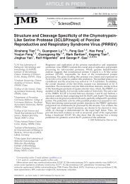
The zymogram <strong>of</strong> culture supernatant shown in Fig. 1 indicated<br />
the presence <strong>of</strong> five b<strong>and</strong>s that showed peptidase activity in the<br />
culture supernatant.<br />
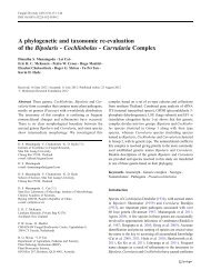
The purification procedures <strong>of</strong> the <strong>four</strong> enzymes is outlined in<br />
Fig. 2. When the precipitated enzyme was loaded onto Sephacryl<br />
S-200 column, <strong>four</strong> major peaks eluted that showed keratinase<br />
activity. The main keratinase in each peak was designated KI, KII,<br />
KIII, <strong>and</strong> KIV based on their order <strong>of</strong> elution. However, the fifth<br />
b<strong>and</strong> appeared on the zymogram gel was not detected from the<br />
elution.<br />
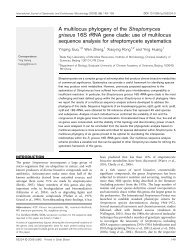
After one to two further steps <strong>of</strong> chromatography, all <strong>four</strong> <strong>keratinases</strong><br />
from Streptomyces sp. strain 16 were ultimately purified<br />
to homogeneity, which was confirmed by SDS–PAGE (Fig. 3).<br />
Molecular weights <strong>of</strong> subunits <strong>of</strong> the <strong>four</strong> enzymes were calculated<br />
as 25 kDa (KI), 50 kDa (KII), 34 kDa (KIII) <strong>and</strong> 19 kDa (KIV).<br />
The determination <strong>of</strong> the molecular weight <strong>of</strong> each <strong>of</strong> the <strong>four</strong><br />
<strong>keratinases</strong> was performed by gel filtration chromatography on a<br />
2.9. Effects <strong>of</strong> protease inhibitors, reducing agents, <strong>and</strong> metal ions on<br />
keratinase activity<br />
To investigate the effects <strong>of</strong> different inhibitors, reducing<br />
agents, <strong>and</strong> metal ions on keratinase activity, the residual activity<br />
after allowing the enzyme to st<strong>and</strong> with the reagent for 30 min<br />
at 30 °C was measured. Phenylmethyl sulphonyl fluoride (PMSF),<br />
para-chloro mercuric benzoate (pCMB), EDAC, <strong>and</strong> ethylene diamine<br />
tetra acetic acid (EDTA) were used at a concentration <strong>of</strong><br />
1 mM as protease inhibitors. Dithiothreitol (DTT) <strong>and</strong> ascorbic acid<br />
were used at a concentration <strong>of</strong> 1 mM, <strong>and</strong> Na 2 SO 3 ,Ca 2+ , <strong>and</strong> Mg 2+<br />
were used at a concentration <strong>of</strong> 10 mM.<br />
2.10. N-terminal amino acid sequencing <strong>of</strong> the purified <strong>keratinases</strong><br />
After SDS–PAGE, each purified ketatinase b<strong>and</strong> was transferred<br />
onto a polyvinylidene fluoride (PVDF) membrane (Pall Corporation,<br />
USA). The keratinase b<strong>and</strong> on the PVDF membrane was cut out <strong>and</strong><br />
dissolved in 20% (v/v) acetonitrile/water containing 0.001% trifluoroacetic<br />
acid (TFA). The N-terminal sequences <strong>of</strong> the purified <strong>keratinases</strong><br />
were determined by automated Edman degradation using a<br />
pulsed-liquid sequence analyzer (ABI PROCISE TM 492cLC protein<br />
sequencing system, Applied Biosystems, USA).<br />
Fig. 1. Zymogram analysis <strong>of</strong> <strong>keratinases</strong> secreted by Streptomyces sp. strain 16.<br />
Arrows indicate the keratinase b<strong>and</strong>s.
F. Xie et al. / Bioresource Technology 101 (2010) 344–350 347<br />
Concentrated culture<br />
Sephacryl S-200<br />
Peak I<br />
DEAE FF<br />
Peak II<br />
DEAE FF<br />
Peak III<br />
Peak IV<br />
Octyl FF<br />
Octyl FF<br />
Octyl FF<br />
KI<br />
KII<br />
KIII<br />
KIV<br />
Fig. 2. <strong>Purification</strong> strategy for extracting <strong>keratinases</strong> from Streptomyces sp. strain 16.<br />
kDa<br />
110<br />
M<br />
1 2 3 4<br />
Table 1<br />
Activity determination in different components treated by ultrafiltration.<br />
1 2 3<br />
66<br />
Protein (lg/ml) 244 256 1.5<br />
Keratinolytic activity (U/ml) 37 42 0<br />
Specific activity (U/mg protein) 151.6 164.1 0<br />
45<br />
35<br />
1. The retentate by 100 kDa ultrafiltration.<br />
2. The retentate by 10 kDa ultrafiltration.<br />
3. Filtrate by 10 kDa ultrafiltration.<br />
25<br />
18<br />
14<br />
Fig. 3. SDS–PAGE analysis <strong>of</strong> the purified <strong>keratinases</strong> (KI, KII, KIII, <strong>and</strong> KIV)<br />
produced by Streptomyces sp. strain 16. The molecular marker, KI, KII, KIII, <strong>and</strong> KIV<br />
are located in lane M, 1, 2, 3, <strong>and</strong> 4, respectively.<br />
Superose 12 10/300 GL column. The results provided a molecular<br />
weight estimate <strong>of</strong> 203.2 kDa for KI, 100.8 kDa for KII, 31.8 kDa<br />
for KIII, <strong>and</strong> 19.2 kDa for KIV. This indicated that KI is a homo-octamer,<br />
KII a homo-dimer, <strong>and</strong> KIII a monomer <strong>and</strong> KIV a monomer.<br />
To further verify that KI is an active homo-octomer, ultrafiltration<br />
was conducted. Retentate with a molecular weight <strong>of</strong> 100 kDa,<br />
retentate with a molecular weight <strong>of</strong> 10 kDa, <strong>and</strong> filtrate with a<br />
molecular weight <strong>of</strong> 10 kDa or less were sequentially collected<br />
<strong>and</strong> analyzed (Table 1). Most <strong>of</strong> the enzyme activity was detected<br />
in retentate with a molecular weight <strong>of</strong> 100 kDa <strong>and</strong> a molecular<br />
weight <strong>of</strong> 10 kDa. However, no activity emerged in the filtrate with<br />
a molecular weight <strong>of</strong> 10 kDa.<br />
3.3. Influence <strong>of</strong> temperature <strong>and</strong> pH on the keratinolytic activity <strong>of</strong> KI,<br />
KII, KIII, <strong>and</strong> KIV<br />
The <strong>four</strong> purified <strong>keratinases</strong> had high activities between pH 8.0<br />
<strong>and</strong> 10.0, with maximal activity at pH 9.0. The <strong>four</strong> <strong>keratinases</strong><br />
showed low activities at pH 6.0 <strong>and</strong> 12.0. The effect <strong>of</strong> pH 7.0 on<br />
each <strong>of</strong> the <strong>four</strong> <strong>keratinases</strong> was different, the keratinase activities<br />
for KI <strong>and</strong> KIV were more than 50% <strong>of</strong> their maximal activities, but<br />
KII <strong>and</strong> KIII activities were less than 30% <strong>of</strong> their maximal activities.<br />
At pH 11.0, KI showed low keratinase activity, while KII, KIII, <strong>and</strong><br />
KIV showed more than 50% <strong>of</strong> their maximal activities. The <strong>four</strong><br />
<strong>keratinases</strong> showed the same pH stability. More than 85% <strong>of</strong> the<br />
maximal activities were retained between pH 8.0 <strong>and</strong> 10.0.<br />
The optimal temperature for <strong>keratinases</strong> KI, KII, <strong>and</strong> KIII was<br />
about 50 °C, while the optimal temperature was about 60 °C for<br />
keratinase KIV. KI, KII, <strong>and</strong> KIII activity dropped fast at temperatures<br />
below 40 °C <strong>and</strong> above 60 °C. For KIV, the activity at 80 °C<br />
was approximately half <strong>of</strong> its maximal activity, which was at<br />
60 °C. All <strong>four</strong> <strong>keratinases</strong> retained more than 70% <strong>of</strong> their maximal<br />
activities in the temperature range <strong>of</strong> 40–60 °C after 1 h incubation.<br />
KIV was more stable than the other three <strong>keratinases</strong>.<br />
3.4. Effects <strong>of</strong> chemicals on keratinolytic activities <strong>of</strong> KI, KII, KIII <strong>and</strong><br />
KIV<br />
Protease inhibitors, reducing agents, <strong>and</strong> metal ions were used<br />
to investigate their effects on the enzyme activities (Table 2). All<br />
<strong>four</strong> enzymes were significantly inhibited by the serine protease<br />
inhibitor PMSF at 1 mM. This indicates that all <strong>of</strong> the enzymes belong<br />
to the serine protease family. Moreover, the residual activities<br />
<strong>of</strong> the <strong>four</strong> enzymes were evidently not affected by cysteine protease<br />
inhibitor pCMB. However, metalloprotease inhibitor EDTA<br />
strongly inhibited KI, KII, <strong>and</strong> KIII, although it did not inhibit the<br />
activity <strong>of</strong> KIV. For all <strong>four</strong> <strong>keratinases</strong>, significant increases in keratinolytic<br />
activity were observed by adding reducing agents DTT,<br />
ascorbic acid, <strong>and</strong> sodium sulfite. Carbonyl group modifier EDAC<br />
had stimulative effects on the activities <strong>of</strong> KI, KIII, <strong>and</strong> KIV. The keratinolytic<br />
activities <strong>of</strong> KI, KIII, <strong>and</strong> KIV increased by 36%, 100%, <strong>and</strong><br />
32%, respectively. The divalent metal ion Ca 2+ showed a stimulative
348 F. Xie et al. / Bioresource Technology 101 (2010) 344–350<br />
Table 2<br />
Effects <strong>of</strong> the different reagents on the keratinolytic activity <strong>of</strong> the <strong>four</strong> <strong>keratinases</strong>.<br />
Compound Concentration (mM) Residual activities (%)<br />
effect on KI, an inhibitory effect on KIII, <strong>and</strong> no effect on KII <strong>and</strong><br />
KIV. The metal ion Mg 2+ had an inhibitory effect on KII <strong>and</strong> KIII,<br />
<strong>and</strong> no effect on KI <strong>and</strong> KIV.<br />
3.5. Substrate specificities <strong>of</strong> KI, KII, KIII, <strong>and</strong> KIV<br />
The ability <strong>of</strong> the <strong>four</strong> enzymes to hydrolyze proteinaceous substrates<br />
was examined with casein, NHFS, keratin azure, collagen,<br />
cock feathers, <strong>and</strong> human hair (Table 3). Results showed that the<br />
<strong>four</strong> enzymes exhibited high activities toward casein <strong>and</strong> NHFS,<br />
however, NHFS could not be solubilized by any one <strong>of</strong> the <strong>four</strong> <strong>keratinases</strong>.<br />
All <strong>four</strong> purified enzymes showed higher activities with<br />
soluble proteinaceous substrates such as casein than with insoluble<br />
proteinaceous substrates such as NHFS. The <strong>four</strong> <strong>keratinases</strong><br />
showed low activities toward keratin azure <strong>and</strong> collagen. For all<br />
<strong>four</strong> <strong>keratinases</strong>, no activity was observed with human hair <strong>and</strong><br />
cock feathers as substrates.<br />
3.6. N-terminal sequence<br />
N-terminal amino acid sequencing <strong>of</strong> the purified <strong>keratinases</strong><br />
resulted in unequivocal determination <strong>of</strong> the first 10 amino acid<br />
residues <strong>of</strong> the <strong>four</strong> <strong>keratinases</strong> (Table 4), with the exception <strong>of</strong> position<br />
10 in KIII, where no assignment could be made. When compared<br />
to the National Center for Biotechnology Information (NCBI)<br />
protein database by BLAST search, the KI, KII, <strong>and</strong> KIII sequences<br />
did not show significant homology to known microbial peptidases.<br />
However, the N-terminal sequence <strong>of</strong> KIV was identical to that <strong>of</strong><br />
the 17.6 kDa protease (Sinha et al., 1991) <strong>and</strong> SFase-2 (Kitadokoro<br />
et al., 1994) from S. fradiae <strong>and</strong> it showed 70% homology to SGPA<br />
from S. griseus (Johnson <strong>and</strong> Smillie, 1974).<br />
4. Discussion<br />
KI KII KIII KIV<br />
Control 0 100 100 100 100<br />
pCMB 1 92 86 96 95<br />
EDAC 1 136 107 200 132<br />
PMSF 1 33 0 0 0<br />
EDTA 1 36 7 0 97<br />
DTT 1 195 157 170 256<br />
Ascorbic acid 10 145 107 370 155<br />
Na 2 SO 3 1 177 160 130 180<br />
CaCl 2 10 114 86 20 82<br />
MgCl 2 10 86 28 60 87<br />
In our previous studies, Streptomyces sp. strain 16 was found to<br />
completely degrade NHFS, <strong>and</strong> <strong>keratinases</strong> were produced when<br />
Table 3<br />
Substrate specificities <strong>of</strong> the <strong>four</strong> <strong>keratinases</strong>.<br />
Substrate Residual activity * (%)<br />
KI KII KIII KIV<br />
Casein 100 100 100 100<br />
NHFS 93 79.1 89 82<br />
Keratin azure 3.7 0.8 1.0 6.5<br />
Collagen 3.7 1.2 1.0 30<br />
Cock feather 0 0 0 0<br />
Human hair 0 0 0 0<br />
* The proteolytic activity toward casein <strong>of</strong> each enzyme was used as 100%. Activity<br />
on other substrate was compared with it.<br />
strain 16 was cultured in a medium that contained NHFS. The addition<br />
<strong>of</strong> yeast extract to the medium was helpful to increase <strong>of</strong> biomass<br />
<strong>and</strong> enzyme production. A zymogram <strong>of</strong> culture filtrates<br />
showed that complete degradation <strong>of</strong> NHFS was due to the presence<br />
<strong>of</strong> multiple <strong>keratinases</strong>. In order to investigate the mechanism<br />
<strong>of</strong> degradation, we purified the <strong>keratinases</strong> from the culture broth.<br />
The results showed that <strong>four</strong> <strong>keratinases</strong> secreted by Streptomyces<br />
sp. strain 16 were responsible for the degradation <strong>of</strong> NHFS in the<br />
medium. In the purification procedure, we also tried to purify the<br />
fifth peptidase appeared on the zymogram gel. Unfortunately, we<br />
failed to isolate this enzyme, perhaps due to its low quantity.<br />
Most microbial <strong>keratinases</strong> are inducible enzymes, as reported<br />
by Cheng et al. (1995). Various keratinous materials such as chicken<br />
feather, feather meal, wool, <strong>and</strong> bovine hair have been used as<br />
inducers <strong>of</strong> <strong>keratinases</strong> (De Toni et al., 2002; Ignatova et al.,<br />
1999; Kumar et al., 2008; Nam et al., 2002). NHFS belongs to s<strong>of</strong>t<br />
keratins from the human sole, which was first used as a keratinase<br />
inducer.<br />
Proteolytic enzymes have dominated the detergent enzyme<br />
market. Nonetheless, there is always a need for new enzymes with<br />
novel properties that can further widen the scope <strong>of</strong> enzyme-based<br />
detergents. Keratinases could help in the removal <strong>of</strong> keratinous<br />
soils that are <strong>of</strong>ten encountered in the laundry <strong>and</strong> on which most<br />
normal peptidases fail to act. In this study, the <strong>keratinases</strong> produced<br />
by Streptomyces sp. strain 16 can efficiently decompose<br />
NHFS, therefore, these <strong>keratinases</strong> may be c<strong>and</strong>idate enzymes to<br />
be used as additives to the detergents.<br />
Similar to most <strong>keratinases</strong> reported in the literature, keratinase<br />
KIV is a monomer. However, KII is composed <strong>of</strong> two subunits in its<br />
native form <strong>and</strong> the molecular mass <strong>of</strong> each subunit is approximately<br />
50 kDa. Interestingly, KI is composed <strong>of</strong> eight subunits in<br />
its native form with a total molecular weight <strong>of</strong> 203.2 kDa. This<br />
is the first report <strong>of</strong> a peptidase with eight subunits. A few<br />
<strong>keratinases</strong> <strong>of</strong> high molecular weight with multimeric structures<br />
<strong>and</strong> multiple domains have been reported. Nam et al. isolated a<br />
native-feather-degrading thermophilic anaerobic bacterium identified<br />
as Fervidobacteium isl<strong>and</strong>icum from a geothermal hot stream<br />
(Nam et al., 2002). A homo-multimeric membrane-bound keratinolytic<br />
peptidase was purified from the cell extracts <strong>of</strong> the anaerobe.<br />
Its molecular weight was estimated to be greater than 200 kDa<br />
with a 97 kDa subunit (Nam et al., 2002). Fervidolysin is a keratinase<br />
(with a molecular weight <strong>of</strong> 73 kDa) as an immature form from<br />
Fervidobacterium pennivorans (Kluskens et al., 2002). The 1.7 Å resolution<br />
crystal structure <strong>of</strong> fervidolysin showed that the peptidase<br />
is composed <strong>of</strong> <strong>four</strong> domains: a catalytic domain (CD), two b-s<strong>and</strong>wich<br />
domains (SDs), <strong>and</strong> the propeptide (PD) domain (Kim et al.,<br />
2004). Keratinases <strong>of</strong> high molecular weight may consist <strong>of</strong> multi-domain<br />
structures or oligomeric proteases, which contribute to<br />
their substrate specificities (Kim et al., 2004). Accordingly, the oligomeric<br />
nature <strong>of</strong> KI <strong>and</strong> KII is also postulated to contribute to the<br />
substrate specificities.<br />
Several Streptomyces <strong>keratinases</strong> have been classified as serine<br />
proteases (Bockle <strong>and</strong> Muller, 1997; Bressollier et al., 1999). Kerotinolytic<br />
activities <strong>of</strong> studied enzymes were significantly inhibited<br />
by PMSF. Consequently, <strong>keratinases</strong> KI, KII, KIII, <strong>and</strong> KIV were classified<br />
as serine proteases. KI was slightly more stable at the same<br />
concentration <strong>of</strong> PMSF, perhaps due to its homo-multimeric form.<br />
Meanwhile, EDTA also showed similar effects on the keratinolytic<br />
activity <strong>of</strong> KI, KII, <strong>and</strong> KIII suggesting that they are metal ion related<br />
enzymes. The activity <strong>of</strong> KIV was not affected by EDTA. Although<br />
KI, KII, <strong>and</strong> KIII shared the same optimal temperature, <strong>and</strong> optimal<br />
pH <strong>of</strong> the <strong>four</strong> <strong>keratinases</strong> were identical, the effects <strong>of</strong> pH <strong>and</strong><br />
temperature on the activities <strong>of</strong> the <strong>four</strong> <strong>keratinases</strong> actually<br />
varied.<br />
According to the difference in molecular mass, characteristics,<br />
<strong>and</strong> the distinct differences in N-terminal sequences, the <strong>four</strong>
F. Xie et al. / Bioresource Technology 101 (2010) 344–350 349<br />
Table 4<br />
N-terminal sequences <strong>of</strong> Streptomyces sp. strain 16 <strong>keratinases</strong> <strong>and</strong> Alignment <strong>of</strong> keratinase KIV, SFase-2, 17.6 kDa protease <strong>and</strong> SGPA.<br />
Microorganism Keratinase N-terminal residues<br />
1 2 3 4 5 6 7 8 9 10<br />
Streptomyces sp. strain 16 KI a A G N S A S E I R V<br />
KII a A A P G D K D V T A<br />
KIII a A P D I I L A N A<br />
KIV a I A G G E A I Y A A<br />
S. fradiae ATCC 14544 SFase-2 b I A G G E A I Y A A<br />
S. fradiae 17.6 kDa c I A G G E A I Y A A<br />
S. griseus SGPA d I A G G E A I T T G<br />
a<br />
This study.<br />
b (Kitadokoro et al., 1994).<br />
c (Sinha et al., 1991).<br />
d (Johnson <strong>and</strong> Smillie, 1974).<br />
<strong>keratinases</strong> <strong>of</strong> Streptomyces sp. strain 16 are apparently different<br />
peptidase. From the identical molecular weight <strong>and</strong> N-terminal sequence,<br />
KIV may be the same peptidase as SFase-2 in S. fradiae, <strong>and</strong><br />
a homologue <strong>of</strong> the 17.6 kDa protease in S. fradiae <strong>and</strong> SGPA in S.<br />
griseus.<br />
Base on the fact that no enzyme among the <strong>four</strong> <strong>keratinases</strong><br />
could solubilize NHFS alone, there must be some cooperation<br />
among them or with some other components in the culture filtrate<br />
in the process <strong>of</strong> degrading NHFS. Therefore, it will be interesting<br />
to study the cooperation among the <strong>keratinases</strong> in further<br />
research.<br />
5. Conclusions<br />
In this work, <strong>four</strong> <strong>keratinases</strong> were purified from the culture<br />
broth <strong>of</strong> Streptomyces sp. strain 16 simultaneously, <strong>and</strong> they differed<br />
from each other in terms <strong>of</strong> molecular weight, oligomeric<br />
nature, <strong>and</strong> some physicochemical characteristics. All <strong>four</strong> <strong>keratinases</strong><br />
showed strong keratinolytic activity toward NHFS. The N-terminal<br />
sequences <strong>of</strong> KI, KII, <strong>and</strong> KIII showed no similarity to<br />
sequences <strong>of</strong> known microbial peptidases, whereas keratinase<br />
KIV shared an identical N-terminal sequence with two other peptidases<br />
<strong>of</strong> Streptomyces. Keratinase KI is a homo-octamer with a<br />
molecular weight <strong>of</strong> 203.2 kDa. Based on the properties <strong>of</strong> the <strong>four</strong><br />
<strong>keratinases</strong>, they might become attractive for potential applications<br />
in laundry detergents.<br />
Acknowledgement<br />
This work was financially supported by the National High Technology<br />
Research <strong>and</strong> Development Program <strong>of</strong> China (863;<br />
2006AA020204).<br />
References<br />
Anbu, P., Hilda, A., Sur, H.W., Hur, B.K., Jayanthi, S., 2008. Extracellular keratinase<br />
from Trichophyton sp. HA-2 isolated from feather dumping soil. Int. Biodeterior.<br />
Biodegrad. 62, 287–292.<br />
Awad Jr., W.M., Soto, A.R., Siegel, S., Skiba, W.E., Bernstrom, G.G., Ochoa, M.S., 1972.<br />
The proteolytic enzymes <strong>of</strong> the K-1 strain <strong>of</strong> Streptomyces griseus obtained from<br />
a commercial preparation (Pronase). I. <strong>Purification</strong> <strong>of</strong> <strong>four</strong> serine<br />
endopeptidases. J. Biol. Chem. 247, 4144–4154.<br />
Bockle, B., Muller, R., 1997. Reduction <strong>of</strong> disulfide bonds by Streptomyces pactum<br />
during growth on chicken feathers. Appl. Environ. Microbiol. 63, 790–792.<br />
Bradford, M.M., 1976. A rapid <strong>and</strong> sensitive method for the quantitation <strong>of</strong><br />
microgram quantities <strong>of</strong> protein utilizing the principle <strong>of</strong> protein-dye binding.<br />
Anal. Biochem. 72, 248–254.<br />
Bressollier, P., Letourneau, F., Urdaci, M., Verneuil, B., 1999. <strong>Purification</strong> <strong>and</strong><br />
<strong>characterization</strong> <strong>of</strong> a keratinolytic serine proteinase from Streptomyces<br />
albid<strong>of</strong>lavus. Appl. Environ. Microbiol. 65, 2570–2576.<br />
Cai, C., Zheng, X., 2009. Medium optimization for keratinase production in hair<br />
substrate by a new Bacillus subtilis KD-N2 using response surface methodology.<br />
J. Ind. Microbiol. Biotechnol. 36, 875–883.<br />
Chao, Y.P., Xie, F.H., Yang, J., Lu, J.H., Qian, S.J., 2007. Screening for a new<br />
Streptomyces strain capable <strong>of</strong> efficient keratin degradation. J. Environ. Sci. 19,<br />
1125–1128.<br />
Cheng, S.W., Hu, H.M., Shen, S.W., Takagi, H., Asano, M., Tsai, Y.C., 1995. Production<br />
<strong>and</strong> <strong>characterization</strong> <strong>of</strong> keratinase <strong>of</strong> a feather-degrading Bacillus licheniformis<br />
PWD-1. Biosci. Biotechnol. Biochem. 59, 2239–2243.<br />
De Toni, C.H., Richter, M.F., Chagas, J.R., Henriques, J.A., Termignoni, C., 2002.<br />
<strong>Purification</strong> <strong>and</strong> <strong>characterization</strong> <strong>of</strong> an alkaline serine endopeptidase from a<br />
feather-degrading Xanthomonas maltophilia strain. Can. J. Microbiol. 48, 342–<br />
348.<br />
Essien, J.P., Umoh, A.A., Akpan, E.J., Eduok, S.I., Umoiyoho, A., 2009. Growth,<br />
keratinolytic proteinase activity <strong>and</strong> thermotolerance <strong>of</strong> dermatophytes<br />
associated with alopecia in Uyo. Nigeria Acta Microbiologica Et Immunologica<br />
Hungarica 56, 61–69.<br />
Gradisar, H., Friedrich, J., Krizaj, I., Jerala, R., 2005. Similarities <strong>and</strong> specificities <strong>of</strong><br />
fungal keratinolytic proteases: comparison <strong>of</strong> <strong>keratinases</strong> <strong>of</strong> Paecilomyces<br />
marqu<strong>and</strong>ii <strong>and</strong> Doratomyces microsporus to some known proteases. Appl.<br />
Environ. Microbiol. 71, 3420–3426.<br />
Gradisar, H., Kern, S., Friedrich, J., 2000. Keratinase <strong>of</strong> Doratomyces microsporus.<br />
Appl. Microbiol. Biotechnol. 53, 196–200.<br />
Ignatova, Z., Gousterova, A., Spassov, G., Nedkov, P., 1999. Isolation <strong>and</strong> partial<br />
characterisation <strong>of</strong> extracellular keratinase from a wool degrading thermophilic<br />
actinomycete strain Thermoactinomyces c<strong>and</strong>idus. Can. J. Microbiol. 45, 217–<br />
222.<br />
Johnson, P., Smillie, L.B., 1974. The amino acid sequence <strong>and</strong> predicted structure <strong>of</strong><br />
Streptomyces griseus protease A. FEBS Lett. 47, 1–6.<br />
Kim, J.S., Kluskens, L.D., de Vos, W.M., Huber, R., van der Oost, J., 2004. Crystal<br />
structure <strong>of</strong> fervidolysin from Fervidobacterium pennivorans, a keratinolytic<br />
enzyme related to subtilisin. J. Mol. Biol. 335, 787–797.<br />
Kitadokoro, K., Tsuzuki, H., Nakamura, E., Sato, T., Teraoka, H., 1994. <strong>Purification</strong>,<br />
<strong>characterization</strong>, primary structure, crystallization <strong>and</strong> preliminary<br />
crystallographic study <strong>of</strong> a serine proteinase from Streptomyces fradiae ATCC<br />
14544. Eur. J. Biochem. 220, 55–61.<br />
Kluskens, L.D., Voorhorst, W.G., Siezen, R.J., Schwerdtfeger, R.M., Antranikian, G., van<br />
der Oost, J., de Vos, W.M., 2002. Molecular <strong>characterization</strong> <strong>of</strong> fervidolysin, a<br />
subtilisin-like serine protease from the thermophilic bacterium<br />
Fervidobacterium pennivorans. Extremophiles 6, 185–194.<br />
Kumar, A.G., Swarnalatha, S., Gayathri, S., Nagesh, N., Sekaran, G., 2008.<br />
Characterization <strong>of</strong> an alkaline active-thiol forming extracellular serine<br />
keratinase by the newly isolated Bacillus pumilus. J. Appl. Microbiol. 104, 411–419.<br />
Laemmli, U.K., 1970. Cleavage <strong>of</strong> structural proteins during assembly <strong>of</strong> head <strong>of</strong><br />
bacteriophage T4. Nature 227, 680–685.<br />
Ledoux, M., Lamy, F., 1986. Determination <strong>of</strong> proteins <strong>and</strong> sulfobetaine with the<br />
folin phenol reagent. Anal. Biochem. 157, 28–31.<br />
Macedo, A.J., da Silva, W.O., Gava, R., Driemeier, D., Henriques, J.A., Termignoni, C.,<br />
2005. Novel keratinase from Bacillus subtilis S14 exhibiting remarkable<br />
dehairing capabilities. Appl. Environ. Microbiol. 71, 594–596.<br />
Margesin, R., Schinner, F., 1991. Characterization <strong>of</strong> a metalloprotease from<br />
phychrophilic Xanthomonas maltophilia. FEMS Microbiol. Lett. 79, 257–262.<br />
Morihara, K., Oka, T., Tsuzuki, H., 1967. Multiple proteolytic enzymes <strong>of</strong><br />
Streptomyces fradiae. Production, isolation, <strong>and</strong> preliminary <strong>characterization</strong>.<br />
Biochimica et Biophysica Acta 139, 382–397.<br />
Nam, G.W., Lee, D.W., Lee, H.S., Lee, N.J., Kim, B.C., Choe, E.A., Hwang, J.K., Suhartono,<br />
M.T., Pyun, Y.R., 2002. Native-feather degradation by Fervidobacterium<br />
isl<strong>and</strong>icum AW-1, a newly isolated keratinase-producing thermophilic<br />
anaerobe. Arch. Microbiol. 178, 538–547.<br />
Onifade, A.A., Al-Sane, N.A., Al-Musallam, A.A., Al-Zarban, S., 1998. A review:<br />
potentials for biotechnological applications <strong>of</strong> keratin-degrading<br />
microorganisms <strong>and</strong> their enzymes for nutritional improvement <strong>of</strong> feathers<br />
<strong>and</strong> other keratins as livestock feed resources. Bioresour. Technol. 66, 1–11.<br />
Pillai, P., Archana, G., 2008. Hide depilation <strong>and</strong> feather disintegration studies with<br />
keratinolytic serine protease from a novel Bacillus subtilis isolate. Appl.<br />
Microbiol. Biotechnol. 78, 643–650.<br />
Simpson, R.J., 2003. Proteins <strong>and</strong> Proteomics: A Laboratory Mannual. Cold Spring<br />
Harbor Laboratory Press, New York.
350 F. Xie et al. / Bioresource Technology 101 (2010) 344–350<br />
Sinha, U., Wolz, S.A., Lad, P.J., 1991. Two new extracellular serine proteases from<br />
Streptomyces fradiae. Int. J. Biochem. 23, 979–984.<br />
Syed, D.G., Lee, J.C., Li, W.J., Kim, C.J., Agasar, D., 2009. Production, <strong>characterization</strong><br />
<strong>and</strong> application <strong>of</strong> keratinase from Streptomyces gulbargensis. Bioresour.<br />
Technol. 100, 1868–1871.<br />
Szabo, I., Benedek, A., Szabo, I.M., Barabas, G., 2000. Feather degradation with a<br />
thermotolerant Streptomyces gramin<strong>of</strong>aciens strain. World J. Microbiol.<br />
Biotechnol. 16, 253–255.<br />
Tatineni, R., Doddapanem, K.K., Potumarthi, R.C., Vellanki, R.N., K<strong>and</strong>athil, M.T.,<br />
Kolli, N., Mangamoori, L.N., 2008. <strong>Purification</strong> <strong>and</strong> <strong>characterization</strong> <strong>of</strong> an<br />
alkaline keratinase from Streptomyces sp. Bioresour. Technol. 99, 1596–<br />
1602.<br />
Yamamura, S., Morita, Y., Hasan, Q., Yokoyama, K., Tamiya, E., 2002. Keratin<br />
degradation: a cooperative action <strong>of</strong> two enzymes from Stenotrophomonas sp.<br />
Biochem. Biophys. Res. Commun. 294, 1138–1143.