NMR Spectroscopy of Polymers
NMR Spectroscopy of Polymers
NMR Spectroscopy of Polymers

Create successful ePaper yourself
Turn your PDF publications into a flip-book with our unique Google optimized e-Paper software.
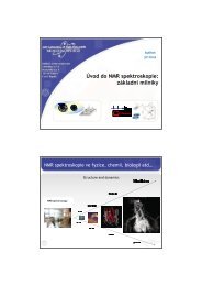
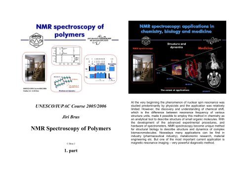
UNESCO/IUPAC Course 2005/2006<br />
Jiri Brus<br />
<strong>NMR</strong> <strong>Spectroscopy</strong> <strong>of</strong> <strong>Polymers</strong><br />
© Brus J<br />
1. part<br />
At the very beginning the phenomenon <strong>of</strong> nuclear spin resonance was<br />
studied predominantly by physicists and the application was relatively<br />
limited. However, the discovery and understanding <strong>of</strong> chemical shift,<br />
which is the difference between resonance frequency <strong>of</strong> various<br />
structure units, made it possible to employ this method in chemistry as<br />
an analytical tool to describe structure <strong>of</strong> small organic molecules. With<br />
the development <strong>of</strong> the advanced experimental procedures, and<br />
hardware <strong>of</strong> spectrometers, <strong>NMR</strong> spectroscopy become unique method<br />
for structural biology to describe structure and dynamics <strong>of</strong> complex<br />
biomacromolecules. Nowadays many applications can be find in<br />
industry (pharmaceutical industry), metabonomic research, material<br />
engineering etc. But one <strong>of</strong> the most important current application is<br />
magnetic resonance imaging – very powerful diagnostic method.
Approximately 13 billion years ago large amount <strong>of</strong> energy was<br />
converted to the matter during the process known as The Big Bang.<br />
And from these times say from the Hadron or Electroweak epoch all<br />
fundamental particles, elementary particles and composite particles<br />
including protons and majority <strong>of</strong> heavier atomic nuclei posses mass,<br />
charge and spin. The internal nuclear spin was for the first time<br />
predicted by Wolfgang Pauli in 1924 as the fourth quantum number.<br />
Although the spin, the property <strong>of</strong> many particles has not entire<br />
macroscopic analogy in many cases it can suggest rotation. Rotation<br />
and spinning is very common motion around us. But in contrast to<br />
macroscopic objects which can rotate around a rotation axis and<br />
gradually and fluently change their angular momentum, the energy<br />
states <strong>of</strong> spins are quantized, and all states and transitions, eigenvalues<br />
and eigenvectors are thoroughly described only by quantum mechanics.
In principle majority <strong>of</strong> all nuclei can be considered as <strong>NMR</strong> active<br />
because they posses nuclear spin, however, we can easily use only<br />
those with nuclear spin one-half. This follows form the fact that these<br />
nuclei can exist only at two energy states (levels), and because we<br />
observe transitions between these levels, the resulting <strong>NMR</strong> spectrum<br />
is relatively narrow and easy to understand. The nuclei with highernumber-spin<br />
can be found at larger number <strong>of</strong> energy levels and<br />
consequently we observe a wide range <strong>of</strong> transitions and the resulting<br />
<strong>NMR</strong> spectrum is complicated and broad.<br />
In quantum mechanics, nuclear spin can be expressed as angular<br />
momentum which is related to nuclear magnetic moment. In the static<br />
magnetic field orientation <strong>of</strong> nuclear magnetic moment is not stable but<br />
it is continuously changing. This leads to the well-known precession <strong>of</strong><br />
spins. The spins rotate around the direction <strong>of</strong> magnetic field on a cone.<br />
Frequency <strong>of</strong> this rotation is the well-known resonant frequency. This<br />
means that the frequency <strong>of</strong> the excitation filed must be exactly the<br />
same to successfully perform <strong>NMR</strong> experiment.<br />
Spins placed into the magnet rapidly orientate into the two possible<br />
directions (parallel and anti-parallel), and originally degenerated energy<br />
level splits. In <strong>NMR</strong> experiment we observe transitions between these<br />
levels. Transition is induced by a pulse <strong>of</strong> rf field with frequency close to<br />
Larmor frequency. The difference in population at the energy levels<br />
determines the sensitivity <strong>of</strong> the experimental method. As you may see<br />
the sensitivity is very low and this is the crucial problem <strong>of</strong> <strong>NMR</strong><br />
spectroscopy.
For instance, the difference given by Boltzmann distribution in a<br />
collection <strong>of</strong> 1 million spins at the magnetic field about 2T is only 16. In<br />
general this difference increases with decreasing temperature and<br />
increasing magnetic filed.<br />
Theoretically it is possible to cool-down the samples, however in<br />
practice this procedure is limited by freezing points <strong>of</strong> solvents or low<br />
solubility, possible phase transitions etc. That is why the second way<br />
how to increase the sensitivity <strong>of</strong> <strong>NMR</strong> experiments is much better and<br />
this is general way which is usually used. On the other hand this way is<br />
very expensive.
Probably it will not be surprising that <strong>NMR</strong> spectroscopy can be also<br />
considered as a certain consequence <strong>of</strong> the research development in<br />
radar technology during the Second War. And that is why one <strong>of</strong> the<br />
first signals <strong>of</strong> water was observed early after the war at 1949 by Felix<br />
Bloch. Approximately ten years after he and E.M. Purclell were awarded<br />
by Nobel Prize.<br />
One <strong>of</strong> the first commercial <strong>NMR</strong> spectrometer equipped by<br />
electromagnet.
A bit better version.<br />
<strong>NMR</strong> spectrometer with a cryomagnet.
A current routine, everything is automated.<br />
Research laboratories are equipped by several huge spectrometers<br />
with very high intensity <strong>of</strong> magnetic fields.
At the beginning <strong>of</strong> any <strong>NMR</strong> experiment there is nothing. There is no<br />
macroscopic magnetization, there is no precession as well. However,<br />
the spins placed into the strong magnetic field became oriented and<br />
start to rotate around the static magnetic field. The sum <strong>of</strong> all the<br />
vectors <strong>of</strong> nuclear magnetic moment produces longitudinal<br />
magnetization. However, this magnetization cannot be detected. That is<br />
why the system must be perturbed. The spin systems is irradiated by a<br />
very short radio-frequency pulse. This leads to the phase coherence in<br />
the rotation <strong>of</strong> spins around the magnetic field. The spins start to rotate<br />
at the same time from the same orientation. Consequently the vector<br />
summation produces transverse magnetization. Right this<br />
magnetization contains required information about the structure <strong>of</strong><br />
molecules.<br />
The transverse magnetization rotates around the magnetic field with the<br />
frequency which is close to theoretical Larmor precession frequency. In<br />
the detection coil a voltage is induced and subsequently recorded as so<br />
called Free Induction Decay. This FID contains all structural<br />
information, however, we are not able to understand it, we cannot read<br />
it. The clue to this problem gave Joseph Fourier in late 18 century. His<br />
mathematic procedure, Fourier transformation, converts the time<br />
functions to the functions <strong>of</strong> frequency. Almost two hundred years later<br />
this transformation was used by Richard Ernst to convert FID in to the<br />
form <strong>of</strong> a classical frequency spectrum. In the resulting spectra it is then<br />
possible to resolve individual structure units in molecules. These<br />
spectra can be understood as unique pictures <strong>of</strong> molecular structure.<br />
The main job <strong>of</strong> <strong>NMR</strong> researchers is a conversion <strong>of</strong> these spectra to a<br />
3D model <strong>of</strong> the molecules.
Why can we resolve individual atoms in molecules?<br />
This possibility follows from the presence <strong>of</strong> electron clouds around<br />
each nucleus. Electrons in magnetic field produce very weak magnetic<br />
fields which can increase or decrease the intensity <strong>of</strong> static magnetic<br />
field produced by the magnet. That is why the nuclei are placed in so<br />
called effective magnetic field and due to this fact frequency <strong>of</strong> the<br />
precession <strong>of</strong> transverse magnetization is slightly affected. We can<br />
observe differences in precession frequencies for different atoms. For<br />
instance, increasing electronegativity <strong>of</strong> a neighboring atom increases<br />
value <strong>of</strong> the chemical shift. Simply speaking position <strong>of</strong> signals in <strong>NMR</strong><br />
spectra strongly depends on the electron density around the<br />
investigated atoms.<br />
However, one crucial problem follows from the fact that the differences<br />
between resonant frequencies <strong>of</strong> various nuclei are very small. In many<br />
cases it is necessary to resolve two signals differing in frequency by<br />
only 0.1Hz. This is very complicated.
Nuclear spins produce very weak signals and magnetization flux<br />
through the detection coil is almost comparable with the noise produced<br />
by electronic components.<br />
Nevertheless solution state <strong>NMR</strong> have become routine technique to<br />
characterize organic molecules. Thanks to fast isotropic molecular<br />
tumbling only isotropic values <strong>of</strong> chemical shift are detected and all<br />
molecules in the solution are equivalent. Consequently the resulting<br />
<strong>NMR</strong> spectra are highly resolved with line-width usually below 0.1 Hz.<br />
Ideally every structure unit in the molecule is resolved and<br />
characterized by its own chemical shift.
Typical 1 H <strong>NMR</strong> spectrum contains four basic parameters. i) The<br />
number <strong>of</strong> signals - this should corresponds to the number <strong>of</strong> basic<br />
structure units. ii) Then the signal intensity - this should reflects the<br />
number <strong>of</strong> equivalent hydrogen atoms in one structural group. iii) The<br />
position <strong>of</strong> signal on <strong>NMR</strong> scale - this is the previously mentioned<br />
chemical shift. Here we can see typical values <strong>of</strong> chemical shifts <strong>of</strong><br />
basic units. iv) And finally this is the multiplicicty <strong>of</strong> signals. This is the<br />
number <strong>of</strong> signals in the multiplet which reflects the number <strong>of</strong> hydrogen<br />
atoms in the neighboring function groups.<br />
The situation should be the same also in polymer systems, however<br />
there are some differences. Clear and well defined multiplicity rapidly<br />
disappears and at the same time signals became broader and broader<br />
as a result <strong>of</strong> increasing molecular weight and viscosity <strong>of</strong> solution.<br />
Narrow signals then reflect low-molecular-weight impurities. However,<br />
shorter oligomers still exhibit high-resolution pattern. Standard onedimensional<br />
experiment performed on copolymer polypeptidepolyethylene<br />
oxide produces traditional 1 H <strong>NMR</strong> spectrum. One can see<br />
residual signal <strong>of</strong> water and signals reflecting traces <strong>of</strong> residual ether.<br />
Polyethylene oxide block is reflected by the main signal and several low<br />
intensive signals correspond to the nonequivalent terminal units. These<br />
terminal units can be found also for polypeptide block. The spectrum<br />
then provide information about the basic structure, composition and<br />
purity <strong>of</strong> the polymer systems.
For peptides and proteins the position <strong>of</strong> signals <strong>of</strong> α-protons also<br />
reflects secondary structure, the conformation <strong>of</strong> the backbone. In<br />
principle it is relatively easy to resolve α-helix or β-sheet. There is a<br />
large database containing values <strong>of</strong> chemical shifts and there are tables<br />
<strong>of</strong> these values for random coil unfolded structures. Deviation from<br />
these random coil range toward higher values indicate formation <strong>of</strong> β-<br />
sheet conformation while deviations toward lower values rather<br />
indicates formation <strong>of</strong> α-helix. This procedure is known as the Chemical<br />
Shifts Indexation (CSI).<br />
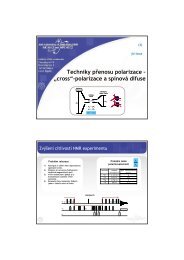
Unambiguous signal assignment can be achieved by two-dimensional<br />
spectroscopy.<br />
Simply speaking, to obtain 1D spectrum one response <strong>of</strong> the sample to<br />
at least single pulse during one detection period must be detected.<br />
The acquisition <strong>of</strong> 2D spectra requires detection <strong>of</strong> the response <strong>of</strong> a<br />
sample to at least two pulses during two detection periods. In practice a<br />
series <strong>of</strong> 1D spectra with gradually prolonging t1 period is recorded. As<br />
magnetization evolves after the first pulse, the intensities <strong>of</strong> signals<br />
detected during the second period are modulated by the length <strong>of</strong> this<br />
period. In fact the intensities <strong>of</strong> these signals oscillate. Consequently<br />
the second FT <strong>of</strong> this oscillation produces a two-dimensional spectrum.<br />
First <strong>of</strong> all this spectrum significantly enhances spectral resolution.<br />
Information originally stored in one dimension are expanded on the<br />
plane.
What can be seen in the 2D spectrum? And what is the interpretation <strong>of</strong><br />
the signals?<br />
Diagonal signals directly correspond to the signals which are detected<br />
in 1D spectrum. However, if information about the polarization state <strong>of</strong> a<br />
hydrogen atom in one structure unit is transferred to the neighboring<br />
hydrogen atom than the <strong>of</strong>f-diagonal signal appears. Such information<br />
or polarization is transferred via bonding electrons and the longest<br />
distance is 3 or 5 chemical bonds. For instance starting from the signal<br />
which probably corresponds to Val methyl protons, the <strong>of</strong>f-diagonal<br />
signal correlates with the structure unit which is connected via three<br />
chemical bonds. So by this way we found out the signal <strong>of</strong> CH beta<br />
hydrogen and subsequently the other correlation signal indicates alpha<br />
proton. This procedure must be repeated for all signals. And by this way<br />
complete assignment <strong>of</strong> all signals in the 1H <strong>NMR</strong> spectra can be<br />
performed.<br />
In general we proceed by the method trial-and-error. At first we must at<br />
least assign one signal for instance the CH proton with double bond.<br />
We find the correlation signal reflecting the neighboring proton number<br />
10 than proton number 9 and so on and so on. It seems to fit well with<br />
our assumption. In general we try to start from well defined signal <strong>of</strong><br />
structure unit with extraordinary chemical shift. Owing to we can trace<br />
proton-proton interaction network with steps which are long only three<br />
chemical bonds. That is why we can describe primary structure or<br />
topology <strong>of</strong> molecules. However, some times the interaction network is<br />
broken and in many cases the resolution in this region is very poor.<br />
That is why we need additional increase in the spectral resolution.
To increase spectral resolution very <strong>of</strong>ten <strong>NMR</strong> spectra <strong>of</strong> other nuclei<br />
especially <strong>of</strong> carbon 13 are measured. As you can see the signals are<br />
well separated and every carbon atom is reflected by a single signal. It<br />
is very good, in general it is true that with increasing number <strong>of</strong><br />
electrons around a nucleus the dispersion <strong>of</strong> chemical shift increases.<br />
On the other hand this experiment is less sensitive because the isotopic<br />
abundance is only 1%<br />
During 70’s and 80’s last century, <strong>NMR</strong> researchers learned to<br />
manipulate with the spin systems so skillfully that they could obtain<br />
detailed information about interatomic distances, local geometry and<br />
segmental dynamics <strong>of</strong> wide range <strong>of</strong> materials and natural products.<br />
Owing to this manipulation which is sometimes called as spingymnastics,<br />
the advanced techniques <strong>of</strong> <strong>NMR</strong> spectroscopy allow us to<br />
determine global structure <strong>of</strong> proteins, nucleic acids and their<br />
complexes in solution.<br />
One <strong>of</strong> the most outstanding scientists which significantly developed<br />
these techniques is Kurt Wüthrich which was awarded by Nobel Prize in<br />
2002. Consequently <strong>NMR</strong> spectroscopy crossed a borderline <strong>of</strong> physics<br />
and chemistry and became inseparable part <strong>of</strong> structural biology.
At present the main research interest is focused to biologically active<br />
macromolecules with limited solubility. At first this is concerned to the<br />
membrane peptides and proteins which control wide range <strong>of</strong> biological<br />
processes in living cells. For instance stabilization <strong>of</strong> membrane<br />
potentials, regulation <strong>of</strong> cell volume or transport <strong>of</strong> ions etc. The second<br />
type <strong>of</strong> the interesting systems provides amyloid proteins and Prionrelated<br />
proteins.<br />
It is very difficult to prepare suitable single crystals <strong>of</strong> theses membrane<br />
proteins or convert these membrane proteins to a solution. In this case<br />
just only solid-state <strong>NMR</strong> spectroscopy can provide required structural<br />
information.