The Properties and Selection of Posterior Direct ... - IneedCE.com
The Properties and Selection of Posterior Direct ... - IneedCE.com
The Properties and Selection of Posterior Direct ... - IneedCE.com

Create successful ePaper yourself
Turn your PDF publications into a flip-book with our unique Google optimized e-Paper software.
Earn<br />
4 CE credits<br />
This course was<br />
written for dentists,<br />
dental hygienists,<br />
<strong>and</strong> assistants.<br />
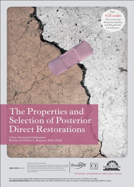
<strong>The</strong> <strong>Properties</strong> <strong>and</strong><br />
<strong>Selection</strong> <strong>of</strong> <strong>Posterior</strong><br />
<strong>Direct</strong> Restorations<br />
A Peer-Reviewed Publication<br />
Written by Robert C. Margeas, DDS, FAGD<br />
PennWell is an ADA CERP recognized provider<br />
ADA CERP is a service <strong>of</strong> the American Dental Association to assist dental pr<strong>of</strong>essionals in identifying<br />
quality providers <strong>of</strong> continuing dental education. ADA CERP does not approve or endorse individual<br />
courses or instructors, nor PennWell does it imply is acceptance an ADA CERP <strong>of</strong> credit Recognized hours by boards Provider <strong>of</strong> dentistry.<br />
Concerns <strong>of</strong> <strong>com</strong>plaints about a CE provider may be directed to the provider or to ADA CERP at<br />
www.ada.org/goto/cerp.<br />
Go Green, Go Online to take your course<br />
This course has been made possible through an unrestricted educational grant. <strong>The</strong> cost <strong>of</strong> this CE course is $59.00 for 4 CE credits.<br />
Cancellation/Refund Policy: Any participant who is not 100% satisfied with this course can request a full refund by contacting PennWell in writing.
Educational Objectives<br />
Overall goal: <strong>The</strong> purpose <strong>of</strong> this article is to provide dental<br />
pr<strong>of</strong>essionals with exp<strong>and</strong>ed information on direct posterior<br />
<strong>com</strong>posites.<br />
Upon <strong>com</strong>pletion <strong>of</strong> this course, the clinician will be able to<br />
do the following:<br />
1. Describe the modes <strong>of</strong> failure, advantages <strong>and</strong> disadvantages<br />
<strong>of</strong> amalgam restorations.<br />
2. Describe the modes <strong>of</strong> failure, advantages <strong>and</strong> disadvantages<br />
<strong>of</strong> <strong>com</strong>posite restorations.<br />
3. Describe the properties <strong>of</strong> an ideal restorative material.<br />
4. Describe the types <strong>of</strong> <strong>com</strong>posite materials <strong>and</strong> recent new<br />
materials <strong>and</strong> their application.<br />
Abstract<br />
Early tooth-colored restorative materials were weak <strong>and</strong><br />
only suitable for anterior teeth. Over time, <strong>com</strong>posites were<br />
developed that <strong>of</strong>fered improved properties enabling their<br />
use in posterior teeth where subject to occlusal loading <strong>and</strong><br />
forces <strong>of</strong> mastication. Secondary caries is the main reason<br />
for failure <strong>of</strong> both amalgam <strong>and</strong> <strong>com</strong>posite restorations.<br />
Amalgam restorations <strong>of</strong>fer ease-<strong>of</strong>-use but poor esthetics.<br />
In the case <strong>of</strong> <strong>com</strong>posite restorations, minimizing polymerization<br />
shrinkage, wear <strong>and</strong> discoloration increase the<br />
longevity <strong>of</strong> these restorations. <strong>Posterior</strong> <strong>com</strong>posite resins<br />
<strong>of</strong>fer excellent esthetics, the main driver for patients who<br />
prefer <strong>com</strong>posite fillings.<br />
Introduction<br />
Historically, posterior direct restorations involved the use <strong>of</strong><br />
amalgam. <strong>The</strong> first modern tooth-colored restorations used<br />
acrylic, which was introduced more than six decades ago.<br />
Subsequently, silicates <strong>and</strong> (di)methacrylate materials were<br />
investigated. Silicate cements <strong>and</strong> early <strong>com</strong>posite materials<br />
were suitable only for anterior restorations due to their weak<br />
physical properties, <strong>and</strong> the silicate cements needed to be<br />
placed in one movement – incremental placement was not<br />
an option. Silicate cements had a high failure rate. Old silicate<br />
restorations were assessed for longevity in a 1986 study<br />
<strong>and</strong> were found to have an estimated 66% replaced due to<br />
marginal discrepancies <strong>and</strong> lost fillings. 1 Early resin-based<br />
<strong>com</strong>posite restorations were an improvement over silicate<br />
cements; however, they were self-curing <strong>and</strong> required mixing<br />
<strong>of</strong> a base <strong>and</strong> a catalyst for curing, resulting in operator<br />
error during mixing <strong>and</strong> difficulties in timely <strong>and</strong> accurate<br />
placement. In addition, strength, bonding <strong>and</strong> retention<br />
were poor. Light-cured dimethacrylate <strong>com</strong>posite restorations<br />
were introduced in the 1970s. 2 By the 1980s, posterior<br />
tooth-colored restorations had been introduced, <strong>and</strong> these<br />
have continued to evolve to <strong>of</strong>fer improved physical properties,<br />
user-friendliness <strong>and</strong> esthetics. Bonding systems <strong>and</strong><br />
techniques have also evolved.<br />
Figure 1. Introduction <strong>of</strong> tooth-colored restorations<br />
1944<br />
1955<br />
1958<br />
1964<br />
1973<br />
1970s<br />
1980s<br />
1990s<br />
2006-2008<br />
Acrylic filling<br />
material introduced<br />
Investigation <strong>of</strong><br />
epoxy filling materials<br />
Introduction <strong>of</strong><br />
Bis–GMA <strong>com</strong>posites<br />
Dimethacrylate–<br />
based fillings investigated<br />
UV-cured resins introduced<br />
Silicate cements <strong>and</strong> early<br />
<strong>com</strong>posites dominate<br />
<strong>Posterior</strong> <strong>com</strong>posites in use<br />
Improved <strong>com</strong>posites<br />
<strong>and</strong> adhesive systems<br />
Investigation <strong>and</strong> introduction<br />
<strong>of</strong> silorane–based material<br />
<strong>The</strong> trend over the last decade has been placement <strong>of</strong> an<br />
increasing number <strong>of</strong> posterior <strong>com</strong>posite restorations <strong>and</strong><br />
a decreasing number <strong>of</strong> amalgams. By 1999, at least 39% <strong>of</strong><br />
direct posterior restorations were <strong>com</strong>posites, <strong>com</strong>pared to<br />
at least 11% in 1990 (in both cases, for the purposes <strong>of</strong> trend<br />
analysis, conservatively making the assumption that all amalgam<br />
placements estimated in the ADA surveys were posterior<br />
restorations) (Table 1). 3<br />
Table 1. Trends in posterior <strong>com</strong>posite placement<br />
<strong>Posterior</strong><br />
<strong>com</strong>posites<br />
1999<br />
Number<br />
placed<br />
% age<br />
<strong>of</strong> total<br />
1990<br />
Number<br />
placed<br />
% age<br />
<strong>of</strong> total<br />
46,116,300 39.38% 13,130,200 11.68%<br />
Amalgams 70,994,700 60.62% 99,256,900 88.32%<br />
2 www.ineedce.<strong>com</strong>
Clinician needs <strong>and</strong> patient dem<strong>and</strong> for esthetic dentistry<br />
continue to drive these trends as well as development <strong>of</strong><br />
products for restorations with improved physical properties<br />
<strong>and</strong> esthetics.<br />
Ideal Restorative Material<br />
<strong>The</strong> ideal posterior restorative material should exhibit a<br />
number <strong>of</strong> features (Table 2). It should be dimensionally<br />
stable, with no expansion or shrinkage either during placement<br />
or subsequent to placement, <strong>and</strong> without any wear<br />
following placement. It must also <strong>of</strong>fer sufficient <strong>com</strong>pressive<br />
<strong>and</strong> flexural strength – in the case <strong>of</strong> posterior Class I <strong>and</strong> II<br />
restorations, it must resist both occlusal forces <strong>and</strong> the forces<br />
<strong>of</strong> mastication. Neither the material nor the tooth should be<br />
subject to stress during loading <strong>of</strong> the material <strong>and</strong>/or tooth.<br />
Bio<strong>com</strong>patibility is important – the material should neither<br />
deteriorate intraorally nor result in any toxic, teratogenic<br />
or other iatrogenic effects. Ideally, the restorative material<br />
should <strong>of</strong>fer antibacterial properties against oral bacteria, <strong>and</strong><br />
preferably should be bactericidal. It should be user-friendly,<br />
<strong>of</strong>fering an appropriate operating time <strong>and</strong> ease <strong>of</strong> placement.<br />
Finally, the material should also be esthetically pleasing to the<br />
patient <strong>and</strong> be color-stable <strong>and</strong> stain-resistant.<br />
Table 2. Ideal Restorative Material <strong>Properties</strong><br />
Dimensionally stable<br />
Resistant to forces <strong>and</strong> stresses<br />
Wear-resistant<br />
Retentive <strong>and</strong> adhesive to the tooth<br />
Requires minimal tooth preparation<br />
Easily placed<br />
Requires minimum time to restore<br />
Cost-effective<br />
Bio<strong>com</strong>patible<br />
Bactericidal<br />
Esthetically pleasing<br />
Color-stable<br />
Stain-resistant<br />
decade up to 2001 found an annual failure rate <strong>of</strong> 1.1% for<br />
amalgams, 2.1% for <strong>com</strong>posites <strong>and</strong> 7.7% for glass ionomer<br />
cements. 7 Reasons for the failure <strong>and</strong> replacement <strong>of</strong> restorations<br />
include secondary caries, fracture, wear, marginal<br />
defects <strong>and</strong> postoperative sensitivity.<br />
<strong>The</strong> primary reason for the replacement <strong>of</strong> direct restorations<br />
has been found to be secondary caries irrespective <strong>of</strong> the<br />
restorative material. 8,9,10,11 While it has been found to be difficult<br />
to reliably diagnose secondary caries, <strong>and</strong> the condition<br />
is responsible for the majority <strong>of</strong> restoration replacements,<br />
the quality <strong>of</strong> the restoration <strong>and</strong> the patient’s (preventive)<br />
home care are important factors in precluding further repeat<br />
replacements. 12 It was found in one study that 65% <strong>of</strong> direct<br />
<strong>and</strong> indirect (5% <strong>of</strong> total) restorations placed were replacement<br />
restorations, with secondary caries the most frequent<br />
reason given, regardless <strong>of</strong> material used. 13 <strong>The</strong> longevity<br />
<strong>of</strong> restorations depends on clinical technique, materials <strong>and</strong><br />
patient care.<br />
Figure 2. Marginal degradation <strong>of</strong> amalgam<br />
Figure 3. Secondary caries<br />
<strong>The</strong> ideal restorative material does not exist, although material<br />
developments have significantly improved how closely<br />
products approach these parameters.<br />
<strong>Direct</strong> Restoration Longevity<br />
Annual failure rates for different materials have been examined<br />
in a number <strong>of</strong> studies. Some studies have found<br />
ranges <strong>of</strong> 0%-7% for amalgams, 0%-9% for direct <strong>com</strong>posites<br />
<strong>and</strong> 1.4%-14.4% for glass ionomer cements in posterior<br />
stress-bearing restorations. 4 A separate, more recent study,<br />
involving only two dentists, found <strong>com</strong>parable failure rates<br />
for <strong>com</strong>posites <strong>and</strong> amalgams assessed as a five-year survival<br />
rate. 5 Annual failure rates in a study conducted on restorations<br />
predominantly placed since 1990 were 3% for amalgams<br />
<strong>and</strong> 2.2% for direct <strong>com</strong>posites, <strong>and</strong> it was also concluded<br />
that more recent studies demonstrated better results. 6 Failure<br />
rates in one study covering restoration placement during the<br />
Amalgam Restorations<br />
Amalgam has been found to be a cost-effective restorative<br />
material <strong>and</strong> to <strong>of</strong>fer good longevity in studies <strong>of</strong> up to a more<br />
than 20-year period. 14 Amalgam restorations are less technique-sensitive<br />
than <strong>com</strong>posites, less sensitive to the presence<br />
<strong>of</strong> moisture <strong>and</strong> easier to place. <strong>The</strong>y require less time to place<br />
than direct <strong>com</strong>posites; an estimated 2.5 times more time is required<br />
for <strong>com</strong>posite placement. 15 While improved materials<br />
www.ineedce.<strong>com</strong> 3
<strong>and</strong> light-curing options may have reduced the time required<br />
for <strong>com</strong>posites, more chairside time is still required than with<br />
amalgams. Amalgam is also bactericidal, which helps to reduce<br />
bacterial colonization <strong>and</strong> bi<strong>of</strong>ilm formation. 16,17<br />
Bulk fractures <strong>and</strong> marginal degradation have been found<br />
to be the main material factors in the replacement <strong>of</strong> amalgam<br />
restorations. 18 Bulk fracture rates have been found to be<br />
similar with or without bonding <strong>of</strong> amalgams (such as with<br />
AmalgamBond Plus) in large restorations, although smaller<br />
restorations benefit from bonding. 19 Bonded amalgam restorations<br />
have been found to <strong>of</strong>fer support <strong>of</strong> undermined<br />
enamel equal to that <strong>of</strong> <strong>com</strong>posites, but inferior marginal<br />
adaptation. 20 Creep-fatigue may be a major factor in marginal<br />
fracture <strong>of</strong> amalgam restorations. 21 Amalgam restorations are<br />
subject to expansion, which can result in cuspal stress over<br />
time, depending upon the design <strong>of</strong> the preparation <strong>and</strong>/or<br />
the location <strong>of</strong> the initial lesion. Expansion <strong>of</strong> amalgam results<br />
from internal phase changes over time, that must be relieved<br />
to reduce stress – it is believed this occurs as a result <strong>of</strong> creep<br />
<strong>of</strong> the amalgam from the confines <strong>of</strong> the restoration <strong>and</strong> its<br />
subsequent extrusion. On the other h<strong>and</strong>, development <strong>of</strong> a<br />
reduced amalgam-tooth margin interface gap size over time<br />
<strong>and</strong> improved marginal seal may occur due to such creep. 22<br />
Amalgam restorations require more tooth preparation<br />
than <strong>com</strong>posites, <strong>and</strong> careful disposal <strong>of</strong> the mercurycontaining<br />
amalgam is m<strong>and</strong>atory. <strong>The</strong> poor esthetic results<br />
provided by amalgams are a major concern for patients, <strong>and</strong><br />
amalgam staining <strong>of</strong> the tooth over time further <strong>com</strong>promises<br />
the appearance. Corrosion is also an issue. Poor esthetics with<br />
amalgam is the main reason why patients increasingly prefer<br />
the use <strong>of</strong> direct posterior <strong>com</strong>posites as well as tooth-colored<br />
indirect restorative materials <strong>and</strong> techniques.<br />
Table 3. Modes <strong>of</strong> failure, advantages <strong>and</strong> disadvantages<br />
<strong>of</strong> amalgams<br />
Modes <strong>of</strong> Failure<br />
Secondary caries<br />
Bulk fracture<br />
Advantages<br />
Ease <strong>of</strong> use<br />
Cost-effective<br />
Disadvantages<br />
More tooth preparation<br />
Poor esthetics<br />
Marginal degradation<br />
Expansion <strong>and</strong> cuspal stress<br />
Can be bonded<br />
Bactericidal<br />
Corrosion<br />
Mercury disposal<br />
Composite Restorations<br />
Material failures accounted for more replacements <strong>of</strong> <strong>com</strong>posites<br />
than amalgams in a review <strong>of</strong> surveys <strong>of</strong> dentists across the<br />
United States, Sc<strong>and</strong>inavia <strong>and</strong> the United Kingdom from the<br />
1980s <strong>and</strong> 1990s. <strong>The</strong>se failures included bulk fracture, marginal<br />
degradation, discoloration <strong>and</strong> loss <strong>of</strong> anatomic shape. 23<br />
Nonetheless, the main reason for replacement is the same<br />
as for amalgam restorations – secondary caries. In addition,<br />
<strong>com</strong>posite restorations have improved over time, <strong>and</strong> recent<br />
studies have shown longevity to more closely reach the longevity<br />
<strong>of</strong> amalgams (albeit over a shorter tested time span).<br />
Table 4. Modes <strong>of</strong> failure, advantages <strong>and</strong> disadvantages<br />
<strong>of</strong> <strong>com</strong>posites<br />
Modes <strong>of</strong> Failure<br />
Secondary caries<br />
Bulk fracture<br />
Marginal degradation<br />
Advantages<br />
Less tooth preparation<br />
Effective bonding<br />
Disadvantages<br />
Technique-sensitive<br />
Increased chairside time<br />
Discoloration<br />
Loss <strong>of</strong> anatomic shape <strong>and</strong> wear<br />
Excellent esthetics<br />
No expansion over time<br />
Polymerization shrinkage<br />
Increased bacterial adhesion<br />
While amalgams exp<strong>and</strong> over time, <strong>com</strong>posite restorations<br />
are subject to polymerization shrinkage. This is regarded as<br />
the largest problem associated with <strong>com</strong>posite use. 24 Polymerization<br />
shrinkage results in stresses that can lead to enamel<br />
cracks, marginal degradation <strong>and</strong> microleakage, <strong>and</strong> postoperative<br />
sensitivity. Other associated problems include potential<br />
debonding <strong>of</strong> the tooth-<strong>com</strong>posite interface. 25 Polymerization<br />
shrinkage occurs due to the affiliation <strong>of</strong> the resin molecules<br />
with one another <strong>and</strong> the formation <strong>of</strong> chemical bonds that<br />
reduce the material’s bulk. Shrinkage <strong>and</strong> occlusal loading<br />
<strong>of</strong> <strong>com</strong>posites result in cuspal deflection, which results in<br />
enamel cracks <strong>and</strong> hypersensitivity. <strong>The</strong> amount <strong>of</strong> deflection<br />
has been found to be greater in larger restorations (MODs)<br />
than smaller ones (MOs). 26 <strong>The</strong> amount <strong>of</strong> shrinkage <strong>and</strong> resulting<br />
stresses also varies with the <strong>com</strong>posite filling material<br />
used. 27,28 It is influenced by the material’s flow, chemistry <strong>and</strong><br />
curing dynamics, <strong>and</strong> the size <strong>and</strong> shape <strong>of</strong> the preparation.<br />
<strong>The</strong> intensity <strong>and</strong> duration <strong>of</strong> light curing have been found to<br />
affect polymerization shrinkage. 29 Shrinkage can be reduced<br />
by increasing the amount <strong>of</strong> filler in <strong>com</strong>posite restorative<br />
materials, as well as by having pre-polymerized clusters in the<br />
material. 30 A recent study by Bouillaguet et al. found that cuspal<br />
deflection (tooth deformation) was statistically similar for<br />
conventional hybrid <strong>com</strong>posites <strong>and</strong> flowable <strong>com</strong>posites. 31<br />
Table 5. Potential effects <strong>of</strong> polymerization shrinkage<br />
Enamel cracks<br />
Marginal degradation<br />
Microleakage<br />
Postoperative sensitivity<br />
Debonding <strong>of</strong> tooth-<strong>com</strong>posite interface<br />
Composite restorations generally <strong>of</strong>fer poor antibacterial<br />
properties <strong>com</strong>pared to amalgam. One in vitro study found a<br />
minimal antibacterial effect with <strong>com</strong>posites that lasted only a<br />
4 www.ineedce.<strong>com</strong>
few days. It was suggested that this might explain the greater<br />
bi<strong>of</strong>ilm growth seen on <strong>com</strong>posites <strong>com</strong>pared to amalgams. 32<br />
A second study assessed the behavior <strong>of</strong> three different <strong>com</strong>posites<br />
(Charisma ® , Heraeus Kulzer; Dyract ® , Dentsply; <strong>and</strong><br />
Pertac, 3M ESPE) in the presence <strong>of</strong> three <strong>com</strong>mon oral<br />
bacteria (S. mutans, S. oralis <strong>and</strong> A. naeslundii) for up to 35<br />
days <strong>and</strong> found that the bacteria colonized the <strong>com</strong>posites in<br />
a matter <strong>of</strong> hours <strong>and</strong> formed deep bi<strong>of</strong>ilms. <strong>The</strong> study also<br />
found, using scanning electron microscopy, that the polyacid<br />
modified <strong>com</strong>posite demonstrated surface damage <strong>and</strong><br />
roughness. 33 Fluoride-releasing <strong>com</strong>posites appear to <strong>of</strong>fer no<br />
benefit over nonfluoride <strong>com</strong>posites. 34<br />
While polymerization shrinkage in particular <strong>and</strong> bi<strong>of</strong>ilm<br />
formation on the surface <strong>of</strong> the restoration are disadvantages<br />
<strong>of</strong> <strong>com</strong>posites <strong>com</strong>pared to amalgams, <strong>com</strong>posites still <strong>of</strong>fer<br />
several advantages over amalgams – superior esthetics, no expansion<br />
over time, as well as highly effective bonding systems<br />
for adhesion <strong>and</strong> retention that enable minimal preparation<br />
<strong>and</strong> improved tooth structure preservation. From the patient’s<br />
perspective, the most obvious advantage <strong>of</strong> <strong>com</strong>posite restorations<br />
is esthetics. Improved color stability, luster <strong>and</strong> stain<br />
resistance have further improved esthetics as <strong>com</strong>posites have<br />
evolved. Improvements in h<strong>and</strong>ling <strong>and</strong> user-friendliness<br />
continue to be developed since the introduction <strong>of</strong> a choice in<br />
bonding agents <strong>and</strong> unit doses, <strong>and</strong> recent developments are<br />
aimed at over<strong>com</strong>ing the physical weaknesses <strong>of</strong> <strong>com</strong>posites.<br />
Recent Composite Material Developments<br />
Composites have been modified to provide greater physical<br />
<strong>and</strong> biological properties. Bi<strong>of</strong>ilm-formation reduction has<br />
been tried by modifying <strong>com</strong>posites as well as dentin bonders,<br />
such as by including glutaraldehyde in the dentin bonder or<br />
incorporating an acidic property. 35<br />
Recent investigations have included researching novel posterior<br />
<strong>com</strong>posite materials with the objective <strong>of</strong> finding materials<br />
that <strong>of</strong>fer reduced polymerization shrinkage <strong>and</strong> improved<br />
esthetic stability. Silsesquioxane (SSQ)-based nano<strong>com</strong>posites<br />
have been found in in vitro testing to <strong>of</strong>fer reduced polymerization<br />
shrinkage <strong>and</strong> rigidity, <strong>of</strong>fering potential solutions for<br />
stresses <strong>and</strong> clinical issues associated with shrinkage. 36 Similarly,<br />
oligomeric thiolene-based materials have been found in<br />
in vitro testing to exhibit up to 92% less polymerization stress<br />
<strong>com</strong>pared to conventional dimethacrylate-based <strong>com</strong>posites. 37<br />
A recently developed <strong>com</strong>posite material based on silorane has<br />
been used <strong>and</strong> tested clinically <strong>and</strong> has been found to result in<br />
reduced polymerization shrinkage <strong>and</strong> stresses. 38<br />
Silorane-based <strong>Posterior</strong> Restorations<br />
Silorane-based posterior <strong>com</strong>posite material (Filtek LS<br />
Low Shrink <strong>Posterior</strong> Restorative, 3M ESPE) has been found<br />
to reduce polymerization shrinkage <strong>and</strong> associated stresses, 39<br />
which would also reduce microleakage <strong>and</strong> postoperative hypersensitivity<br />
while demonstrating other physical properties<br />
<strong>com</strong>parable to leading <strong>com</strong>posites in in vitro testing. 40 Shrinkage<br />
is decreased due to the material’s chemical <strong>com</strong>position<br />
<strong>and</strong> polymerization dynamics. Silorane is derived from the<br />
<strong>com</strong>bination <strong>of</strong> siloxane <strong>and</strong> oxirane <strong>and</strong> has a <strong>com</strong>pact ring<br />
structure (Figure 4a) that unlinks during polymerization.<br />
When polymerization shrinkage begins, the silorane ring simultaneously<br />
opens up <strong>and</strong> <strong>com</strong>pensates for material shrinkage<br />
by exp<strong>and</strong>ing its molecular volume <strong>and</strong> bulking up the<br />
material. Shrinkage has been found to be less than 1% using<br />
this material (Figures 4b–d). 41 An initiator included in the material<br />
starts the ring-opening process in a controlled manner<br />
<strong>and</strong>, according to the manufacturer, increases operating time.<br />
Figure 4a. Silorane molecule<br />
Figure 4b. Application <strong>of</strong> primer<br />
Figure 4c. Silorane-based material in preparation after separate<br />
applications <strong>and</strong> curing <strong>of</strong> both primer <strong>and</strong> adhesive<br />
Figure 4d. Light-curing <strong>of</strong> silorane-based material opens silorane<br />
ring structure, reduces shrinkage<br />
www.ineedce.<strong>com</strong> 5
In vitro testing has found lower polymerization shrinkage<br />
<strong>and</strong> reduced polymerization stress <strong>and</strong> tooth deformation<br />
<strong>com</strong>pared to leading methacrylate-based conventional <strong>and</strong><br />
flowable <strong>com</strong>posite resin materials. 42,43,44 At the same time,<br />
adhesion <strong>and</strong> shear bond strength have not been <strong>com</strong>promised,<br />
<strong>and</strong> reduced shrinkage helps preserve the tooth<br />
bond-<strong>com</strong>posite adhesive interfaces. Other desired physical<br />
properties, such as <strong>com</strong>pressive <strong>and</strong> flexural strength,<br />
have been found to be similar to those <strong>of</strong> leading <strong>com</strong>posite<br />
materials. <strong>The</strong> silorane-based restorative is a microhybrid<br />
<strong>com</strong>posite that contains fine silane-coated quartz filler<br />
with yttrium fluoride for radiopacity. Bacterial adhesion<br />
<strong>of</strong> <strong>com</strong>mon oral bacteria has been found to be reduced in<br />
in vitro testing using silorane-based <strong>com</strong>posite, associated<br />
with its hydrophobic chemistry. 45 One-year clinical testing<br />
has found good clinical performance using this new material<br />
<strong>com</strong>pared to other posterior <strong>com</strong>posite material.<br />
Case Study<br />
<strong>The</strong> case shown here demonstrated the use <strong>of</strong> posterior <strong>com</strong>posite<br />
material (Filtek LS restorative) in the restoration <strong>of</strong><br />
a carious upper left first bicuspid. On examination, a distal<br />
lesion was identified (Figure 5a). A rubber dam was placed<br />
prior to the DO preparation.<br />
Figure 5c. Cavity preparation <strong>and</strong> Composi-Tight® matrix <strong>and</strong><br />
wedge placed<br />
After the matrix <strong>and</strong> wedge (Composi-Tight ® , Garrison<br />
Dental) were placed around the distal box, a thin layer <strong>of</strong><br />
self-etching primer (LS System Adhesive Self-Etch Primer,<br />
3M ESPE) was placed on the dentin in the preparation using a<br />
microbrush for 15 seconds, dispersed using air, then cured for<br />
10 seconds. <strong>The</strong> primer has a pH <strong>of</strong> 2.7, produces mild etching<br />
<strong>and</strong> increases the hydrophobicity <strong>of</strong> the area prior to placement<br />
<strong>of</strong> the adhesive (LS System Adhesive Bond, 3M ESPE).<br />
Figure 5d. Self-etching primer placed<br />
Figure 5a. Distal lesion in upper left first bicuspid<br />
Figure 5e. Curing <strong>of</strong> self-etching primer<br />
Figure 5b. Rubber dam placement prior to preparation<br />
<strong>The</strong> next step is to place a thin layer <strong>of</strong> the adhesive in the<br />
preparation over the cured primer, <strong>and</strong> to light-cure the adhesive<br />
for 10 seconds before placing any <strong>com</strong>posite material<br />
in the preparation. Filtek LS restorative is highly hydrophobic<br />
6 www.ineedce.<strong>com</strong>
<strong>and</strong> the LS System Adhesive must function as a bridging<br />
mechanism between the primer <strong>and</strong> the <strong>com</strong>posite. Only the<br />
LS System Adhesive Self-Etch Primer <strong>and</strong> Bond are <strong>com</strong>patible<br />
with Filtek LS restorative chemistry (the use <strong>of</strong> other<br />
primers <strong>and</strong> adhesives is contraindicated).<br />
<strong>The</strong> <strong>com</strong>posite shade is selected <strong>and</strong> injected first as a 2 mm<br />
increment in the distal box, where it is condensed using a #9<br />
Garrison. <strong>The</strong> remainder <strong>of</strong> the void is filled by injecting more<br />
<strong>com</strong>posite, taking care not to overfill the area, <strong>and</strong> the #9 <strong>com</strong>posite<br />
instrument is used to remove flash prior to light-curing<br />
the <strong>com</strong>posite for 20 seconds (note: plasma lights, lasers <strong>and</strong><br />
other high-power curing lights should not be used with Filtek<br />
LS restorative). A long working time under operatory light<br />
aids detailed shaping <strong>and</strong> flash removal prior to curing.<br />
Figure 5f. Injecting distal box with Filtek LS restorative<br />
Figure 5i. Cured <strong>com</strong>posite after removal <strong>of</strong> matrix <strong>and</strong> wedge<br />
Figure 5j. High polish created using Jiffy® polisher<br />
Figure 5g. Condensing <strong>com</strong>posite with #9 Garrison<br />
Figure 5k. Final polished restoration<br />
Figure 5h. Flash being removed from filled preparation prior to curing<br />
After removal <strong>of</strong> the matrix <strong>and</strong> wedge, the restoration is<br />
polished using a S<strong>of</strong>-Lex disk (Ultradent) used to remove<br />
any flash <strong>and</strong> a Jiffy® Polisher (Ultradent) is then used to create<br />
a high shine. <strong>The</strong> final restoration using the low shrinkage<br />
posterior <strong>com</strong>posite <strong>of</strong>fers excellent esthetics <strong>and</strong> function.<br />
Case Study<br />
<strong>The</strong> second case here shows replacement <strong>of</strong> a degrading<br />
<strong>and</strong> fractured amalgam restoration with a silorane-based<br />
posterior <strong>com</strong>posite. After preparation <strong>and</strong> application <strong>of</strong><br />
a liner, the primer <strong>and</strong> adhesive were separately applied<br />
<strong>and</strong> separately cured. <strong>The</strong> restorative material was then<br />
www.ineedce.<strong>com</strong> 7
injected, condensed <strong>and</strong> light-cured prior to finishing <strong>and</strong><br />
polishing the restoration.<br />
Figure 6e. Finished restoration<br />
Figure 6a. Fractured, degrading amalgam<br />
Figure 6b. Preparation with liner mesially<br />
Figure 6c. Application <strong>of</strong> primer<br />
Figure 6d. Application <strong>of</strong> adhesive after primer was cured<br />
Summary<br />
Increasingly, <strong>com</strong>posites are being placed in preference to<br />
amalgams in large part due to patient dem<strong>and</strong>s for esthetics<br />
as well as the clinical desire to do minimal preparation where<br />
possible <strong>and</strong> provide patients with bonded, esthetic restorations.<br />
Since their introduction, the properties <strong>of</strong> <strong>com</strong>posites<br />
have improved dramatically. Amalgam <strong>and</strong> <strong>com</strong>posite restorations<br />
both have advantages <strong>and</strong> disadvantages. While<br />
amalgam restorations fail by secondary caries <strong>and</strong> are subject<br />
to expansion, <strong>com</strong>posite restorations fail by secondary caries<br />
<strong>and</strong> are subject to shrinkage. Recent developments <strong>and</strong> investigations<br />
<strong>of</strong> materials are aimed at reducing polymerization<br />
shrinkage <strong>of</strong> <strong>com</strong>posites to increase the longevity <strong>of</strong> these<br />
restorations <strong>and</strong> reduce the potential for failure.<br />
References<br />
1 Qvist V, Thylstrup A, Mjör IA. Restorative treatment<br />
pattern <strong>and</strong> longevity <strong>of</strong> resin restorations in Denmark. Acta<br />
Odontol Sc<strong>and</strong>. 1986;44(6):351-6.<br />
2 Small BW. <strong>Direct</strong> resin <strong>com</strong>posites for 2002 <strong>and</strong> beyond.<br />
Gen Dent. 2002;50(1):30-3.<br />
3 ADA Survey <strong>of</strong> Services Rendered, 2002.<br />
4 Hickel R, Manhart J. Longevity <strong>of</strong> restorations in<br />
posterior teeth <strong>and</strong> reasons for failure. J Adhes Dent.<br />
2001;3(1):45-64.<br />
5 Opdam NJ, Bronkhorst EM, Roeters JM, Loomans BA.<br />
A retrospective clinical study on longevity <strong>of</strong> posterior<br />
<strong>com</strong>posite <strong>and</strong> amalgam restorations. Dent Mater.<br />
2007;23(1):2-8. Epub 2006 Jan 18.<br />
6 Manhart J, Chen H, Hamm G, Hickel R. Buonocore<br />
Memorial Lecture. Review <strong>of</strong> the clinical survival <strong>of</strong> direct<br />
<strong>and</strong> indirect restorations in posterior teeth <strong>of</strong> the permanent<br />
dentition. Oper Dent. 2004;29(5):481-508.<br />
7 Hickel R, Manhart J, García-Godoy F. Clinical results <strong>and</strong><br />
new developments <strong>of</strong> direct posterior restorations. Am J<br />
Dent. 2000;13(Spec No):41D-54D.<br />
8 Hickel R, Manhart J. Longevity <strong>of</strong> restorations in<br />
posterior teeth <strong>and</strong> reasons for failure. J Adhes Dent.<br />
2001;3(1):45-64.<br />
9 Deligeorgi V, Mjör IA, Wilson NH. An overview <strong>of</strong> reasons<br />
for the placement <strong>and</strong> replacement <strong>of</strong> restorations. Prim<br />
Dent Care. 2001;8(1):5-11.<br />
10 Mjör IA, Moorhead JE, Dahl JE. Reasons for replacement<br />
<strong>of</strong> restorations in permanent teeth in general dental practice.<br />
Int Dent J. 2000;50(6):361-6.<br />
11 All<strong>and</strong>er L, Birkhed D, Bratthall D. Reasons for replacement<br />
<strong>of</strong> Class II amalgam restorations in private practice. Swed<br />
Dent J. 1990;14(4):179-84.<br />
8 www.ineedce.<strong>com</strong>
12 Kidd EA, T<strong>of</strong>fenetti F, Mjör IA. Secondary caries. Int Dent<br />
J. 1992;42(3):127-38.<br />
13 Forss H, Widström E. Reasons for restorative therapy <strong>and</strong><br />
the longevity <strong>of</strong> restorations in adults. Acta Odontol Sc<strong>and</strong>.<br />
2004;62(2):82-6.<br />
14 Roulet JF. Benefits <strong>and</strong> disadvantages <strong>of</strong> tooth-coloured<br />
alternatives to amalgam. J Dent. 1997;25(6):459-73.<br />
15 Ibid.<br />
16 Beyth N, Domb AJ, Weiss EI. An in vitro quantitative<br />
antibacterial analysis <strong>of</strong> amalgam <strong>and</strong> <strong>com</strong>posite resins. J<br />
Dent. 2007;35(3):201-6. Epub 2006 Sep 25.<br />
17 Willershausen B, Callaway A, Ernst CP, Stender E. <strong>The</strong><br />
influence <strong>of</strong> oral bacteria on the surfaces <strong>of</strong> resin-based<br />
dental restorative materials: an in vitro study. Int Dent J.<br />
1999;49(4):231-9.<br />
18 Qvist V, Thylstrup A, Mjör IA. Restorative treatment pattern<br />
<strong>and</strong> longevity <strong>of</strong> amalgam restorations in Denmark. Acta<br />
Odontol Sc<strong>and</strong>. 1986;44(6):343-9.<br />
19 Lindemuth JS, Hagge MS, Broome JS. Effect <strong>of</strong> restoration<br />
size on fracture resistance <strong>of</strong> bonded amalgam restorations.<br />
Oper Dent. 2000;25(3):177-81.<br />
20 Franchi M, Breschi L, Ruggeri O. Cusp fracture resistance<br />
in <strong>com</strong>posite-amalgam <strong>com</strong>bined restorations. J Dent.<br />
1999;27(1):47-52.<br />
21 Williams PT, Hedge GL. Creep-fatigue as a possible<br />
cause <strong>of</strong> dental amalgam margin failure. J Dent Res.<br />
1985;64(3):470-5.<br />
22 Osborne JW. Creep as a mechanism for sealing amalgams.<br />
Oper Dent. 2006;31(2):161-4.<br />
23 Deligeorgi V, Mjör IA, Wilson NH. An overview <strong>of</strong> reasons<br />
for the placement <strong>and</strong> replacement <strong>of</strong> restorations. Prim<br />
Dent Care. 2001;8(1):5-11.<br />
24 Giachetti L, Scaminaci Russo D, Bambi C, Gr<strong>and</strong>ini<br />
R. A review <strong>of</strong> polymerization shrinkage stress: current<br />
techniques for posterior direct resin restorations. J Contemp<br />
Dent Pract. 2006;7(4):79-88.<br />
25 van Dijken JW. A 6-year clinical evaluation <strong>of</strong> Class I polyacid<br />
modified resin <strong>com</strong>posite/resin <strong>com</strong>posite laminate<br />
restorations cured with a two-step curing technique. Dent<br />
Mater. 2003;19(5):423-8.<br />
26 González-López S, Vilchez Díaz MA, de Haro-Gasquet<br />
F, Ceballos L, de Haro-Muñoz C. Cuspal flexure <strong>of</strong> teeth<br />
with <strong>com</strong>posite restorations subjected to occlusal loading. J<br />
Adhes Dent. 2007 Feb;9(1):11-5.<br />
27 Rüttermann S, Krüger S, Raab WH, J<strong>and</strong>a R. Polymerization<br />
shrinkage <strong>and</strong> hygroscopic expansion <strong>of</strong> contemporary<br />
posterior resin-based filling materials: a <strong>com</strong>parative study.<br />
J Dent. 2007;35(10):806-13. Epub 2007 Sep 10.<br />
28 Cadenaro M, Biasotto M, Scuor N, Breschi L, Davidson<br />
CL, Di Lenarda R. Assessment <strong>of</strong> polymerization<br />
contraction stress <strong>of</strong> three <strong>com</strong>posite resins. Dent Mater.<br />
2008;24(5):681-5. Epub 2007 Aug 31.<br />
29 Visvanathan A, Ilie N, Hickel R, Kunzelmann KH. <strong>The</strong><br />
influence <strong>of</strong> curing times <strong>and</strong> light curing methods on the<br />
polymerization shrinkage stress <strong>of</strong> a shrinkage-optimized<br />
<strong>com</strong>posite with hybrid-type prepolymer fillers. Dent Mater.<br />
2007;23(7):777-84. Epub 2006 Aug 17.<br />
30 Kleverlaan CJ, Feilzer AJ. Polymerization shrinkage <strong>and</strong><br />
contraction stress <strong>of</strong> dental resin <strong>com</strong>posites. Dent Mater.<br />
2005;21(12):1150-7. Epub 2005 Jul 22.<br />
31 Bouillaguet S, Gamba J, Forchelet J, Krejci I, Wataha<br />
JC. Dynamics <strong>of</strong> <strong>com</strong>posite polymerization mediates<br />
the development <strong>of</strong> cuspal strain. Dent Mater.<br />
2006;22(10):896-902. Epub 2005 Dec 20.<br />
32 Beyth N, Domb AJ, Weiss EI. An in vitro quantitative<br />
antibacterial analysis <strong>of</strong> amalgam <strong>and</strong> <strong>com</strong>posite resins. J<br />
Dent. 2007;35(3):201-6. Epub 2006 Sep 25.<br />
33 Willershausen B, Callaway A, Ernst CP, Stender E. <strong>The</strong><br />
influence <strong>of</strong> oral bacteria on the surfaces <strong>of</strong> resin-based<br />
dental restorative materials: an in vitro study. Int Dent J.<br />
1999;49(4):231-9.<br />
34 Imazato S. Antibacterial properties <strong>of</strong> resin <strong>com</strong>posites <strong>and</strong><br />
dentin bonding systems. Dent Mater. 2003;19(6):449-57.<br />
35 Ibid.<br />
36 Soh MS, Yap AU, Sellinger A. Physi<strong>com</strong>echanical<br />
evaluation <strong>of</strong> low-shrinkage dental nano<strong>com</strong>posites based<br />
on silsesquioxane cores. Eur J Oral Sci. 2007;115(3):230-8.<br />
37 Carioscia JA, Lu H, Stanbury JW, Bowman CN. Thiolene<br />
oligomers as dental restorative materials. Dent Mater.<br />
2005;21(12):1137-43. Epub 2005 Jul 25.<br />
38 Bouillaguet S, Gamba J, Forchelet J, Krejci I, Wataha<br />
JC. Dynamics <strong>of</strong> <strong>com</strong>posite polymerization mediates<br />
the development <strong>of</strong> cuspal strain. Dent Mater.<br />
2006;22(10):896-902. Epub 2005 Dec 20.<br />
39 Ilie N, Jelen E, Clementino-Luedemann T, Hickel R. Lowshrinkage<br />
<strong>com</strong>posite for dental application. Dent Mater J.<br />
2007;26(2):149-55.<br />
40 Ilie N, Hickel R. Silorane-based dental <strong>com</strong>posite: behavior<br />
<strong>and</strong> abilities. Dent Mater J. 2006;25(3):445-54.<br />
41 Weinmann W, Thalacker C, Guggenberger R. Siloranes in<br />
dental <strong>com</strong>posites. Dent Mater. 2005 Jan;21(1):68-74.<br />
42 Musanje L, Sakaguchi RL, Ferracane JL et al. Light-source,<br />
material <strong>and</strong> measuring-device effects on contraction stress<br />
in <strong>com</strong>posites. IADR 2005;Abstract 0294.<br />
43 Bouillaguet S, Gamba J, Forchelet J, Krejci I, Wataha<br />
JC. Dynamics <strong>of</strong> <strong>com</strong>posite polymerization mediates<br />
the development <strong>of</strong> cuspal strain. Dent Mater.<br />
2006;22(10):896-902. Epub 2005 Dec 20.<br />
44 Ernst CP, Meyer GR, Klöcker K, Willershausen B.<br />
Determination <strong>of</strong> polymerization shrinkage stress by<br />
means <strong>of</strong> a photoelastic investigation. Dent Mater.<br />
2004;20(4):313-21.<br />
45 Lang R, Groeger G, Rosentritt M, H<strong>and</strong>el G. Adhesion <strong>of</strong><br />
S. mutans to dental restorations. CED 2005, abstract 0426.<br />
Author Pr<strong>of</strong>ile<br />
Robert C. Margeas, DDS, FAGD<br />
Dr. Robert Margeas currently serves as Adjunct<br />
Pr<strong>of</strong>essor in the Department <strong>of</strong> Operative Dentistry<br />
at the University <strong>of</strong> Iowa College <strong>of</strong> Dentistry. He is<br />
also the Clinical <strong>Direct</strong>or <strong>and</strong> Instructor at the Center<br />
for Esthetic Excellence, Chicago, IL. Dr. Margeas<br />
has published numerous articles on esthetic dentistry <strong>and</strong> is a highly<br />
sought after international lecturer on the subject. His credentials<br />
include board certification by the American Board <strong>of</strong> Operative Dentistry<br />
<strong>and</strong> he is a Fellow <strong>of</strong> the Academy <strong>of</strong> General Dentistry (AGD).<br />
Dr. Margeas is a consultant in Oral Health matters for the country <strong>of</strong><br />
Canada. He maintains a very successful private practice, with a focus<br />
on <strong>com</strong>prehensive esthetic restorative dentistry, in Des Moines, IA.<br />
Disclaimer<br />
Dr. Margeas has been a speaker on behalf <strong>of</strong> 3M ESPE as well<br />
as other <strong>com</strong>posite manufacturers.<br />
Reader Feedback<br />
We encourage your <strong>com</strong>ments on this or any PennWell course.<br />
For your convenience, an online feedback form is available at<br />
www.ineedce.<strong>com</strong>.<br />
www.ineedce.<strong>com</strong> 9
1. Historically, posterior direct restorations<br />
involved the use <strong>of</strong> _________.<br />
a. filaments<br />
b. amalgams<br />
c. <strong>com</strong>posites<br />
d. all <strong>of</strong> the above<br />
2. Old silicate restorations were found<br />
in a 1986 study to be replaced due to<br />
_________ <strong>and</strong> _________.<br />
a. expansion, microleakage<br />
b. expansion, lost fillings<br />
c. marginal discrepancies, lost fillings<br />
d. expansion, contraction<br />
3. <strong>Posterior</strong> tooth-colored restorations<br />
had been introduced _________.<br />
a. by the 1960s<br />
b. by the 1970s<br />
c. by the 1980s<br />
d. none <strong>of</strong> the above<br />
4. By 1999, at least 59% <strong>of</strong> direct posterior<br />
restorations were <strong>com</strong>posites.<br />
a. True<br />
b. False<br />
5. <strong>The</strong> ideal posterior restorative material<br />
should <strong>of</strong>fer _________.<br />
a. ease <strong>of</strong> placement<br />
b. bio<strong>com</strong>patibility<br />
c. appropriate flexural <strong>and</strong> <strong>com</strong>pressive<br />
strength<br />
d. all <strong>of</strong> the above<br />
6. <strong>Posterior</strong> Class I <strong>and</strong> II restorations<br />
must resist _________ <strong>and</strong> _________.<br />
a. occlusal forces, buccal forces<br />
b. occlusal forces, forces <strong>of</strong> mastication<br />
c. buccal forces, forces <strong>of</strong> dysphagia<br />
d. none <strong>of</strong> the above<br />
7. Annual failure rates in a study <strong>of</strong> direct<br />
posterior restorations predominantly<br />
placed since 1990 were _________<br />
<strong>and</strong> _________.<br />
a. 2% for amalgams, 4.5% for <strong>com</strong>posites<br />
b. 1% for <strong>com</strong>posites, 3% for amalgams<br />
c. 3% for amalgams, 2.2% for <strong>com</strong>posites<br />
d. none <strong>of</strong> the above<br />
8. As a result <strong>of</strong> recent developments, the<br />
ideal restorative material now exists.<br />
a. True<br />
b. False<br />
9. <strong>The</strong> quality <strong>of</strong> a restoration <strong>and</strong> the<br />
patient’s (preventive) home care are<br />
important factors in precluding repeat<br />
replacement <strong>of</strong> restorations.<br />
a. True<br />
b. False<br />
10. <strong>The</strong> main material factors in the<br />
replacement <strong>of</strong> amalgam restorations<br />
have been found to be _________<br />
<strong>and</strong> _________.<br />
a. bulk fractures, marginal degradation<br />
b. polymerization shrinkage, microsopic<br />
fractures<br />
c. bulk fractures, polymerization shrinkage<br />
d. all <strong>of</strong> the above<br />
Questions<br />
11. <strong>The</strong> longevity <strong>of</strong> restorations depends<br />
only on clinical technique.<br />
a. True<br />
b. False<br />
12. Bonded amalgam restorations<br />
have been found to <strong>of</strong>fer support <strong>of</strong><br />
undermined enamel equal to that <strong>of</strong><br />
<strong>com</strong>posites, with _________.<br />
a. superior marginal adaptation<br />
b. inferior marginal adaptation<br />
c. inferior obtusion<br />
d. none <strong>of</strong> the above<br />
13. Creep-fatigue may be a factor<br />
in _________.<br />
a. marginal fracture <strong>of</strong> amalgam restorations<br />
b. bulk fracture <strong>of</strong> amalgam restorations<br />
c. reducing stress caused by expansion <strong>of</strong><br />
amalgam restorations<br />
d. a <strong>and</strong> c<br />
14. Poor esthetics with amalgam is the<br />
main reason why patients increasingly<br />
prefer direct posterior <strong>com</strong>posites over<br />
amalgams.<br />
a. True<br />
b. False<br />
15. Reasons for <strong>com</strong>posite restoration<br />
failure include _________.<br />
a. marginal degradation<br />
b. discoloration <strong>and</strong> loss <strong>of</strong> anatomic shape<br />
c. bulk fracture<br />
d. all <strong>of</strong> the above<br />
16. Secondary caries is the single most<br />
<strong>com</strong>mon reason for the replacement<br />
<strong>of</strong> both amalgam <strong>and</strong> posterior<br />
<strong>com</strong>posite restorations.<br />
a. True<br />
b. False<br />
17. Polymerization shrinkage <strong>of</strong><br />
<strong>com</strong>posites results in stresses that can<br />
lead to _________.<br />
a. enamel cracks<br />
b. postoperative sensitivity<br />
c. marginal degradation<br />
d. all <strong>of</strong> the above<br />
18. Polymerization shrinkage occurs due<br />
to the affiliation <strong>of</strong> resin molecules<br />
with one another <strong>and</strong> the formation<br />
<strong>of</strong> chemical bonds that reduce the<br />
material’s bulk.<br />
a. True<br />
b. False<br />
19. Polymerization shrinkage is<br />
influenced by the _________.<br />
a. intensity <strong>and</strong> duration <strong>of</strong> light curing<br />
b. material’s shade<br />
c. material’s chemistry <strong>and</strong> curing dynamics<br />
d. a <strong>and</strong> c<br />
20. A recent study by _________<br />
found that cuspal deflection (tooth<br />
deformation) was statistically similar<br />
for conventional hybrid <strong>com</strong>posites<br />
<strong>and</strong> flowable <strong>com</strong>posites.<br />
a. Bourguignon et al.<br />
b. Bouillaguet et al.<br />
c. Black et al.<br />
d. Bellman et al.<br />
21. Composite restorations generally<br />
<strong>of</strong>fer superior antibacterial properties<br />
<strong>com</strong>pared to amalgam.<br />
a. True<br />
b. False<br />
22. Fluoride-releasing <strong>com</strong>posites appear<br />
to <strong>of</strong>fer substantial benefits over<br />
nonfluoride <strong>com</strong>posites.<br />
a. True<br />
b. False<br />
23. Currently-available <strong>com</strong>posites <strong>of</strong>fer<br />
_________ <strong>com</strong>pared to the earliest<br />
<strong>com</strong>posites.<br />
a. improved color stability <strong>and</strong> esthetics<br />
b. improved physical properties<br />
c. improved h<strong>and</strong>ling<br />
d. all <strong>of</strong> the above<br />
24. Bi<strong>of</strong>ilm-formation reduction on<br />
<strong>com</strong>posites has been tried by _________.<br />
a. modifying <strong>com</strong>posites<br />
b. modifying dentin bonders<br />
c. including glutaraldehyde in the dentin<br />
bonder<br />
d. all <strong>of</strong> the above<br />
25. Silsesquioxane-based nano<strong>com</strong>posites<br />
<strong>and</strong> oligomeric thiolene-based<br />
materials have been investigated for<br />
reductions in shrinkage.<br />
a. True<br />
b. False<br />
26. Silorane-based posterior <strong>com</strong>posite<br />
material has been found to reduce<br />
polymerization shrinkage to
ANSWER SHEET<br />
<strong>The</strong> <strong>Properties</strong> <strong>and</strong> <strong>Selection</strong> <strong>of</strong> <strong>Posterior</strong> <strong>Direct</strong> Restorations<br />
Name: Title: Specialty:<br />
Address:<br />
E-mail:<br />
City: State: ZIP:<br />
Telephone: Home ( ) Office ( )<br />
Requirements for successful <strong>com</strong>pletion <strong>of</strong> the course <strong>and</strong> to obtain dental continuing education credits: 1) Read the entire course. 2) Complete all<br />
information above. 3) Complete answer sheets in either pen or pencil. 4) Mark only one answer for each question. 5) A score <strong>of</strong> 70% on this test will earn<br />
you 4 CE credits. 6) Complete the Course Evaluation below. 7) Make check payable to PennWell Corp.<br />
Educational Objectives<br />
1. Describe the modes <strong>of</strong> failure, advantages <strong>and</strong> disadvantages <strong>of</strong> amalgam restorations<br />
2. Describe the modes <strong>of</strong> failure, advantages <strong>and</strong> disadvantages <strong>of</strong> <strong>com</strong>posite restorations<br />
3. Describe the properties <strong>of</strong> an ideal restorative material<br />
4 Describe the types <strong>of</strong> <strong>com</strong>posite materials <strong>and</strong> recent new materials <strong>and</strong> their application<br />
Course Evaluation<br />
Please evaluate this course by responding to the following statements, using a scale <strong>of</strong> Excellent = 5 to Poor = 0.<br />
1. Were the individual course objectives met? Objective #1: Yes No Objective #3: Yes No<br />
Objective #2: Yes No Objective #4: Yes No<br />
2. To what extent were the course objectives ac<strong>com</strong>plished overall? 5 4 3 2 1 0<br />
3. Please rate your personal mastery <strong>of</strong> the course objectives. 5 4 3 2 1 0<br />
Mail <strong>com</strong>pleted answer sheet to<br />
Academy <strong>of</strong> Dental <strong>The</strong>rapeutics <strong>and</strong> Stomatology,<br />
A Division <strong>of</strong> PennWell Corp.<br />
P.O. Box 116, Chesterl<strong>and</strong>, OH 44026<br />
or fax to: (440) 845-3447<br />
For immediate results, go to www.ineedce.<strong>com</strong><br />
<strong>and</strong> click on the button “Take Tests Online.” Answer<br />
sheets can be faxed with credit card payment to<br />
(440) 845-3447, (216) 398-7922, or (216) 255-6619.<br />
Payment <strong>of</strong> $59.00 is enclosed.<br />
(Checks <strong>and</strong> credit cards are accepted.)<br />
If paying by credit card, please <strong>com</strong>plete the<br />
following: MC Visa AmEx Discover<br />
Acct. Number: _______________________________<br />
Exp. Date: _____________________<br />
Charges on your statement will show up as PennWell<br />
4. How would you rate the objectives <strong>and</strong> educational methods? 5 4 3 2 1 0<br />
5. How do you rate the author’s grasp <strong>of</strong> the topic? 5 4 3 2 1 0<br />
6. Please rate the instructor’s effectiveness. 5 4 3 2 1 0<br />
7. Was the overall administration <strong>of</strong> the course effective? 5 4 3 2 1 0<br />
8. Do you feel that the references were adequate? Yes No<br />
9. Would you participate in a similar program on a different topic? Yes No<br />
10. If any <strong>of</strong> the continuing education questions were unclear or ambiguous, please list them.<br />
___________________________________________________________________<br />
11. Was there any subject matter you found confusing? Please describe.<br />
___________________________________________________________________<br />
___________________________________________________________________<br />
12. What additional continuing dental education topics would you like to see?<br />
___________________________________________________________________<br />
___________________________________________________________________ AGD Code 253<br />
PLEASE PHOTOCOPY ANSWER SHEET FOR ADDITIONAL PARTICIPANTS.<br />
AUTHOR DISCLAIMER<br />
Dr. Margeas has been a speaker on behalf <strong>of</strong> 3M ESPE as well as other <strong>com</strong>posite manufacturers.<br />
SPONSOR/PROVIDER<br />
This course was made possible through an unrestricted educational grant from 3M ESPE.<br />
No manufacturer or third party has had any input into the development <strong>of</strong> course content.<br />
All content has been derived from references listed, <strong>and</strong> or the opinions <strong>of</strong> clinicians. Please<br />
direct all questions pertaining to PennWell or the administration <strong>of</strong> this course to Machele<br />
Galloway, 1421 S. Sheridan Rd., Tulsa, OK 74112 or macheleg@pennwell.<strong>com</strong>.<br />
COURSE EVALUATION <strong>and</strong> PARTICIPANT FEEDBACK<br />
We encourage participant feedback pertaining to all courses. Please be sure to <strong>com</strong>plete the<br />
survey included with the course. Please e-mail all questions to: macheleg@pennwell.<strong>com</strong>.<br />
INSTRUCTIONS<br />
All questions should have only one answer. Grading <strong>of</strong> this examination is done<br />
manually. Participants will receive confirmation <strong>of</strong> passing by receipt <strong>of</strong> a verification<br />
form. Verification forms will be mailed within two weeks after taking an examination.<br />
EDUCATIONAL DISCLAIMER<br />
<strong>The</strong> opinions <strong>of</strong> efficacy or perceived value <strong>of</strong> any products or <strong>com</strong>panies mentioned<br />
in this course <strong>and</strong> expressed herein are those <strong>of</strong> the author(s) <strong>of</strong> the course <strong>and</strong> do not<br />
necessarily reflect those <strong>of</strong> PennWell.<br />
Completing a single continuing education course does not provide enough information<br />
to give the participant the feeling that s/he is an expert in the field related to the course<br />
topic. It is a <strong>com</strong>bination <strong>of</strong> many educational courses <strong>and</strong> clinical experience that<br />
allows the participant to develop skills <strong>and</strong> expertise.<br />
COURSE CREDITS/COST<br />
All participants scoring at least 70% (answering 21 or more questions correctly) on the<br />
examination will receive a verification form verifying 4 CE credits. <strong>The</strong> formal continuing<br />
education program <strong>of</strong> this sponsor is accepted by the AGD for Fellowship/Mastership<br />
credit. Please contact PennWell for current term <strong>of</strong> acceptance. Participants are urged to<br />
contact their state dental boards for continuing education requirements. PennWell is a<br />
California Provider. <strong>The</strong> California Provider number is 3274. <strong>The</strong> cost for courses ranges<br />
from $49.00 to $110.00.<br />
Many PennWell self-study courses have been approved by the Dental Assisting National<br />
Board, Inc. (DANB) <strong>and</strong> can be used by dental assistants who are DANB Certified to meet<br />
DANB’s annual continuing education requirements. To find out if this course or any other<br />
PennWell course has been approved by DANB, please contact DANB’s Recertification<br />
Department at 1-800-FOR-DANB, ext. 445.<br />
RECORD KEEPING<br />
PennWell maintains records <strong>of</strong> your successful <strong>com</strong>pletion <strong>of</strong> any exam. Please contact our<br />
<strong>of</strong>fices for a copy <strong>of</strong> your continuing education credits report. This report, which will list<br />
all credits earned to date, will be generated <strong>and</strong> mailed to you within five business days<br />
<strong>of</strong> receipt.<br />
CANCELLATION/REFUND POLICY<br />
Any participant who is not 100% satisfied with this course can request a full refund by<br />
contacting PennWell in writing.<br />
© 2008 by the Academy <strong>of</strong> Dental <strong>The</strong>rapeutics <strong>and</strong> Stomatology, a division<br />
<strong>of</strong> PennWell<br />
REST0807PAT<br />
www.ineedce.<strong>com</strong> 11