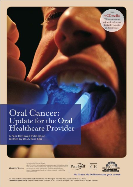
Oral Cancer: Update for the Oral Healthcare Provider - IneedCE.com
Oral Cancer: Update for the Oral Healthcare Provider - IneedCE.com
Oral Cancer: Update for the Oral Healthcare Provider - IneedCE.com

You also want an ePaper? Increase the reach of your titles
YUMPU automatically turns print PDFs into web optimized ePapers that Google loves.
Earn<br />
4 CE credits<br />
This course was<br />
written <strong>for</strong> dentists,<br />
dental hygienists,<br />
and assistants.<br />
<strong>Oral</strong> <strong>Cancer</strong>:<br />
<strong>Update</strong> <strong>for</strong> <strong>the</strong> <strong>Oral</strong><br />
<strong>Healthcare</strong> <strong>Provider</strong><br />
A Peer-Reviewed Publication<br />
Written by Dr. A. Ross Kerr<br />
PennWell is an ADA CERP recognized provider<br />
ADA CERP is a service of <strong>the</strong> American Dental Association to assist dental professionals in identifying<br />
quality providers of continuing dental education. ADA CERP does not approve or endorse individual<br />
courses or instructors, nor does it imply acceptance of credit hours by boards of dentistry.<br />
PennWell is an ADA CERP Recognized <strong>Provider</strong><br />
Concerns of <strong>com</strong>plaints about a CE provider may be directed to <strong>the</strong> provider or to ADA CERP at<br />
www.ada.org/goto/cerp.<br />
Go Green, Go Online to take your course<br />
This course has been made possible through an unrestricted educational grant. The cost of this CE course is $59.00 <strong>for</strong> 4 CE credits.<br />
Cancellation/Refund Policy: Any participant who is not 100% satisfied with this course can request a full refund by contacting PennWell in writing.
Educational Objectives<br />
Upon <strong>com</strong>pletion of this course, <strong>the</strong> clinician will be able to<br />
do <strong>the</strong> following:<br />
1. Understand <strong>the</strong> diverse demographic of patients who are<br />
affected by oral cancer.<br />
2. Understand <strong>the</strong> risk factors that directly relate in culmination<br />
of oral cancer, especially alcohol and tobacco use.<br />
3. Comprehend healthcare professional methods <strong>for</strong> detection,<br />
(ie., types of cancer screening), and provide proper<br />
protocol <strong>for</strong> prevention.<br />
Abstract<br />
Over 30,000 new cases are expected to be diagnosed in <strong>the</strong><br />
United States this year alone. With proper intra-oral and<br />
extra-oral examinations conducted by a trained healthcare<br />
professional, early diagnosis can make all <strong>the</strong> difference in<br />
<strong>the</strong> management of <strong>the</strong> patient’s condition, although prevention<br />
remains <strong>the</strong> healthcare provider’s best option. To help<br />
avert future incidence, at-risk patients need to be educated<br />
by healthcare providers, including on nutritional in<strong>for</strong>mation<br />
and, in <strong>the</strong> case of tobacco users, <strong>the</strong> U.S. Public Health<br />
Service’s “Five A’s.” Armed with a full understanding of <strong>the</strong><br />
disease, <strong>the</strong> oral healthcare professional is <strong>the</strong> most likely<br />
frontline clinician to prevent, diagnose and manage a patient’s<br />
oral cancer.<br />
Introduction<br />
For <strong>the</strong> purpose of this article, <strong>the</strong> term “oral cancer” applies<br />
to malignancies occurring distal to <strong>the</strong> vermillion border of<br />
<strong>the</strong> lips in <strong>the</strong> oral cavity, and some pharyngeal sites, such as<br />
<strong>the</strong> oropharynx and tonsils, that are readily visible or palpable<br />
during a <strong>com</strong>prehensive examination by an oral healthcare<br />
provider (OHCP). <strong>Oral</strong> squamous cell carcinoma (SCCA),<br />
arising from <strong>the</strong> epi<strong>the</strong>lium, <strong>com</strong>prises 90 percent of oral cancers,<br />
with <strong>the</strong> remaining 10 percent attributed to rare cancers,<br />
including salivary gland malignancies, sar<strong>com</strong>as, and o<strong>the</strong>rs.<br />
SCCA is <strong>the</strong> endpoint of an accumulation of alterations in<br />
genes responsible <strong>for</strong> maintaining <strong>the</strong> integrity of <strong>the</strong> genome<br />
(“caretaker” genes) and/or those responsible <strong>for</strong> controlling<br />
cellular proliferation (“gatekeeper” genes). Such alterations<br />
vary and range from subtle changes in DNA sequences to<br />
gross chromosomal gains or losses. The precise mechanisms<br />
by which <strong>the</strong>se alterations lead to <strong>the</strong> genetic instability predicting<br />
malignant trans<strong>for</strong>mation, proliferation, and metastasis<br />
remains hotly debated. In general it seems clear that <strong>the</strong><br />
progression follows a distinct temporal pattern from normal<br />
mucosa to precancer (oral epi<strong>the</strong>lial dysplasia) to SCCA<br />
(<strong>com</strong>mensurate with invasion into submucosal tissues), and<br />
that <strong>the</strong> majority of <strong>the</strong> genetic alterations found in SCCA<br />
are present in lesions preceding malignant trans<strong>for</strong>mation. 1,2<br />
Although <strong>the</strong> time line <strong>for</strong> pathogenesis is variable, <strong>the</strong>re is<br />
an opportunity in most cases to visualize this disease during<br />
<strong>the</strong> premalignant stage where <strong>the</strong> changes are confined to <strong>the</strong><br />
epi<strong>the</strong>lial layers, or while <strong>the</strong> SCCA is at an early stage be<strong>for</strong>e<br />
it has spread from <strong>the</strong> primary site. Indeed, in <strong>the</strong> future it<br />
may be possible to detect molecular markers <strong>for</strong> <strong>the</strong>se genetic<br />
mutations be<strong>for</strong>e any visual changes are evident. This article<br />
provides an update to OHCPs to facilitate early detection,<br />
which should translate into a reduction in both <strong>the</strong> morbidity<br />
and mortality of this terrible disease.<br />
Epidemiology<br />
United States estimates <strong>for</strong> oral cavity and pharyngeal cancers<br />
in 2006 indicate that 30,990 new cases will be diagnosed,<br />
7,430 of which will be terminal. 3 From 1975–2002 this data<br />
represents a modest yet significant decrease in <strong>the</strong> annual percentage<br />
changes in both incidence and mortality rates. That<br />
said, <strong>the</strong> 2006 data represents 1.5 percent and 5.5 percent increases<br />
in incidence and mortality, respectively. The lifetime<br />
risk <strong>for</strong> developing oral and pharyngeal cancer is one in 98.<br />
With respect to incidence this translates to an age-adjusted<br />
rate of 10.5 in 100,000 men and women per year. Incidence<br />
rates increase with age, with 89 percent over <strong>the</strong> age<br />
of 35 and an average age at diagnosis of 63. There has been a<br />
significant increase in oral cancer in <strong>the</strong> under-40 age group. 4<br />
In men, oral cavity and pharynx <strong>com</strong>prise <strong>the</strong> eighth leading<br />
site <strong>for</strong> cancer in <strong>the</strong> U.S., with incidence rates (15.5/100,000)<br />
that are more than twice those of women (6.4/100,000). 5 By<br />
race, African American men have <strong>the</strong> highest incidence rates<br />
(19.0/100,000) and <strong>the</strong> poorest survival out<strong>com</strong>es. Oropharyngeal<br />
cancers can occur at any subsite, with <strong>the</strong> tongue<br />
presenting <strong>the</strong> greatest number or occurences.<br />
With respect to mortality, <strong>the</strong>re is a similar age, gender,<br />
and racial trend: an overall age-adjusted mortality rate of 2.8<br />
in 100,000 men and women per year with higher rates in men<br />
(4.2/100,000 versus 1.6/100,000 in women), and <strong>the</strong> highest<br />
rates in African American men (7.1/100,000).<br />
However, <strong>the</strong> most important epidemiologic data is<br />
related to survival. Given <strong>the</strong> easy screening access, it is remarkable<br />
that <strong>the</strong> overall five-year relative survival rate <strong>for</strong> a<br />
patient diagnosed with oral and pharyngeal cancer during <strong>the</strong><br />
period from 1995–2001 has been estimated to be about 59.4<br />
percent, in stark contrast to breast or prostate cancer which<br />
have overall five-year relative survival rates of 88 percent<br />
and 100 percent respectively. Only 34 percent of oral and<br />
pharyngeal cancers are diagnosed with localized stage disease<br />
(i.e., no detectable spread of <strong>the</strong> primary cancer to regional<br />
lymph nodes). 5 The importance of early detection is evident<br />
when survival rates are stratified by stage: localized disease<br />
is associated with <strong>the</strong> highest five-year relative survival rates<br />
(82.1 percent), <strong>com</strong>pared to advanced disease that has spread<br />
to regional lymph nodes (51.3 percent) or to distant sites (27.6<br />
percent). In addition to <strong>the</strong> high mortality of this disease if<br />
detected in <strong>the</strong> advanced stages, it is also important to note<br />
that quality of life <strong>for</strong> patients who do survive is greatly diminished<br />
due to <strong>the</strong> nature of <strong>the</strong> main treatment options,<br />
which include disfiguring surgery and radiation <strong>the</strong>rapy that<br />
predict numerous <strong>com</strong>plications.<br />
2 www.ineedce.<strong>com</strong>
Risk Factors<br />
Seventy-five percent of all cases of oral cancer in <strong>the</strong> United<br />
States are associated with tobacco smoking and heavy alcohol<br />
use. 6 It is <strong>the</strong> long-term carcinogenic exposure to such risk<br />
factors that, in <strong>the</strong> context of individual host susceptibility,<br />
causes <strong>the</strong> accumulation of genetic abnormalities leading to<br />
oral cancer. Cigarette smokers older than 40 years of age with<br />
a history of heavy alcohol use are at highest risk. Use of all<br />
<strong>for</strong>ms of tobacco has been associated with oral cancer, including<br />
cigar and pipe smoking, smokeless or chewing tobacco,<br />
and o<strong>the</strong>r <strong>for</strong>ms of tobacco used by certain ethnic groups such<br />
as bidi smoking, clove cigarettes, hookahs, or chewing a <strong>com</strong>bination<br />
of areca nut and tobacco (paan masala or gutkha). 7<br />
O<strong>the</strong>r avoidable risk factors include sun exposure (<strong>for</strong> lip<br />
cancer), a diet low in fruits and vegetables, and recent data<br />
suggests a possible association with marijuana use, although<br />
<strong>the</strong>re is conflicting data. 8,9,10 Patients with a past history of<br />
upper aerodigestive tract cancer, particularly oral cancer, are<br />
at a much higher risk <strong>for</strong> recurrence. Immuno<strong>com</strong>promised<br />
patients, including but not limited to those with HIV infection,<br />
may also be at heightened risk. 11<br />
Risk factors <strong>for</strong> <strong>the</strong> remaining 25 percent of <strong>the</strong> population<br />
who develop oral cancer remain enigmatic. O<strong>the</strong>r than<br />
inherited genetic mutations, researchers speculate that oral<br />
infection with oncogenic viruses, particularly certain strains<br />
of <strong>the</strong> human papilloma virus, may confer a higher risk and<br />
may explain <strong>the</strong> increase in oral cancer within <strong>the</strong> under-40<br />
age group.<br />
Role of <strong>Oral</strong> <strong>Healthcare</strong> <strong>Provider</strong>s in Early<br />
Detection and Prevention<br />
Of <strong>the</strong> various frontline clinicians, <strong>the</strong> OHCPs’ training<br />
arguably makes <strong>the</strong>m <strong>the</strong> best qualified to examine <strong>the</strong> oral<br />
cavity and intercept oral premalignancy or early oral cancers.<br />
Given <strong>the</strong> devastating consequences of delayed diagnosis,<br />
<strong>the</strong>y have a professional obligation to optimize <strong>the</strong>ir ability<br />
to detect disease earlier by per<strong>for</strong>ming opportunistic oral<br />
cancer screenings <strong>for</strong> every patient presenting to <strong>the</strong> dental<br />
clinic, whe<strong>the</strong>r <strong>for</strong> routine or emergency care. In addition,<br />
preventive counseling about avoidable risk factors and proper<br />
nutrition must also be considered standard care. The practice<br />
of early detection and prevention constitutes an oral cancer<br />
screening, and <strong>the</strong> first step towards inculcating this practice<br />
is to ensure that you are updated in this area. Indeed, some<br />
U.S. states are now mandating continuing education in oral<br />
cancer early detection and prevention. Fur<strong>the</strong>rmore, <strong>the</strong>re is<br />
an increase in malpractice suits stemming from late diagnoses<br />
of oral cancer. 12<br />
<strong>Oral</strong> <strong>Cancer</strong> Screening<br />
This can take fewer than five minutes and could save a life.<br />
It involves risk factor assessment, physical examination, and<br />
preventive counseling. In addition to visualizing oral abnormalities,<br />
screening exams can be used as an opportunity to<br />
educate patients about <strong>the</strong> symptoms of oral cancer and help<br />
<strong>the</strong>m understand <strong>the</strong> factors that put <strong>the</strong>m at risk. Step 1 is a<br />
risk factor assessment that includes eliciting current and past<br />
history of tobacco and alcohol use, a medical history (or family<br />
history) of previous cancers (particularly upper aerodigestive<br />
tract cancers) or indicative of a <strong>com</strong>promised immune system<br />
(e.g., HIV infection, chronic steroid use), and a nutritional<br />
history. Step 2, applied regardless of <strong>the</strong> results of <strong>the</strong> risk factor<br />
assessment, is a physical examination of both extra-oral<br />
and intra-oral tissues. In order <strong>for</strong> <strong>the</strong> examination to have<br />
both high sensitivity and specificity <strong>for</strong> detecting an abnormal<br />
finding, <strong>the</strong> examiner must have knowledge of <strong>the</strong> range<br />
of “normal” anatomy (including <strong>the</strong> anatomy of edentulous<br />
patients) and <strong>the</strong> ability to visualize or gain access to all regions<br />
to be examined. Many patients are unfamiliar with such<br />
an examination, so it is important to explain and demonstrate<br />
<strong>the</strong> process and encourage self-examination between visits.<br />
There are no current guidelines <strong>for</strong> <strong>the</strong> frequency of oral<br />
cancer screenings in <strong>the</strong> general population, although given<br />
<strong>the</strong> minimal time required and <strong>the</strong> potential benefits of early<br />
detection, screening all new patients (including emergencies)<br />
and at recall seems prudent. Remember, if you bill <strong>the</strong> patient<br />
or a third party <strong>for</strong> a <strong>com</strong>prehensive or periodontal evaluation<br />
(CDT D0150 or D0180), you are obligated to per<strong>for</strong>m an oral<br />
cancer screening.<br />
Extra-<strong>Oral</strong> Examination<br />
The extra-oral examination should include inspection and<br />
palpation of both <strong>the</strong> midline and lateral structures of <strong>the</strong><br />
head and neck primarily to detect abnormal lymph nodes<br />
(lymphadenopathy) that may indicate regional spread.<br />
Indeed, <strong>the</strong> most <strong>com</strong>mon reason <strong>for</strong> a lateral neck swelling<br />
in an adult is a metastatic lymph node. However, this is<br />
also an opportunity to intercept head and neck skin cancers<br />
or o<strong>the</strong>r abnormalities in this region. Clothing or long hair<br />
restricting direct visualization should be removed or pushed<br />
aside respectively. Under proper lighting, assess <strong>the</strong> patient’s<br />
general features <strong>for</strong> swelling and asymmetry. Throughout<br />
<strong>the</strong> examination, carefully inspect <strong>the</strong> skin <strong>for</strong> color changes,<br />
growths, or ulcerations. In addition to o<strong>the</strong>r structures such<br />
as <strong>the</strong> TMJs, muscles of mastication, and <strong>the</strong> parotid glands,<br />
<strong>the</strong> examiner should pay close attention to <strong>the</strong> structures as<br />
described below.<br />
Thyroid gland<br />
A bilobed structure attached to <strong>the</strong> trachea, with <strong>the</strong> isthmus<br />
at a level just inferior to <strong>the</strong> cricoid cartilage (Figure 1). An<br />
abnormal thyroid is consistent with glandular enlargement,<br />
ei<strong>the</strong>r diffuse (which may be obvious by visual inspection<br />
alone) or as a discrete nodule or nodules. Standing behind<br />
<strong>the</strong> patient, <strong>the</strong> examiner should locate <strong>the</strong> isthmus, using<br />
<strong>the</strong> prominent thyroid cartilage as a landmark and moving<br />
inferior to <strong>the</strong> cricoid cartilage (Figure 2). Pushing <strong>the</strong> sternocleidomastoid<br />
muscle laterally and posteriorly with two<br />
www.ineedce.<strong>com</strong> 3
Figure 1 Figure 2 Figure 3 Figure 4<br />
Figure 5 Figure 6<br />
fingers, <strong>the</strong> examiner may palpate <strong>the</strong> lobes (in turn) by applying<br />
light pressure (Figure 3). Asking <strong>the</strong> patient to swallow<br />
some water may also facilitate palpation of <strong>the</strong> thyroid.<br />
Trachea<br />
An abnormal trachea may be consistent with tracheal fixation<br />
(axial immobility) or displacement from its usual midline<br />
position. With <strong>the</strong> patient’s neck relaxed, <strong>the</strong> examiner<br />
should grasp <strong>the</strong> tracheal rings and gently move <strong>the</strong> trachea<br />
laterally (Figure 4). Free and symmetrical axial movement<br />
with mild crepitus is normal. Tracheal displacement may<br />
be evaluated by palpating its relationship to <strong>the</strong> midline<br />
suprasternal notch.<br />
Lymph Nodes of <strong>the</strong> Head and Neck<br />
The examiner must be familiar with <strong>the</strong> lymphatic drainage<br />
of <strong>the</strong> head and neck (Figure 5), and understand how to<br />
recognize lymphadenopathy. The lymphatic system of <strong>the</strong><br />
head and neck is both <strong>com</strong>plex and highly variable. Superficial<br />
lymph nodes drain into deep lymph nodes, and lymph<br />
nodes of <strong>the</strong> head drain into <strong>the</strong> descending nodes of <strong>the</strong><br />
neck. In a healthy individual, some of <strong>the</strong> superficial nodes<br />
may be palpable. When lymph drainage contains mediators<br />
of inflammation or <strong>for</strong>eign antigens (e.g., microorganisms<br />
or tumors), lymphadenitis or lymph node hyperplasia may<br />
result. These lymph nodes are enlarged, freely movable,<br />
and often tender. Metastatic lymph nodes occur following<br />
seeding of malignant cells (especially carcinomas) into <strong>the</strong><br />
regional lymph node, and, depending upon time and tumor<br />
type, this usually manifests clinically as a firm and nonpainful<br />
nodal enlargement that may be bound to surrounding tissues.<br />
This bonding is known as “fixation.” Lymphomas, especially<br />
Hodgkin’s disease, may manifest as rubbery enlarged cervical<br />
lymph nodes. While enlargement of superficial nodes is easier<br />
to detect, deep nodes are only palpable when larger than 2 cm<br />
in diameter. Depending on <strong>the</strong> region, most lymph drainage<br />
from <strong>the</strong> oral cavity will enter <strong>the</strong> submental, submandibular,<br />
or jugulodigastric lymph nodes ei<strong>the</strong>r directly or indirectly.<br />
In oral cancer, metastatic disease will usually involve <strong>the</strong>se<br />
lymph nodes and spread inferiorly. If <strong>the</strong> examiner prefers to<br />
start <strong>the</strong> extra-oral examination with <strong>the</strong> thyroid and trachea,<br />
it makes sense to examine <strong>the</strong> lymph nodes of <strong>the</strong> neck be<strong>for</strong>e<br />
those of <strong>the</strong> head. Since skin is elastic, it is more efficient to<br />
palpate underlying structures by digitally moving a section<br />
of skin ra<strong>the</strong>r than sliding fingers over <strong>the</strong> skin surface. The<br />
sternocleidomastoid (SCM) muscles separate <strong>the</strong> anterior and<br />
posterior triangles of <strong>the</strong> neck, serving as an ideal landmark<br />
<strong>for</strong> examination of <strong>the</strong> neck lymph nodes since <strong>the</strong> cervical<br />
lymph nodes are found in close proximity to <strong>the</strong>m. Position<br />
<strong>the</strong> patient’s head to reveal <strong>the</strong> outline of <strong>the</strong> SCM. This can<br />
4 www.ineedce.<strong>com</strong>
e ac<strong>com</strong>plished by having <strong>the</strong> patient lift his or her head<br />
off <strong>the</strong> headrest or by turning his or her head away from <strong>the</strong><br />
side being examined. Once <strong>the</strong> SCM is visible, starting superiorly,<br />
palpate along <strong>the</strong> entire anterior border of <strong>the</strong> SCM,<br />
pushing under <strong>the</strong> body of <strong>the</strong> muscle to facilitate detection<br />
of enlarged deep cervical nodes (Figure 6). Although rarely<br />
a site <strong>for</strong> metastatic lymphadenopathy, you can also palpate<br />
<strong>the</strong> posterior neck triangle along <strong>the</strong> entire posterior border<br />
of <strong>the</strong> SCM from <strong>the</strong> suboccipital region superiorly to <strong>the</strong><br />
supraclavicular region inferiorly. Repeat <strong>the</strong> process <strong>for</strong> <strong>the</strong><br />
contralateral neck lymph nodes and <strong>com</strong>pare findings. The<br />
submandibular node can easily be palpated in most patients<br />
and allows <strong>the</strong> examiner to experience <strong>the</strong> tactile characteristics<br />
of a healthy node. Have <strong>the</strong> patient position his chin<br />
down and, with <strong>the</strong> fingers cupped, drag <strong>the</strong> loose skin under<br />
<strong>the</strong> chin laterally across <strong>the</strong> inferior border of <strong>the</strong> mandible<br />
(Figure 7). In this way it is possible to capture and palpate <strong>the</strong><br />
node against <strong>the</strong> mandible. In a similar fashion, <strong>the</strong> submental<br />
node can be palpated by dragging tissue under <strong>the</strong> chin<br />
anteriorly (Figure 8).<br />
Figure 7 Figure 8<br />
Intra-<strong>Oral</strong> Examination<br />
<strong>Oral</strong> abnormal findings may range from an asymptomatic<br />
and small (less than 1 cm in diameter) red, white, or mixed<br />
red/white patch with or without ulceration as <strong>the</strong> earliest<br />
visible premalignant epi<strong>the</strong>lial change to a massive (greater<br />
than 4 cm in diameter) exophytic tumor with induration and<br />
associated pain with an advanced SCCA. No awards go to<br />
oral healthcare providers who detect advanced cancers; <strong>the</strong><br />
odds of survival do not favor <strong>the</strong> patient. However, <strong>the</strong> critical<br />
interception of early disease requires <strong>the</strong> examiner to be able<br />
to carefully visualize all soft tissues. <strong>Oral</strong> cancer may develop<br />
on any mucosal surface, although <strong>the</strong> highest risk sites in <strong>the</strong><br />
U.S. are <strong>the</strong> tongue, followed by <strong>the</strong> oropharynx, lower lip,<br />
and floor of <strong>the</strong> mouth.<br />
Patients should be asked to remove lipstick and removable<br />
dental appliances and to rinse out food particles. It is<br />
important <strong>for</strong> <strong>the</strong> examiner to develop a consistent examination<br />
sequence. The order of this is not important, as long as all<br />
elements are <strong>com</strong>pleted. An adequate light source is critical; if<br />
a standard overhead halogen dental light is not available, <strong>the</strong><br />
clinician is advised to use a portable headlight in order to keep<br />
both hands free. An air syringe, mouth mirror, and gauze are<br />
helpful adjuncts to <strong>the</strong> standard intra-oral examination.<br />
1. Lips: Abnormal lip findings include a loss of vermillion<br />
border, ulcerations that have not healed within two<br />
weeks, and o<strong>the</strong>r surface changes or color irregularities.<br />
Bimanual palpation of <strong>the</strong> lips is essential to rule out<br />
submucosal growths.<br />
2. Labial Mucosae: Reflecting <strong>the</strong> lips (Figure 9), look <strong>for</strong><br />
any surface changes.<br />
3. Buccal Mucosae: Use <strong>the</strong> fingers or <strong>the</strong> mirror to retract<br />
and visualize all aspects of <strong>the</strong> buccal mucosae (Figure<br />
10). Palpate <strong>the</strong> parotid extra-orally to ensure that<br />
salivary flow through <strong>the</strong> Stensen’s duct orifice is normal.<br />
4. Gingivae: Both palpation and visualization<br />
are important.<br />
5. Tongue: All surfaces of <strong>the</strong> anterior two-thirds of <strong>the</strong><br />
tongue (dorsal, lateral, and ventral) are generally easy to<br />
visualize and palpate. A piece of gauze may be wrapped<br />
around <strong>the</strong> tongue to allow access to <strong>the</strong> posterior aspect<br />
of <strong>the</strong> dorsum and posterolateral border of <strong>the</strong> tongue<br />
where <strong>the</strong> foliate papillae can be visualized (Figure 11).<br />
The posterior third of <strong>the</strong> tongue is more difficult to<br />
visualize directly, digital palpation and indirect visualization<br />
by mirror are encouraged (Figure 12).<br />
6. Floor of Mouth: Since surface changes may be subtle,<br />
air-drying this region facilitates examination (Figure<br />
13). Bimanual palpation <strong>for</strong> any growths or asymmetry<br />
can be achieved by moving two opposing fingers, one<br />
extra-orally and <strong>the</strong> o<strong>the</strong>r intra-orally, from posterior to<br />
anterior, palpating <strong>the</strong> interposing soft tissue (Figure 14).<br />
7. Palate: Palpation and direct visualization of both <strong>the</strong> hard<br />
and soft palate is important (Figure 15).<br />
8. Oropharynx: Often overlooked, this area includes <strong>the</strong><br />
soft palate and uvula, <strong>the</strong> anterior and posterior pillars (or<br />
fauces), <strong>the</strong> palatine tonsils (or scars from <strong>the</strong>ir removal),<br />
and <strong>the</strong> posterior pharyngeal wall (Figure 16).<br />
<strong>Oral</strong> <strong>Cancer</strong> Screening Adjuncts<br />
In recent years new screening adjuncts have emerged (and<br />
continue to emerge) with <strong>the</strong> principal aim being not to replace<br />
<strong>the</strong> standard screening examination but to improve its<br />
“yield.” An ideal screening adjunct should be highly sensitive<br />
(i.e., it should detect oral premalignant lesions or early oral<br />
cancers), inexpensive, noninvasive, and simple to use. Currently<br />
<strong>the</strong>re is only one <strong>com</strong>mercially available screening adjunct<br />
in <strong>the</strong> United States. ViziLite ® (Zila Inc., Phoenix, AZ)<br />
is FDA-cleared as an adjunct to <strong>the</strong> standard oral examination<br />
to improve identification, evaluation, and monitoring of<br />
white oral mucosal abnormalities in populations at increased<br />
risk <strong>for</strong> oral cancer. There is a CDT code <strong>for</strong> this test (D0431)<br />
that may be covered by some insurance carriers. Based on<br />
chemiluminescence (a low-energy blue-white light), it is an<br />
www.ineedce.<strong>com</strong> 5
Figure 9<br />
Figure 10<br />
Figure 11<br />
Figure 12<br />
Figure 13<br />
Figure 14<br />
Figure 15<br />
Figure 16<br />
immediate and noninvasive screening<br />
adjunctive technique adapted <strong>for</strong> oral<br />
use following demonstration of its ability<br />
in gynecology to enhance <strong>the</strong> direct<br />
visualization of cervical lesions suspicious<br />
<strong>for</strong> neoplasia. 13 The technique<br />
involves a customary standard visual<br />
intra-oral examination (recording any<br />
abnormalities), and <strong>the</strong>n <strong>the</strong> patient is<br />
asked to rinse <strong>for</strong> 60 seconds with dilute<br />
acetic acid. The chemiluminescent<br />
“wand” is <strong>the</strong>n activated, and a second<br />
visual examination is per<strong>for</strong>med in<br />
a darkened room to detect any abnormalities,<br />
specifically “acetowhite<br />
changes” (Figure 17). Two preliminary<br />
screening studies carried out in highrisk<br />
populations by oral medicine<br />
experts have demonstrated that this<br />
technology provides modest additional<br />
visual in<strong>for</strong>mation to <strong>the</strong> standard examination,<br />
although <strong>the</strong> utility of this<br />
adjunct to specifically detect oral cancer<br />
and precancer was not tested. 14,15 White<br />
lesions, particularly nonhomogeneous<br />
ones, are more likely to be visualized by<br />
ViziLite. 15 A recent study in a small cohort<br />
of patients with suspicious lesions<br />
suggests that this technology has high<br />
accuracy in detecting oral cancer and<br />
precancer. 16 More studies are needed<br />
to better define <strong>the</strong> role of ViziLite in<br />
<strong>the</strong> hands of frontline screeners, and at<br />
<strong>the</strong> very least its use may benefit oral<br />
cancer awareness in both <strong>the</strong> public and<br />
professional domain.<br />
Recently, ViziLitePlus (Zila Inc.,<br />
Phoenix, AZ) has been introduced<br />
into <strong>the</strong> marketplace. This product<br />
includes a TBlue 630 <strong>Oral</strong> Lesion Mark<br />
ing System, a toluidine blue dye used<br />
to assist in <strong>the</strong> fur<strong>the</strong>r evaluation of<br />
ViziLite-identified white oral mucosal<br />
lesions <strong>for</strong> patients at increased risk <strong>for</strong><br />
oral cancer.<br />
Toluidine blue is well established<br />
as both a screening and diagnostic<br />
adjunct. It has been used “off-label”<br />
<strong>for</strong> many years and it is <strong>com</strong>mercially<br />
available as a screening adjunct<br />
(OraTest, Zila Inc., Phoenix, AZ) in 14<br />
countries outside of <strong>the</strong> United States.<br />
As of <strong>the</strong> publication of this article,<br />
studies are currently under way to sup-<br />
6 www.ineedce.<strong>com</strong>
port its FDA clearance. Toluidine blue is a vital stain that is<br />
rapidly absorbed into actively dividing (e.g., neoplastic) cells.<br />
There are numerous studies demonstrating toluidine blue’s<br />
extremely high accuracy <strong>for</strong> detecting SCCA, carcinoma in<br />
situ, and higher-grade dysplastic lesions via an intense blue<br />
stain, but it is less accurate in <strong>the</strong> detection of lower-grade<br />
dysplastic lesions, as demonstrated by false negative rates of<br />
greater than 20 percent. 17,18 In addition, since toluidine blue<br />
can also adhere mechanically to tissue crevices or bind to<br />
actively dividing nonneoplastic cells, such as inflammatory<br />
conditions or tissues undergoing repair, false positive results<br />
are possible unless <strong>the</strong> clinician restains tissues after a waiting<br />
period of two to three weeks or has adequate experience in<br />
interpretation. Recently, it has been demonstrated that positive<br />
toluidine blue staining predicts a high risk of malignant<br />
trans<strong>for</strong>mation in premalignant lesions, suggesting its utility<br />
as a prognostic test. 19<br />
O<strong>the</strong>r emerging screening technologies include tissue autofluorescence<br />
and salivary molecular diagnostics. <strong>Oral</strong> cancer<br />
and precancerous lesions show different autofluorescence<br />
profiles <strong>com</strong>pared to normal oral mucosa and technology<br />
<strong>for</strong> chairside visualization of tissue autofluorescence offers<br />
ano<strong>the</strong>r novel approach to screening. 20 The VELscope (LED<br />
Medical Diagnostics Inc., Vancouver, British Columbia,<br />
Canada) has approval in Canada <strong>for</strong> use and FDA clearance<br />
is pending.<br />
Diagnostic Procedures<br />
Upon an abnormal finding during <strong>the</strong> standard examination,<br />
<strong>the</strong> oral health provider should attempt to develop a<br />
provisional or differential diagnosis. Diagnosing an advanced<br />
SCCA requires very little diagnostic acumen because of its<br />
obvious clinical presentation: large; usually exophytic; ulcerated;<br />
nodular; indurated/fixated; bleeds easily upon slight<br />
provocation; lymphadenopathic; and possessed of associated<br />
symptoms such as pain, dysphagia, or limitation of function<br />
(Figure 18). These patients have a high likelihood of metastatic<br />
disease and a poor prognosis. The diagnostic challenge<br />
of early detection be<strong>com</strong>es more difficult as one regresses in<br />
<strong>the</strong> lifetime of <strong>the</strong> cancer through metastasis and malignant<br />
trans<strong>for</strong>mation, and back to <strong>the</strong> time when <strong>the</strong> earliest precancerous<br />
changes are evident, or potentially even earlier.<br />
The terms leukoplakia, erythroplakia, and erythroleukoplakia<br />
are clinical diagnoses given to persistent, most<br />
often unilateral, white, red, or mixed red/white patches<br />
respectively, and <strong>for</strong> which an etiology remains unclear. 21<br />
Leukoplakias cannot be wiped off and are much less likely to<br />
undergo malignant trans<strong>for</strong>mation <strong>com</strong>pared to patches with<br />
a red <strong>com</strong>ponent. These and chronically ulcerated lesions of<br />
unclear etiology represent <strong>the</strong> clinical spectrum of premalignant<br />
(or early cancer) presentations and should be suspected<br />
as such until proven o<strong>the</strong>rwise. There should be a high index<br />
of suspicion when a clinician detects such lesions with one or<br />
more of <strong>the</strong> following features:<br />
Figure 17 Figure 18<br />
Figure 19<br />
1. High-risk site (such as <strong>the</strong> lateral/ventral tongue, floor of<br />
mouth, or oropharynx).<br />
2. Larger size (greater than 1 cm in diameter) or multiple<br />
site involvement.<br />
3. Containing a red <strong>com</strong>ponent (erythroplakic area),<br />
nonhomogeneous leukoplakias, or leukoplakias with<br />
irregular surface changes (Figure 19). Patients over 40<br />
with a positive risk factor history are at higher risk <strong>for</strong><br />
oral cancer, although a younger patient presenting with<br />
<strong>the</strong>se features is even more worrisome and may indicate<br />
highly aggressive disease. Such patients who also display<br />
overtly suspicious epi<strong>the</strong>lial lesions should be promptly<br />
referred to an expert <strong>for</strong> scalpel or punch biopsy and<br />
definitive histopathologic diagnosis.<br />
However, earlier stage precancerous or cancerous lesions<br />
are often smaller, show subtler features, and can easily be<br />
mistaken <strong>for</strong> benign-looking <strong>com</strong>mon inflammatory or<br />
traumatically induced epi<strong>the</strong>lial changes (Figure 20). When<br />
faced with such benign-looking lesions, a decision must be<br />
made about how to proceed. The first step should be to go<br />
back through <strong>the</strong> patient’s data: demographics, a careful history<br />
and risk factor assessment, and <strong>the</strong> results of a thorough<br />
clinical examination. This data may reveal possible etiologies<br />
<strong>for</strong> <strong>the</strong> lesion. The removal or modification of a putative<br />
etiologic factor, such as <strong>the</strong> influence of a sharp tooth cusp<br />
or habit followed by a period of waiting and watching (two<br />
to three weeks is usually sufficient time <strong>for</strong> an inflammatory<br />
or traumatic lesion to heal), is an acceptable way to proceed<br />
unless <strong>the</strong> patient fails to return <strong>for</strong> follow-up in a timely<br />
manner, or if <strong>the</strong> oral health provider <strong>for</strong>gets to watch <strong>the</strong><br />
www.ineedce.<strong>com</strong> 7
Figure 20<br />
Figure 21<br />
Figure 22<br />
Figure 23<br />
lesion. Take a photographic record of <strong>the</strong> lesion at baseline,<br />
and if <strong>the</strong> lesion persists, fur<strong>the</strong>r investigation is warranted.<br />
There are a number of diagnostic options available today.<br />
The gold standard diagnostic “test” applied to an overtly suspicious<br />
epi<strong>the</strong>lial lesion is a scalpel/punch biopsy followed<br />
by histopathological examination by an oral pathologist to<br />
rule out a squamous (or verrucous) carcinoma or epi<strong>the</strong>lial<br />
dysplasia (premalignancy). Technically, a tissue biopsy usually<br />
requires very little expertise, particularly with <strong>the</strong> use<br />
of a punch (CDT code D07286). Biopsy site selection may<br />
require expertise, and adjunctive toluidine blue stain applied<br />
to <strong>the</strong> lesion can direct <strong>the</strong> clinician to <strong>the</strong> highest-risk<br />
site (Figure 21). Few general dentists feel adequately trained<br />
to per<strong>for</strong>m <strong>the</strong>m and <strong>the</strong>re is hesitation to send patients with<br />
a benign-looking lesion <strong>for</strong> an invasive and costly procedure<br />
that will likely confirm a benign result. In addition, faced<br />
with <strong>the</strong> thought of <strong>the</strong> associated dis<strong>com</strong><strong>for</strong>t of undergoing<br />
a biopsy, many patients refuse referral and opt to wait.<br />
New diagnostic adjuncts are available to assess <strong>the</strong><br />
significance of epi<strong>the</strong>lial lesions that are not highly suspicious<br />
(i.e., benign-looking lesions) without subjecting<br />
<strong>the</strong> patient to a standard biopsy. Exfoliative cytology, a<br />
technique traditionally used in cervical cancer screening,<br />
has limitations <strong>for</strong> use in <strong>the</strong> oral cavity by <strong>the</strong> general<br />
dentist because <strong>the</strong> cells collected are superficial and may<br />
not be representative of atypical cells found in deeper<br />
layers of <strong>the</strong> epi<strong>the</strong>lium. The <strong>Oral</strong> CDX brush biopsy is<br />
a simple, painless, and accurate cytopathologic technique<br />
developed by CDX Laboratories (Sufferin, NY). A number<br />
of studies support its indicated use. The sampling<br />
problems associated with exfoliative cytology have been<br />
solved by using a brush designed to sample cells from all<br />
layers of <strong>the</strong> epi<strong>the</strong>lium (Figure 22). In general practices,<br />
<strong>the</strong> brush biopsy is indicated <strong>for</strong> small, benign-looking<br />
epi<strong>the</strong>lial lesions of unclear etiology. Although overtly<br />
suspicious epi<strong>the</strong>lial lesions may be tested using this technique,<br />
referral of patients with such lesions to a specialist<br />
with more experience is prudent, particularly in <strong>the</strong> case<br />
of larger lesions where <strong>the</strong>re may be a risk of sampling<br />
error. Without local anes<strong>the</strong>sia, <strong>the</strong> brush is rotated<br />
repeatedly against <strong>the</strong> lesion. A cell sample is <strong>the</strong>n transported<br />
and fixed onto a glass slide, which is <strong>the</strong>n mailed<br />
to <strong>the</strong> laboratory (Figure 22). Upon receipt, <strong>the</strong> slide is<br />
stained and <strong>the</strong> cells are analyzed using a neural network<br />
software that can identify cells with atypical morphological<br />
characteristics to facilitate diagnosis by an oral<br />
cytopathologist. A CDT code <strong>for</strong> a transepi<strong>the</strong>lial brush<br />
biopsy (D7288) now exists and is covered by a number<br />
of insurance carriers. There are four results: inadequate<br />
sample, benign, atypical, and positive. CDX laboratories<br />
will fax <strong>the</strong> result within a week and later send a color<br />
hard copy depicting <strong>the</strong> cellular morphology, a powerful<br />
way to explain <strong>the</strong> significance of <strong>the</strong> result to <strong>the</strong> patient<br />
and <strong>the</strong>reby increase appropriate follow-up <strong>com</strong>pliance.<br />
Eighty-five to 90 percent of all brush biopsy results are<br />
benign, meaning that <strong>the</strong> sample submitted contained no<br />
atypical cells. However, because a negative result could<br />
lead to a false sense of <strong>com</strong>placency in <strong>the</strong> patient, <strong>the</strong><br />
dentist should use a benign test result as an opportunity<br />
to promote risk factor behavior modification and regular<br />
follow-up. All persistent benign lesions should be serially<br />
tested or referred to a specialist <strong>for</strong> definitive diagnosis.<br />
An atypical result (approximately 10–15 percent of all<br />
results) indicates that <strong>the</strong> lesion requires fur<strong>the</strong>r evaluation<br />
and warrants prompt referral to a specialist. Several<br />
studies show that an atypical result has a positive predictive<br />
value of 20–40 percent. In o<strong>the</strong>r words, two to four<br />
8 www.ineedce.<strong>com</strong>
out of every 10 atypical results represent a premalignant<br />
or malignant change. 22,23,24 A positive result, although<br />
rare, is almost guaranteed to represent a malignant or<br />
premalignant lesion.<br />
With <strong>the</strong> advent of <strong>the</strong> sophisticated technology available<br />
today, <strong>the</strong>re is hope that we can employ biomarkers<br />
to detect early disease, predict <strong>the</strong> likelihood <strong>for</strong> malignant<br />
trans<strong>for</strong>mation in premalignant disease, or indicate<br />
optimal management strategies. Biomarkers include<br />
gross analysis of DNA content (DNA ploidy), classic<br />
cytogenetics (karyotyping), molecular cytogenetics such<br />
as fluorescence in-situ hybridization (FISH), <strong>com</strong>parative<br />
genomic hybridization (CGH), microsatellite analyses to<br />
detect loss of heterozygosity or microsatellite instability,<br />
gene expression microarrays, serial analysis of gene expression<br />
(SAGE), and <strong>the</strong> identification of mutations of various<br />
genes (e.g., telomerase, p53, ras, COX-2).<br />
Management of <strong>Oral</strong> <strong>Cancer</strong> and Precancer<br />
Management strategies <strong>for</strong> patients with oral cancer and<br />
precancer are variable. The most important issue is that<br />
once diagnosed, patients must avoid all risky behaviors and<br />
be placed under <strong>the</strong> close surveillance of an expert over <strong>the</strong><br />
long term.<br />
OHCPs can play an important role in <strong>the</strong> management<br />
of this patient population. Each of <strong>the</strong> current<br />
cancer treatment modalities of surgery, radio<strong>the</strong>rapy, and<br />
chemo<strong>the</strong>rapy may cause both short-term and long-term<br />
oral <strong>com</strong>plications that can influence morbidity and, when<br />
severe, even mortality. Such <strong>com</strong>plications may include<br />
mucositis, taste alterations, dysphagia, difficulties in mastication,<br />
xerostomia, radiation caries, trismus, periodontal<br />
deterioration, decreased resiliency in perioral tissue,<br />
temporomandibular dysfunction, oral and/or systemic<br />
infections, bleeding, soft tissue radionecrosis and osteoradionecrosis,<br />
and <strong>the</strong> potential <strong>for</strong> ei<strong>the</strong>r a recurrence or<br />
a new malignancy. 25 The OHCP should participate in <strong>the</strong><br />
prevention, <strong>the</strong> early interception, and treatment of <strong>the</strong>se<br />
<strong>com</strong>plications. The maintenance of oral health during and<br />
after cancer <strong>the</strong>rapy requires an investment of time and<br />
ef<strong>for</strong>t beyond that needed <strong>for</strong> normal oral care.<br />
Prevention<br />
Early detection is only part of <strong>the</strong> responsibility of <strong>the</strong><br />
OHCP. Our role in treating patients with tobacco dependence<br />
is be<strong>com</strong>ing more important, not only <strong>for</strong> cancer<br />
prevention, but also because of <strong>the</strong> strong association<br />
with loss of periodontal attachment in tobacco users. The<br />
U.S. Public Health Service guidelines using “<strong>the</strong> 5 A’s”<br />
(Ask, Advise, Assess, Assist, and Arrange) constitute an<br />
evidence-based strategy to help patients quit and even<br />
minimal interventions by OHCPs are effective. 26 Promoting<br />
use of alcohol in moderation and nutrition high in fresh<br />
fruits and vegetables should also be part of our message.<br />
Case Study<br />
• 62-year-old male with history of smoking tobacco<br />
• Location-ascending ramus up to <strong>the</strong> level of <strong>the</strong><br />
retromolar pad<br />
• Tissue biospy revealed squamous cell carcinoma<br />
Under incandescent lighting<br />
Under ViziLite illumination<br />
TBlue 630 marking extent of <strong>the</strong> lesion<br />
Case<br />
A 58-year-old Hispanic male presents <strong>for</strong> a dental check up.<br />
He has numerous broken teeth but experiences no pain. His<br />
medical history is pertinent <strong>for</strong> HIV infection (viral load <<br />
1000 viral particles/mL3, CD4 count 350 cells/mL3, on<br />
three antiretroviral medications). He has a past history of tobacco<br />
use (two packs per day <strong>for</strong> 30 years, quit six years ago:<br />
60-pack per year history) and alcohol use (four drinks per day<br />
on average <strong>for</strong> 30 years, quit 10 years ago). His examination<br />
reveals no lymphadenopathy. His soft tissue examination<br />
reveals multiple broken teeth and a mixed red/white patch<br />
(erythroplakia) measuring approximately 2 cm in diameter<br />
on <strong>the</strong> left oropharynx that bleeds easily and has an exophytic<br />
<strong>com</strong>ponent (inferior left aspect) that is firm to palpation.<br />
www.ineedce.<strong>com</strong> 9
Your Approach<br />
His history reveals several risk factors. He is an immuno<strong>com</strong>promised<br />
male over <strong>the</strong> age of 40 with a lengthy history<br />
of heavy tobacco and alcohol use. His exam should raise<br />
your index of suspicion <strong>for</strong> an oral premalignant lesion<br />
or SCCA because it is on a high-risk site (oropharynx),<br />
is more than 1 cm in diameter, has a red <strong>com</strong>ponent, has<br />
a surface irregularity, is firm in a site that should be soft,<br />
and bleeds easily. You should photograph <strong>the</strong> lesion, document<br />
your findings, and immediately refer this patient <strong>for</strong><br />
a scalpel/punch biopsy. Toluidine blue staining fur<strong>the</strong>r<br />
confirms <strong>the</strong> suspicious nature of this lesion, and biopsy at<br />
Site 1 revealed carcinoma in situ and at Site 2, SCCA. He<br />
was staged T2N0M0 (tumor of greater than 2 cm diameter,<br />
with no lymph nodes and no metastatic spread), and<br />
was treated surgically with no radiation.<br />
References<br />
1. Califano, J., et al., Genetic progression model <strong>for</strong> head and neck cancer:<br />
implications <strong>for</strong> field cancerization. <strong>Cancer</strong> Research, 1996. 56(11): p. 2488–92.<br />
2. Ha, P.K., et al., A transcriptional progression model <strong>for</strong> head and neck cancer.<br />
Clinical <strong>Cancer</strong> Research., 2003. 9(8): p. 3058–64.<br />
3. <strong>Cancer</strong> Facts & Figures. 2006, American <strong>Cancer</strong> Society.<br />
4. Schantz, S.P., Yu, G.P. Head and neck cancer incidence trends in young Americans,<br />
1973–1997, with a special analysis <strong>for</strong> tongue cancer. Archives of Otolaryngology<br />
— Head & Neck Surgery, 2002. 128(3): p. 268–74.<br />
5. Ries, L.A.G., et al. (eds). SEER <strong>Cancer</strong> Statistics Review, 1975–2002 (based on<br />
November 2004 SEER data submission, posted to <strong>the</strong> SEER web site 2005),<br />
National <strong>Cancer</strong> Institute. Be<strong>the</strong>sda, MD.<br />
6. Blot, W.J., et al., Smoking and drinking in relation to oral and pharyngeal cancer.<br />
<strong>Cancer</strong> Research., 1988. 48(11): p. 3282–7.<br />
7. Thomas, S., Kearsley, J. Betel quid and oral cancer: a review. European Journal of<br />
<strong>Cancer</strong>. Part B, <strong>Oral</strong> Oncology., 1993. 29B(4): p. 251–5.<br />
8. Winn, D.M., Diet and nutrition in <strong>the</strong> etiology of oral cancer. American Journal of<br />
Clinical Nutrition., 1995. 61(2): p. 437S–445S.<br />
9. Zhang, Z.F., et al., Marijuana use and increased risk of squamous cell carcinoma<br />
of <strong>the</strong> head and neck. <strong>Cancer</strong> Epidemiology, Biomarkers & Prevention., 1999.<br />
8(12): p. 1071–8.<br />
10. Llewellyn, C.D., et al., An analysis of risk factors <strong>for</strong> oral cancer in young people:<br />
a case-control study. <strong>Oral</strong> Oncology, 2004. 40(3): p. 304–13.<br />
11. Epstein, J.B., Silverman, S. Jr., Head and neck malignancies associated with HIV<br />
infection. <strong>Oral</strong> Surgery, <strong>Oral</strong> Medicine, <strong>Oral</strong> Pathology, 1992. 73(2): p. 193–200.<br />
12. Lydiatt, D.D., <strong>Cancer</strong> of <strong>the</strong> oral cavity and medical malpractice. Laryngoscope.,<br />
2002. 112(5): p. 816–9.<br />
13. Parham, G.P., et al., Comparison of immediate and deferred colposcopy in a<br />
cervical screening program. Obstetrics & Gynecology., 2000. 95(3): p. 340–4.<br />
14. Huber, M.A., et al, Acetic acid wash and chemiluminescent illumination as<br />
an adjunct to conventional oral soft tissue examination <strong>for</strong> <strong>the</strong> detection of<br />
dysplasia: a pilot study. Quintessence International, 2004. 35(5): p. 378–84.<br />
15. Kerr, A.R., Sirois D.A., Clinical Evaluation of a New Adjunctive Technique <strong>for</strong> <strong>Oral</strong><br />
Mucosal Examinations: <strong>Oral</strong> Speculoscopy. <strong>Oral</strong> Surg <strong>Oral</strong> Med <strong>Oral</strong> Pathol <strong>Oral</strong><br />
Radiol Endod, 2004. 97(4): p. 451.<br />
16. Ram, S., et al., Chemiluminescence as a diagnostic aid in <strong>the</strong> detection of oral<br />
cancer and potentially malignant epi<strong>the</strong>lial lesions. International Journal of <strong>Oral</strong><br />
& Maxillofacial Surgery, 2005. 34(5): p. 521–7.<br />
17. Warnakulasuriya, K.A., Johnson, N.W., Sensitivity and specificity of OraScan (R)<br />
toluidine blue mouthrinse in <strong>the</strong> detection of oral cancer and precancer. Journal<br />
of <strong>Oral</strong> Pathology & Medicine, 1996. 25(3): p. 97–103.<br />
18. Epstein, J.B., et al., The utility of toluidine blue application as a diagnostic aid<br />
in patients previously treated <strong>for</strong> upper oropharyngeal carcinoma. <strong>Oral</strong> Surgery,<br />
<strong>Oral</strong> Medicine, <strong>Oral</strong> Pathology, <strong>Oral</strong> Radiology, & Endodontics, 1997. 83(5): p.<br />
537–47.<br />
19. Zhang, L., et al., Toluidine blue staining identifies high-risk primary oral<br />
premalignant lesions with poor out<strong>com</strong>e. <strong>Cancer</strong> Research, 2005. 65(17): p.<br />
8017–21.<br />
20. Gillenwater, A., et al., Noninvasive diagnosis of oral neoplasia based on<br />
fluorescence spectroscopy and native tissue autofluorescence. Archives of<br />
Otolaryngology — Head & Neck Surgery, 1998. 124(11): p. 1251–8.<br />
21. Axell, T., et al., <strong>Oral</strong> white lesions with special reference to precancerous and<br />
tobacco-related lesions: conclusions of an international symposium held in<br />
Uppsala, Sweden, May 18–21 1994. International Collaborative Group on <strong>Oral</strong><br />
White Lesions. Journal of <strong>Oral</strong> Pathology & Medicine, 1996. 25(2): p. 49–54.<br />
22. Sciubba, J.J., Improving detection of precancerous and cancerous oral lesions.<br />
Computer-assisted analysis of <strong>the</strong> oral brush biopsy. U.S. Collaborative <strong>Oral</strong>CDx<br />
Study Group. Journal of <strong>the</strong> American Dental Association, 1999. 130(10): p.<br />
1445–57.<br />
23. Scheifele, C., et al., The sensitivity and specificity of <strong>the</strong> <strong>Oral</strong>CDx technique:<br />
evaluation of 103 cases.[see <strong>com</strong>ment]. <strong>Oral</strong> Oncology, 2004. 40(8): p. 824–8.<br />
24. Poate, T.W., et al., An audit of <strong>the</strong> efficacy of <strong>the</strong> oral brush biopsy technique in<br />
a specialist <strong>Oral</strong> Medicine unit.[see <strong>com</strong>ment]. <strong>Oral</strong> Oncology, 2004. 40(8): p.<br />
829–34.<br />
25. Vissink, A., et al., <strong>Oral</strong> sequelae of head and neck radio<strong>the</strong>rapy. Critical Reviews<br />
in <strong>Oral</strong> Biology & Medicine, 2003. 14(3): p. 199–212.<br />
26. Anderson, J.E., et al., Treating tobacco use and dependence: an evidence-based<br />
clinical practice guideline <strong>for</strong> tobacco cessation.[see <strong>com</strong>ment]. Chest, 2002.<br />
121(3): p. 932–41.<br />
Author Profile<br />
Dr. A. Ross Kerr<br />
Dr. Kerr received his DDS from McGill<br />
University in Montreal, Canada, and his<br />
MSD and certificate in oral medicine at<br />
<strong>the</strong> University of Washington. He is a<br />
diplomate of <strong>the</strong> American Board of <strong>Oral</strong><br />
Medicine and clinical associate professor<br />
in <strong>the</strong> department of oral and maxillofacial pathology,<br />
radiology and medicine at New York University College<br />
of Dentistry, where he is <strong>the</strong> director of <strong>the</strong> <strong>Oral</strong> Mucosal<br />
Disease Service. He is <strong>the</strong> chair of <strong>the</strong> <strong>Oral</strong> <strong>Cancer</strong><br />
Consortium and is an investigator on two ongoing NIHfunded<br />
grants, one evaluating current and emerging<br />
technologies <strong>for</strong> <strong>the</strong> early detection of oral cancer, and<br />
<strong>the</strong> o<strong>the</strong>r evaluating tobacco cessation in a dental practice<br />
setting. He has lectured nationally and internationally<br />
on oral-medicine-related topics, and has contributed to<br />
<strong>the</strong> dental and medical literature. His private practice is<br />
focused on <strong>the</strong> dental management of medically <strong>com</strong>plex<br />
patients and <strong>the</strong> management of patients with oral mucosal<br />
disease, salivary hypofunction, and orofacial pain.<br />
Disclaimer<br />
The author of this course has no <strong>com</strong>mercial ties with <strong>the</strong><br />
sponsors or <strong>the</strong> providers of <strong>the</strong> unrestricted educational<br />
grant <strong>for</strong> this course.<br />
Reader Feedback<br />
We encourage your <strong>com</strong>ments on this or any PennWell course.<br />
For your convenience, an online feedback <strong>for</strong>m is available at<br />
www.ineedce.<strong>com</strong>.<br />
10 www.ineedce.<strong>com</strong>
1. Which type of carcinoma accounts <strong>for</strong><br />
over 90 percent of all oral cancers?<br />
a. Sar<strong>com</strong>a<br />
b. Basal cell carcinoma<br />
c. Leukoplakia<br />
d. Squamous cell carcinoma<br />
2. Genes responsible <strong>for</strong> maintaining <strong>the</strong><br />
integrity of <strong>the</strong> genome are <strong>com</strong>monly<br />
referred to as<br />
a. Caretaker genes<br />
b. Proliferation genes<br />
c. Keymaster genes<br />
d. Gatekeeper genes<br />
3. According to <strong>the</strong> article, approximately<br />
how many Americans will be diagnosed<br />
with oral/pharyngeal cancer in 2006?<br />
a. 40,000<br />
b. 30,000<br />
c. 50,000<br />
d. 20,000<br />
4. The average age of diagnosis <strong>for</strong><br />
oral/pharyngeal cancer is<br />
a. 40<br />
b. 35<br />
c. 63<br />
d. 89<br />
5. According to <strong>the</strong> article, <strong>the</strong> lifetime<br />
risk <strong>for</strong> developing oral/pharyngeal<br />
cancer is<br />
a. One in 40<br />
b. One in 98<br />
c. One in 89<br />
d. One in 100,000<br />
6. How does <strong>the</strong> average rate of<br />
incidence <strong>for</strong> African American<br />
men <strong>com</strong>pare to <strong>the</strong> average rate of<br />
incidence <strong>for</strong> women?<br />
a. Half<br />
b. Twice<br />
c. Approximately <strong>the</strong> same as<br />
d. Nearly three times<br />
7. What is <strong>the</strong> highest risk site <strong>for</strong><br />
oral cancer?<br />
a. Gingiva<br />
b. Tongue<br />
c. Buccal mucosa<br />
d. Lower lip<br />
8. The overall five-year relative survival<br />
rate <strong>for</strong> a patient with oral/pharyngeal<br />
cancer is _______ that of a patient with<br />
prostate cancer.<br />
a. Approximately <strong>the</strong> same as<br />
b. Nearly twice<br />
c. Nearly half<br />
d. One-third<br />
9. Seventy-five percent of all oral<br />
cancer cases in <strong>the</strong> United States<br />
are associated with<br />
a. Tobacco and alcohol<br />
b. A diet low in fruit<br />
c. Genetic risk factors<br />
d. Papilloma virus<br />
10. Which of <strong>the</strong> following noted during<br />
an oral cancer screening carries <strong>the</strong><br />
strongest association <strong>for</strong> <strong>the</strong> development<br />
of oral cancer?<br />
a. Patient has a poor diet<br />
b. Patient has a history of cancer in his family<br />
c. Patient <strong>com</strong>plains of frequent canker sores<br />
d. Patient reports heavy alcohol and tobacco use<br />
Questions<br />
11. The first step in an oral cancer<br />
screening should be<br />
a. Physical examination<br />
b. Risk factor assessment<br />
c. Preventative counseling<br />
d. Periodontal evaluation<br />
12. Which of <strong>the</strong>se does <strong>the</strong> author<br />
suggest is a prerequisite to conducting a<br />
physical examination?<br />
a. The results of a risk factor assessment<br />
b. The patient’s attitude during<br />
preventative counseling<br />
c. A knowledge of <strong>the</strong> range of normal anatomy<br />
d. The patient’s prior familiarity with<br />
<strong>the</strong> examination<br />
13. Current guidelines suggest that<br />
an oral cancer screening should be<br />
conducted every three months.<br />
a. True<br />
b. False<br />
14. The most <strong>com</strong>mon cause of lateral<br />
neck swelling in an adult is<br />
a. A metastatic lymph node<br />
b. The parotid gland<br />
c. An abnormal trachea<br />
d. None of <strong>the</strong> above<br />
15. A firm and nonpainful nodal enlargement,<br />
possibly bound to surrounding<br />
tissues, is a probable symptom of<br />
a. Lymphoma, particularly Hodgkin’s disease<br />
b. Lymphadenitis<br />
c. A metastatic lymph node<br />
d. Lymph node hyperplasia<br />
16. Deep nodes are only palpable when<br />
_____ or more in diameter.<br />
a. 1 cm<br />
b. 2 mm<br />
c. 2 cm<br />
d. 3 cm<br />
17. In oral cancer, metastatic disease will<br />
usually spread<br />
a. Interiorly<br />
b. Anteriorly<br />
c. Posteriorly<br />
d. Inferiorly<br />
18. Which lymph nodes are found in close<br />
proximity to <strong>the</strong> SCM muscles?<br />
a. Cervical<br />
b. Submandibular<br />
c. Jugulodigastric<br />
d. Submental<br />
19. The anterior and posterior pillars, or<br />
fauces, are part of <strong>the</strong><br />
a. Palate<br />
b. Floor of mouth<br />
c. Oropharynx<br />
d. Buccal mucosae<br />
20. According to <strong>the</strong> article, an ideal<br />
screening adjunct should be all of <strong>the</strong><br />
following. except<br />
a. Highly sensitive<br />
b. Easy to use<br />
c. Fast<br />
d. Inexpensive<br />
21. Which of <strong>the</strong> following is true<br />
about chemiluminescence?<br />
a. It is intended <strong>for</strong> stand-alone use to detect<br />
oral cancer<br />
b. The patient must rinse with dilute acetic acid<br />
be<strong>for</strong>e examination<br />
c. Red changes are visualized better than<br />
white changes<br />
d. It is a diagnostic test<br />
22. Toluidine blue is <strong>the</strong> preferred screening<br />
adjunct in <strong>the</strong> United States.<br />
a. True<br />
b. False<br />
23. An oral leukoplakia is a white<br />
patch that<br />
a. Is found on <strong>the</strong> oral mucosa<br />
b. Cannot be characterized as any o<strong>the</strong>r definable<br />
lesion<br />
c. Has a high risk <strong>for</strong> malignant trans<strong>for</strong>mation<br />
d. Can easily be rubbed off <strong>the</strong> mucosa<br />
24. The “gold standard” diagnostic<br />
method applied to epi<strong>the</strong>lial lesions is<br />
a. The transepi<strong>the</strong>lial brush biopsy technique<br />
b. Exfoliative cytology<br />
c. The scalpel/punch biopsy<br />
d. FISH<br />
25. Which of <strong>the</strong> following is NOT<br />
true of <strong>the</strong> transepi<strong>the</strong>lial brush<br />
biopsy technique?<br />
a. It can be used by any trained oral healthcare<br />
provider<br />
b. It excels at detecting squamous cell carcinoma<br />
c. It samples <strong>the</strong> superficial cell layers only<br />
d. It does not require local anes<strong>the</strong>sia<br />
26. As much as ____ percent of all brush<br />
biopsy results are benign.<br />
a. 75<br />
b. 80<br />
c. 85<br />
d. 90<br />
27. As much as ____ percent of all<br />
atypical brush biopsy results represent<br />
a premalignant or malignant change.<br />
a. 30<br />
b. 40<br />
c. 50<br />
d. 15<br />
28. <strong>Oral</strong> <strong>com</strong>plications that can<br />
result from current cancer treatment<br />
modalities include<br />
a. Taste alterations<br />
b. Radiation caries<br />
c. Temporomandibular dysfunction<br />
d. All of <strong>the</strong> above<br />
29. Which of <strong>the</strong>se is not one of <strong>the</strong> U.S.<br />
Public Health Service’s “Five A’s”?<br />
a. Advise<br />
b. Ask<br />
c. Attend<br />
d. Arrange<br />
30. The most appropriate diagnostic<br />
pathway <strong>for</strong> a patient with a 1 x 1.5<br />
cm painless unilateral red patchof<br />
unknown duration located on <strong>the</strong> floor<br />
of <strong>the</strong> mouth is<br />
a. Prompt referral <strong>for</strong> scalpel/punch biopsy<br />
b. Watch and wait <strong>for</strong> three weeks and, if it hasn’t<br />
resolved, referral <strong>for</strong> biopsy<br />
c. Brush biopsy<br />
d. a or b<br />
www.ineedce.<strong>com</strong> 11
ANSWER SHEET<br />
<strong>Oral</strong> <strong>Cancer</strong>: <strong>Update</strong> <strong>for</strong> <strong>the</strong> <strong>Oral</strong> <strong>Healthcare</strong> <strong>Provider</strong><br />
Name: Title: Specialty:<br />
Address:<br />
E-mail:<br />
City: State: ZIP:<br />
Telephone: Home ( ) Office ( )<br />
Requirements <strong>for</strong> successful <strong>com</strong>pletion of <strong>the</strong> course and to obtain dental continuing education credits: 1) Read <strong>the</strong> entire course. 2) Complete all<br />
in<strong>for</strong>mation above. 3) Complete answer sheets in ei<strong>the</strong>r pen or pencil. 4) Mark only one answer <strong>for</strong> each question. 5) A score of 70% on this test will earn<br />
you 4 CE credits. 6) Complete <strong>the</strong> Course Evaluation below. 7) Make check payable to PennWell Corp.<br />
Educational Objectives<br />
1. Understand <strong>the</strong> diverse demographic of patients who are affected by oral cancer.<br />
2. Understand <strong>the</strong> risk factors that directly relate in culmination of oral cancer, especially alcohol and tobacco use.<br />
3. Comprehend healthcare professional methods <strong>for</strong> detection, (ie., types of cancer screening), and provide proper protocol<br />
<strong>for</strong> prevention.<br />
Course Evaluation<br />
Please evaluate this course by responding to <strong>the</strong> following statements, using a scale of Excellent = 5 to Poor = 0.<br />
1. Were <strong>the</strong> individual course objectives met? Objective #1: Yes No Objective #3: Yes No<br />
Objective #2: Yes No<br />
2. To what extent were <strong>the</strong> course objectives ac<strong>com</strong>plished overall? 5 4 3 2 1 0<br />
3. Please rate your personal mastery of <strong>the</strong> course objectives. 5 4 3 2 1 0<br />
Mail <strong>com</strong>pleted answer sheet to<br />
Academy of Dental Therapeutics and Stomatology,<br />
A Division of PennWell Corp.<br />
P.O. Box 116, Chesterland, OH 44026<br />
or fax to: (440) 845-3447<br />
For immediate results, go to www.ineedce.<strong>com</strong><br />
and click on <strong>the</strong> button “Take Tests Online.” Answer<br />
sheets can be faxed with credit card payment to<br />
(440) 845-3447, (216) 398-7922, or (216) 255-6619.<br />
Payment of $59.00 is enclosed.<br />
(Checks and credit cards are accepted.)<br />
If paying by credit card, please <strong>com</strong>plete <strong>the</strong><br />
following: MC Visa AmEx Discover<br />
Acct. Number: _______________________________<br />
Exp. Date: _____________________<br />
Charges on your statement will show up as PennWell<br />
4. How would you rate <strong>the</strong> objectives and educational methods? 5 4 3 2 1 0<br />
5. How do you rate <strong>the</strong> author’s grasp of <strong>the</strong> topic? 5 4 3 2 1 0<br />
6. Please rate <strong>the</strong> instructor’s effectiveness. 5 4 3 2 1 0<br />
7. Was <strong>the</strong> overall administration of <strong>the</strong> course effective? 5 4 3 2 1 0<br />
8. Do you feel that <strong>the</strong> references were adequate? Yes No<br />
9. Would you participate in a similar program on a different topic? Yes No<br />
10. If any of <strong>the</strong> continuing education questions were unclear or ambiguous, please list <strong>the</strong>m.<br />
___________________________________________________________________<br />
11. Was <strong>the</strong>re any subject matter you found confusing? Please describe.<br />
___________________________________________________________________<br />
___________________________________________________________________<br />
12. What additional continuing dental education topics would you like to see?<br />
___________________________________________________________________<br />
___________________________________________________________________<br />
AGD Code 734<br />
PLEASE PHOTOCOPY ANSWER SHEET FOR ADDITIONAL PARTICIPANTS.<br />
AUTHOR DISCLAIMER<br />
The author of this course has no <strong>com</strong>mercial ties with <strong>the</strong> sponsors or <strong>the</strong> providers of<br />
<strong>the</strong> unrestricted educational grant <strong>for</strong> this course.<br />
SPONSOR/PROVIDER<br />
This course was made possible through an unrestricted educational grant. No<br />
manufacturer or third party has had any input into <strong>the</strong> development of course content.<br />
All content has been derived from references listed, and or <strong>the</strong> opinions of clinicians.<br />
Please direct all questions pertaining to PennWell or <strong>the</strong> administration of this course to<br />
Machele Galloway, 1421 S. Sheridan Rd., Tulsa, OK 74112 or macheleg@pennwell.<strong>com</strong>.<br />
COURSE EVALUATION and PARTICIPANT FEEDBACK<br />
We encourage participant feedback pertaining to all courses. Please be sure to <strong>com</strong>plete <strong>the</strong><br />
survey included with <strong>the</strong> course. Please e-mail all questions to: macheleg@pennwell.<strong>com</strong>.<br />
INSTRUCTIONS<br />
All questions should have only one answer. Grading of this examination is done<br />
manually. Participants will receive confirmation of passing by receipt of a verification<br />
<strong>for</strong>m. Verification <strong>for</strong>ms will be mailed within two weeks after taking an examination.<br />
EDUCATIONAL DISCLAIMER<br />
The opinions of efficacy or perceived value of any products or <strong>com</strong>panies mentioned<br />
in this course and expressed herein are those of <strong>the</strong> author(s) of <strong>the</strong> course and do not<br />
necessarily reflect those of PennWell.<br />
Completing a single continuing education course does not provide enough in<strong>for</strong>mation<br />
to give <strong>the</strong> participant <strong>the</strong> feeling that s/he is an expert in <strong>the</strong> field related to <strong>the</strong> course<br />
topic. It is a <strong>com</strong>bination of many educational courses and clinical experience that<br />
allows <strong>the</strong> participant to develop skills and expertise.<br />
COURSE CREDITS/COST<br />
All participants scoring at least 70% (answering 21 or more questions correctly) on <strong>the</strong><br />
examination will receive a verification <strong>for</strong>m verifying 4 CE credits. The <strong>for</strong>mal continuing<br />
education program of this sponsor is accepted by <strong>the</strong> AGD <strong>for</strong> Fellowship/Mastership<br />
credit. Please contact PennWell <strong>for</strong> current term of acceptance. Participants are urged to<br />
contact <strong>the</strong>ir state dental boards <strong>for</strong> continuing education requirements. PennWell is a<br />
Cali<strong>for</strong>nia <strong>Provider</strong>. The Cali<strong>for</strong>nia <strong>Provider</strong> number is 3274. The cost <strong>for</strong> courses ranges<br />
from $49.00 to $110.00.<br />
Many PennWell self-study courses have been approved by <strong>the</strong> Dental Assisting National<br />
Board, Inc. (DANB) and can be used by dental assistants who are DANB Certified to meet<br />
DANB’s annual continuing education requirements. To find out if this course or any o<strong>the</strong>r<br />
PennWell course has been approved by DANB, please contact DANB’s Recertification<br />
Department at 1-800-FOR-DANB, ext. 445.<br />
RECORD KEEPING<br />
PennWell maintains records of your successful <strong>com</strong>pletion of any exam. Please contact our<br />
offices <strong>for</strong> a copy of your continuing education credits report. This report, which will list<br />
all credits earned to date, will be generated and mailed to you within five business days<br />
of receipt.<br />
CANCELLATION/REFUND POLICY<br />
Any participant who is not 100% satisfied with this course can request a full refund by<br />
contacting PennWell in writing.<br />
© 2008 by <strong>the</strong> Academy of Dental Therapeutics and Stomatology, a division<br />
of PennWell<br />
12 www.ineedce.<strong>com</strong>