

Pelvic & Acetabular Fracture Treatment - Stryker
Pelvic & Acetabular Fracture Treatment - Stryker
Pelvic & Acetabular Fracture Treatment - Stryker

Create successful ePaper yourself
Turn your PDF publications into a flip-book with our unique Google optimized e-Paper software.
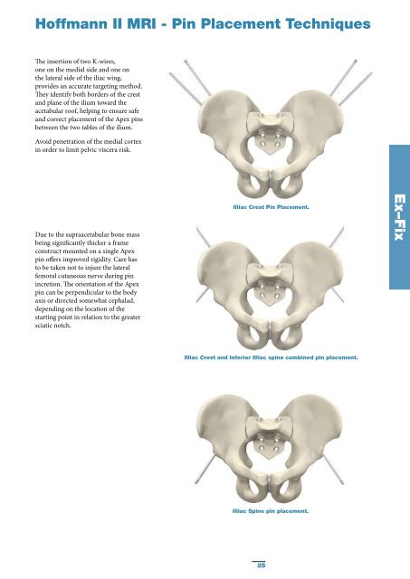
Hoffmann II MRI - Pin Placement Techniques<br />
The insertion of two K-wires,<br />
one on the medial side and one on<br />
the lateral side of the iliac wing,<br />
provides an accurate targeting method.<br />
They identify both borders of the crest<br />
and plane of the ilium toward the<br />
acetabular roof, helping to ensure safe<br />
and correct placement of the Apex pins<br />
between the two tables of the ilium.<br />
Avoid penetration of the medial cortex<br />
in order to limit pelvic viscera risk.<br />
Due to the supraacetabular bone mass<br />
being significantly thicker a frame<br />
construct mounted on a single Apex<br />
pin offers improved rigidity. Care has<br />
to be taken not to injure the lateral<br />
femoral cutaneous nerve during pin<br />
incretion. The orientation of the Apex<br />
pin can be perpendicular to the body<br />
axis or directed somewhat cephalad,<br />
depending on the location of the<br />
starting point in relation to the greater<br />
sciatic notch.<br />
Illiac Crest Pin Placement.<br />
Ex–Fix<br />
Illiac Crest and Inferior Illiac spine combined pin placement.<br />
Illiac Spine pin placement.<br />
25


