address editor in chief co-editors secretary editorial board
address editor in chief co-editors secretary editorial board
address editor in chief co-editors secretary editorial board

You also want an ePaper? Increase the reach of your titles
YUMPU automatically turns print PDFs into web optimized ePapers that Google loves.
JURNALUL PEDIATRULUI – Year XV, X<br />
, Vol. XV, Nr. 59-60<br />
60, , july<br />
j<br />
uly-december 2012<br />
KLIPPEL-TRENAUNAY SYNDROME WITH RARE<br />
ASSOCIATED COMPLICATIONS – A CASE REPORT<br />
Maria-Cor<strong>in</strong>a Stănciulescu 1 , Emanuela Verenca 2 , CM Popoiu 1,2 , ES Boia 1,2 ,<br />
Anca Popoiu 1,2 , VL David 1,2 , Camelia Dăescu 1,2 , Simona Cerbu 1 , Maria Puiu 1,2<br />
Abstract<br />
Klippel-Trenaunay Syndrome (KTS) is a rare<br />
<strong>co</strong>ngenital malformation <strong>co</strong>nsist<strong>in</strong>g of venous, capillary and<br />
lymphatic abnormalities, <strong>in</strong> addition to hypertrophy and<br />
overgrowth of the bony and/or soft tissue of the affected<br />
limbs. We report a unique case of KTS due to its <strong>co</strong>mplex<br />
association of <strong>co</strong>mplications (hydronephrosis, rectorrhagia<br />
and splenomegaly).<br />
Key words: Klippel-Trenaunay Syndrome (KTS),<br />
hydronephrosis, rectorrhagia, splenomegaly<br />
Introduction<br />
Klippel-Trenaunay Syndrome (KTS) was first<br />
described <strong>in</strong> 1900 by physicians Klippel and Trenaunay,<br />
who evidenced the <strong>co</strong>nstellation of three major cl<strong>in</strong>ical<br />
features: capillary, venous and lymphatic vascular<br />
malformations; vari<strong>co</strong>se ve<strong>in</strong>s with an early onset; and bony<br />
and/or soft tissue hypertrophy and overgrowth [1]. One of<br />
the lower limbs is the most frequent <strong>in</strong>volved site and it is<br />
usually hypertrophied [2].<br />
Other <strong>co</strong>mmon features of KTS <strong>in</strong>clude:<br />
hyperhydrosis, sk<strong>in</strong> atrophy, verrucae, dermatitis,<br />
thrombophlebitis, and cellulitis [3]. Deep ve<strong>in</strong> thrombosis,<br />
pulmonary embolism, gram-negative sepsis and<br />
<strong>co</strong>agulopathy are sometimes present and represent lifethreaten<strong>in</strong>g<br />
<strong>co</strong>mplications [4]. While most KTS cases are<br />
sporadic, theories argu<strong>in</strong>g for an autosomal dom<strong>in</strong>ant (AD)<br />
mode of <strong>in</strong>heritance have been formulated. It was noticed<br />
that <strong>in</strong> the case of some affected <strong>in</strong>dividuals, first-degree<br />
relatives presented a high <strong>in</strong>cidence of capillary<br />
malformations and vari<strong>co</strong>se ve<strong>in</strong>s [5]. KTS has been l<strong>in</strong>ked<br />
to two balanced reciprocal translocations, namely<br />
46,XX,t(5;11) (q13.3;p15.1) [6] and 46,XY,t(8;14)(q22.3;<br />
q13) [7]. Moreover, a genetic predisposition for the<br />
development of KTS has been established follow<strong>in</strong>g the<br />
dis<strong>co</strong>very of a susceptibility gene for the syndrome,<br />
specifically the angiogenic factor gene VG5Q, the upregulation<br />
of which results <strong>in</strong> <strong>in</strong>creased angiogenesis [8]. As<br />
these tests are unavailable <strong>in</strong> most cl<strong>in</strong>ics, a diagnosis of<br />
KTS, be it idiopathic or genetic, is usually based on cl<strong>in</strong>ical<br />
signs, ultrasound and imagistic studies.<br />
Case presentation<br />
An 8 year and 7 month old child, first newborn of<br />
healthy, non<strong>co</strong>nsangu<strong>in</strong>eous parents aged 20 and 24 was<br />
referred to us. The patient’s mother had an uneventful<br />
pregnancy, with no history of medication <strong>in</strong>take. However,<br />
both parents worked <strong>in</strong> a toxic environment (car cable<br />
factory) both prior and throughout pregnancy. Family<br />
history is unremarkable. Exam<strong>in</strong>ation of the <strong>in</strong>fant after<br />
birth revealed multiple diffuse cutaneous hemangiomatosis<br />
of the port-w<strong>in</strong>e type (<strong>co</strong>ver<strong>in</strong>g 60% of body surface and<br />
<strong>in</strong>volv<strong>in</strong>g <strong>co</strong>mpletely both legs and the left arm, and part of<br />
the gluteal region, genitalia, trunk and face); edema and<br />
hypertrophy of the above-mentioned limbs; and bilateral<br />
syndactyly of the se<strong>co</strong>nd and third toes. Ultrasound of the<br />
abdomen showed bilateral <strong>co</strong>ngenital hydronephrosis<br />
(stenosis of the vesi<strong>co</strong>ureteral junction on the right and<br />
stenosis of the pielo-ureteral junction on the left).<br />
Chromosomal analysis revealed the 46,XY karyotype.<br />
Over the years, the patient underwent surgery for the<br />
follow<strong>in</strong>g <strong>co</strong>nditions: hydronephrosis (right ureterostomy at<br />
5 months of age followed by ureter reimplantation at 10<br />
months of age; left Hynes-Anderson pyeloplasty at 5 months<br />
of age); left <strong>in</strong>gu<strong>in</strong>al hernia; <strong>co</strong>ngenital verru<strong>co</strong>us lesions of<br />
the endobucal and perianal cavity; and periodontal abscess.<br />
Patient history is significant for the follow<strong>in</strong>g: pneumocystis<br />
pneumonia; rectorrhagia; recurrent bronchiolitis; and<br />
profound venous thrombosis and thrombophlebitis (for<br />
which he is tak<strong>in</strong>g anti<strong>co</strong>agulant treatment).<br />
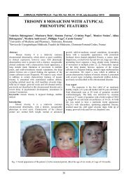
At the moment (Figure 1) the patient presents,<br />
throughout the entire length of the lower extremities, edema<br />
and hypertrophy, with multiple dilated and tortuous venous<br />
vari<strong>co</strong>sities. The left lower extremity is more severely<br />
affected than its <strong>co</strong>unterpart (50 cm versus 43 cm <strong>in</strong> girth at<br />
the level of the hip and 34 cm versus 31 cm at the level of<br />
the thigh). Moreover, the left leg presents an overgrowth <strong>in</strong><br />
length of 4 cm <strong>co</strong>mpared to the right leg; this difference was<br />
2 cm two years ago. The patient’s left upper extremity<br />
presents muscular hypoplasia. Doppler echography<br />
demonstrates normal arterial and venous blood flow all<br />
throughout the affected members; however, there is<br />
destruction of the venous valves and higher blood flow rate<br />
<strong>in</strong> the left leg versus the right leg.<br />
1 Emergency Hospital for Children “Louis Ţurcanu” Timişoara, România<br />
2 University of Medic<strong>in</strong>e and Pharmacy “Victor Babeş” Timişoara, România<br />
E-mail: stanciulescu<strong>co</strong>r<strong>in</strong>a@yahoo.<strong>co</strong>m, emma_ver@yahoo.<strong>co</strong>m, mcpopoiu@yahoo.<strong>co</strong>m, boiaeugen@yahoo.<strong>co</strong>m,<br />
david.vlad@yahoo.<strong>co</strong>m, camidaescu@yahoo.<strong>co</strong>m, cerbusimona@yahoo.<strong>co</strong>m, maria_puiu@umft.ro<br />
10