address editor in chief co-editors secretary editorial board
address editor in chief co-editors secretary editorial board
address editor in chief co-editors secretary editorial board

Create successful ePaper yourself
Turn your PDF publications into a flip-book with our unique Google optimized e-Paper software.
JURNALUL PEDIATRULUI – Year XV, X<br />
, Vol. XV, Nr. 59-60<br />
60, , july<br />
j<br />
uly-december 2012<br />
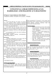
We decide to perform native and <strong>co</strong>ntrast CT<br />
exam<strong>in</strong>ation that dur<strong>in</strong>g early and late stages <strong>in</strong>dicates the<br />
follow<strong>in</strong>g:<br />
- At the pelvis level, anterior to rectum a space-replac<strong>in</strong>g<br />
formation is evidenced, with mixed structure <strong>co</strong>mpris<strong>in</strong>g<br />
lipid densities and <strong>in</strong>cluded calcareous formations, as well<br />
as multiple cystic formations, with moderate absorption of<br />
<strong>co</strong>ntrast medium <strong>in</strong> the parenchyma, without <strong>co</strong>ntrast<br />
medium absorption at the cystic and calcareous <strong>co</strong>mponents<br />
level. The lesion measures 3.5/5 cm <strong>in</strong> maxim axial<br />
diameters, well delimited, <strong>in</strong>volv<strong>in</strong>g the right ovary, but<br />
neatly demarcated of the uter<strong>in</strong>e <strong>co</strong>rpus and anterior rectal<br />
wall, with ur<strong>in</strong>ary bladder <strong>co</strong>mpression and displacement.<br />
- The ur<strong>in</strong>ary bladder presents walls with neat borders,<br />
homogenously opacified after 10 m<strong>in</strong>utes.<br />
- No pathological adenopathies at the retroperitoneal or<br />
pelvic levels are evidenced.<br />
- No free fluid <strong>in</strong> the abdom<strong>in</strong>al pelvic cavity is visualised.<br />
Conclusions of CT Exam<br />
Retrovesical space-replac<strong>in</strong>g formation, <strong>in</strong>volv<strong>in</strong>g the<br />
right ovary with aspect <strong>in</strong>dicat<strong>in</strong>g most probably a dermoid<br />
cyst, present<strong>in</strong>g mixed <strong>co</strong>ntent, lipomatous, liquid and<br />
multiple calcifications, neatly delimited of the uter<strong>in</strong>e<br />
<strong>co</strong>rpus and anterior rectal wall, impress<strong>in</strong>g the ur<strong>in</strong>ary<br />
bladder, without <strong>in</strong>vad<strong>in</strong>g the neighbour<strong>in</strong>g structures<br />
(figure 1).<br />
Fig. 1. CT aspect of cystic teratoma of the right ovary with calcifications.<br />
Treatment<br />
Surgery is performed on January 26, 2010 and<br />
subumbilical m<strong>in</strong>ilaparotomy under general anaesthesia is<br />
carried out. A right ovarian tumour formation, torsioned,<br />
extended <strong>in</strong>to the Pouch of Douglas, with maximum<br />
diameter of approximately 5 cm, relatively round <strong>in</strong> shape,<br />
violaceous <strong>in</strong> <strong>co</strong>lour, with neat and bright surface is<br />
evidenced. Ablation of tumour formation, preventive<br />
appendectomy and dra<strong>in</strong>age of Pouch of Douglas are<br />
performed. No post-operative <strong>co</strong>mplication occurred,<br />
48