FETAL MEMBRANES Placenta Nutrition
FETAL MEMBRANES Placenta Nutrition
FETAL MEMBRANES Placenta Nutrition

You also want an ePaper? Increase the reach of your titles
YUMPU automatically turns print PDFs into web optimized ePapers that Google loves.
<strong>FETAL</strong> <strong>MEMBRANES</strong><br />
<strong>Placenta</strong><br />
<strong>Nutrition</strong><br />
2011
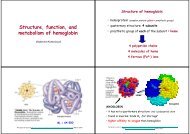
Fetal membranes<br />
<br />
<br />
<br />
<br />
<br />
Chorion<br />
Amnion<br />
Yolk sac<br />
Allantois<br />
They develop from zygote, however they<br />
are not involved in embryo formation<br />
except for small part of yolk sac, that<br />
participate on gut formation
Amnion<br />
<br />
<br />
<br />
<br />
<br />
Amnionic fluid<br />
Amnioblasts,<br />
interstitial fluid form<br />
endometrium<br />
Embryo (before skin<br />
keratinization) –<br />
transudation from<br />
body<br />
Umbilical cord<br />
Respiratory system,<br />
urine<br />
<br />
<br />
10th week – 30ml<br />
20<br />
th<br />
week – 350ml<br />
37<br />
th<br />
week - 700-<br />
1000ml
Function of amniotic fluid<br />
<br />
<br />
<br />
<br />
<br />
<br />
<br />
<br />
It allows symetric growth<br />
Protects from infection<br />
Facilitates normal development of lung<br />
Protects from adhesion<br />
Protects from injury<br />
Keeps stabile temperature<br />
Alows free movement<br />
Takes place in homeostasis (electrolytes)
Failures of amnionic fluid<br />
volume<br />
Oligohydramnion – less than 400ml -<br />
rupture of fetal membranes, renal<br />
agenesis – (Potter syndrome) -<br />
pulmonary hypoplasia, pes equinovarus,<br />
face dysmorfy<br />
<br />
Polyhydramnion – more than 2000 ml –<br />
malformed CNS, esofageal atresia, twins,<br />
idiopatic
Yolk sac<br />
<br />
<br />
<br />
<br />
Transfer and metabolism of nutrients – as<br />
liver<br />
Development of blood cells and vessels<br />
-vitelline vascular system<br />
Participation on formation of primitive<br />
gut<br />
Primordial gamets in endoderm of yolk<br />
sac during 3 rd week
Allantois<br />
<br />
<br />
<br />
Development form hindgut – evagination<br />
into embryonic stalk - only transient<br />
Vessels – umbilical arteries and veins for<br />
placenta supply<br />
Intraembryonic part – urachus, urinary<br />
bladder ( ligamentum umbilicale<br />
medianum)
<strong>Placenta</strong><br />
<br />
<br />
Fetal organ providing nutrition and many<br />
other functions:<br />
Function:<br />
− Metabolism (synthesis, for example glycogen)<br />
− Transport of gases and nutrients<br />
− Excretion of waste products<br />
− Production of hormones (hCG)
Embryo nutrition<br />
<br />
<br />
<br />
<br />
Nutrients supply in yolk sac (AA, lipids)<br />
Histiotrophe – secret from glandular cells<br />
in fallopian tube, uterus, digestion of<br />
endometrium<br />
Haematotrophe – maternal blood<br />
Organ that provide nutrition of embryo in<br />
mammals - chorion/placenta
<strong>Placenta</strong> - structure<br />
<br />
<br />
Fetal part – chorion – chorionic plate and<br />
chorionic villi<br />
Maternal part – endometrium – pars<br />
functionalis – decidua basalis
Decidua<br />
<br />
<br />
Zona functionalis that is changing during<br />
pregnancy<br />
<br />
<br />
<br />
Decidua basalis<br />
Decidua capsularis<br />
Decidua parietalis<br />
Progesteron – cell in stroma (fibroblasts)<br />
are changed in decidual cells (content of<br />
glycogen and lipids)+ changes in vascular<br />
supply = decidual reaction
Implantation
Implantation<br />
<br />
<br />
<br />
During implantation embryo invades in<br />
zona functionalis of endometrium<br />
Trophoblast diferentiates into<br />
syncytiotrophoblast and cytotrophoblast<br />
Extraembryonic mesoderm adds to<br />
cytotrophoblast<br />
<br />
= CHORION
Development of chorionic villi<br />
<br />
<br />
<br />
<br />
Primary: Syncytiotrophoblast and<br />
cytotrophoblast<br />
Secondary: Syncytiotrophoblast,<br />
cytotrophoblast and extraembryonic<br />
mesoderm<br />
Tertiary: Vessels occur in mesoderm<br />
Terctiary villi are all from 3<br />
rd<br />
week of<br />
development
<strong>Placenta</strong> development<br />
<br />
<br />
<br />
Chorion laeve<br />
Chorion frondosum<br />
Decidua capsularis get thiner, later<br />
disappears, chorion laeve is on the<br />
surface, it unites with decidua parietalis<br />
and obliterates uterine cavity ( week 22<br />
-24)
Intervillous space<br />
<br />
<br />
It develops from lacunae in sytiotrophoblast<br />
It is divided by placental septa<br />
<br />
Maternal blood – spiral arteries in decidua basalis –<br />
uteroplacental vessels<br />
<br />
<br />
It wash up villi – it is drained into placental veins. Fetal<br />
and maternal blood do not mix !!!<br />
Hemocytoblasts (stem cells) may cross from embryonic<br />
to maternal blood and stay there for relatively long time<br />
(several years) -chimera
<strong>Placenta</strong>l circulation<br />
<br />
<br />
<br />
Umbilical arteries<br />
-deoxygenated<br />
blood from<br />
embryonic body<br />
Chorionic arteries<br />
branching in<br />
chorionic plate<br />
Capillary network<br />
in chorionic villi
<strong>Placenta</strong>l membrana<br />
<br />
<br />
Interface between maternal and fetal<br />
blood<br />
• Syncytiotrophoblast<br />
• Cytotrophoblast<br />
• Connective tissue<br />
• Endothelium of fetal capillary<br />
After week 12 cytotrophoblast gradually<br />
disappears, vessels come near to surface<br />
and get in contact with<br />
syncytiotrophoblast
3rd trimester<br />
<br />
<br />
Nuclei of<br />
syncytiotrophoblast<br />
form aggregations –<br />
syncytial knots that<br />
may set free<br />
Formation of<br />
fibrinoid – it reduces<br />
placental transfer
Syncytiotrophoblast<br />
– microvilli, SER,<br />
RER, GC, mito –<br />
active synthesis<br />
Cytotrophoblast –<br />
undifferentiated<br />
cells – mitoses<br />
<br />
<br />
Basal membrane<br />
Continuous<br />
endothelium
Feto-maternal junction<br />
<br />
Different genotype – necessity to supress<br />
imunity:<br />
<br />
Maternal and fetal tissue are separted by the<br />
cells that do not have typical superficial<br />
antigens. Hormonal changes (progesteron,<br />
glucocorticods)<br />
– Blood - Syncytiotrophoblast<br />
– Connective tissue – Cytotrophoblastic shell<br />
<br />
Stem - anchoring villi attach chorion to the<br />
decidua basalis – inside cytotrophoblastic<br />
plug
<strong>Placenta</strong><br />
<strong>Placenta</strong>l shape – discoid (olliformis) +<br />
<br />
<br />
<br />
haemochorial<br />
<strong>Placenta</strong>l septa – rests of decidua basalis.<br />
They separate placenta from maternal<br />
side in lobes - cotyledons<br />
Cotyledons – contain 2 and more<br />
anchoring villi<br />
Diameter – 15 -20 cm, thickness 2-3 cm,<br />
weight 500 to 600 g
<strong>Placenta</strong>l transfer<br />
<br />
<br />
<br />
<br />
<br />
Difusion<br />
Facilitated difusion<br />
Active transport<br />
Pinocytosis<br />
Other types of transfer:<br />
<br />
<br />
<br />
Damage of placetal barrier – blood cellas<br />
Own activity – Treponema pallidum<br />
Damage due to infection - toxoplasmosa
Transfer<br />
<br />
<br />
<br />
<br />
Many substances from maternal blood<br />
may transfer placental barrier including<br />
drugs<br />
Nutrients – glucose, AK, fatty acids,<br />
water, vitamines, electrolytes<br />
Hormones – only steroid unconjugates<br />
Maternal antibodies, transferin+ iron
Syntesis<br />
<br />
<br />
<br />
<br />
<br />
hCG – human chorionic gonadotropin<br />
hCS – human chorionic somatomammotropin/placental<br />
lactogen<br />
hCT human chorionic thyrotropin<br />
hCACTH human chorionic corticotropin<br />
Progesteron and estrogens
<strong>Placenta</strong>l abnormalities<br />
<br />
<br />
<br />
<br />
<br />
<br />
<br />
Atypical implatation:<br />
<strong>Placenta</strong> previa<br />
<strong>Placenta</strong> accreta<br />
<strong>Placenta</strong> percreta<br />
<strong>Placenta</strong> membranacea<br />
<strong>Placenta</strong> accessoria<br />
Atypical attachment of umbilical cord--<br />
marginal, velamentous
Development of mbilical cord<br />
<br />
<br />
<br />
<br />
Connective stalk with umbilical arteries<br />
and veins and allantois<br />
Ductus omphaloentericus connecting gut<br />
with yolk sac<br />
Extraembryonic coelom<br />
Umbilicus
Development<br />
of umbilical<br />
cord<br />
• Length 50 cm