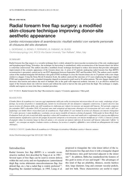
Radial forearm free flap surgery: a modified skin-closure technique ...
Radial forearm free flap surgery: a modified skin-closure technique ...
Radial forearm free flap surgery: a modified skin-closure technique ...

Create successful ePaper yourself
Turn your PDF publications into a flip-book with our unique Google optimized e-Paper software.
ACTA otorhinolaryngologica italica 2012;32:158-163<br />
Head and neck<br />
<strong>Radial</strong> <strong>forearm</strong> <strong>free</strong> <strong>flap</strong> <strong>surgery</strong>: a <strong>modified</strong><br />
<strong>skin</strong>-<strong>closure</strong> <strong>technique</strong> improving donor-site<br />
aesthetic appearance<br />
Lembo microvascolare di avambraccio: risultati estetici con variante personale<br />
di chiusura del sito donatore<br />
L. Giordano, S. Bondi, F. Ferrario, B. Fabiano, M. Bussi<br />
Otorhinolaryngology Unit, IRCCS Vita-Salute University “San Raffaele”, Milan, Italy<br />
Summary<br />
<strong>Radial</strong> <strong>forearm</strong> <strong>free</strong> <strong>flap</strong> <strong>surgery</strong> is a versatile <strong>technique</strong> that is widely adopted for microvascular reconstruction of the oral, oropharyngeal<br />
and hypopharyngeal lining. Nowadays, the <strong>technique</strong> for harvesting is standardized, while reconstruction of the <strong>forearm</strong> donor site defect<br />
is somewhat controversial. The authors describe a <strong>modified</strong> <strong>closure</strong> <strong>technique</strong> developed to reduce <strong>skin</strong> tension that provides subsequent<br />
improvement of the cosmetic appearance of the <strong>forearm</strong> donor site. A series of 43 patients undergoing radial <strong>forearm</strong> <strong>free</strong> <strong>flap</strong> (RFFF)<br />
reconstruction is presented, carried out by our ENT department between September 2007 and December 2010. The authors used a modification<br />
of the standard triangular full-thickness <strong>skin</strong> graft (FTSG) <strong>technique</strong> to close the <strong>forearm</strong> donor site on 23 patients with a new shape<br />
similar to a dagger. Using the Stony Brook Evaluation Scale, the authors analyzed the outcomes of 23 cases employing the dagger-shaped<br />
FTSG and compared these with a standard (triangular shaped) reconstructive graft used in 20 earlier patients. The new dagger-shaped graft<br />
decreases <strong>skin</strong> tension and reduces the need of multiple slits in the graft with improved aesthetic outcome; it is an effective method for<br />
repair of the <strong>forearm</strong> donor site with low tension and without the need to harvest the <strong>skin</strong> graft from the thigh. The <strong>technique</strong> is simple,<br />
reliable and requires no more time than a standard procedure.<br />
Key words: <strong>Radial</strong> <strong>forearm</strong> <strong>free</strong> <strong>flap</strong> • Reconstructive <strong>surgery</strong> • Cosmetic appearance • Skin graft<br />
Riassunto<br />
Il lembo libero di avambraccio è ancora oggi ampiamente utilizzato nella ricostruzione microvascolare di cavo orale, orofaringe ed ipofaringe.<br />
La tecnica di prelievo è standardizzata, mentre la ricostruzione del sito donatore è alquanto controversa. L’autore descrive una<br />
nuova tecnica di chiusura della cute dell’avambraccio elaborata per ridurre la tensione sui margini dell’innesto. Nel periodo compreso<br />
fra settembre 2007 e dicembre 2010, 43 pazienti sono stati sottoposti ad intervento chirurgico di ricostruzione del cavo orale e dell’oroipofaringe<br />
con lembo libero rivascolarizzato di avambraccio. Negli ultimi 23 casi trattati la tecnica di chiusura con innesto triangolare<br />
V-Y a tutto spessore è stata modificata e sostituita con un nuovo modello di innesto a forma di pugnale. Utilizzando la Stony Brook Scar<br />
Evaluation Scale gli esiti cicatriziali della superficie volare dell’avambraccio sono stati analizzati e confrontati ed è emersa una differenza<br />
statisticamente significativa a favore del gruppo di pazienti sottoposti a ricostruzione con innesto modificato (p < 0,01). L’innesto a forma<br />
di pugnale riduce la tensione lungo la linea di sutura e ciò consente un numero inferiore di incisioni sull’innesto riducendo l’effetto “a<br />
maglie”; ne consegue un miglior risultato estetico a distanza. La tecnica è semplice, affidabile e non richiede un allungamento dei tempi<br />
chirurgici.<br />
parole chiave: Lembo libero di avambraccio • Chirurgia ricostruttiva • Risultato estetico • Innesto cutaneo<br />
Acta Otorhinolaryngol Ital 2012;32:158-163<br />
Introduction<br />
<strong>Radial</strong> <strong>forearm</strong> <strong>free</strong> <strong>flap</strong> (RFFF) is a versatile reconstructive<br />
method, first described by Yang et al. in 1981 1 .<br />
Several types of <strong>free</strong> <strong>flap</strong>s can be used to restore defects<br />
within the oral cavity and oro-hypopharynx (anterolateral<br />
thigh <strong>flap</strong>, rectus abdominis musculocutaneous <strong>flap</strong>, <strong>free</strong><br />
deltoid <strong>flap</strong>, etc.), but a radial <strong>flap</strong> is widely preferred 2-5 .<br />
Regarding the standard <strong>technique</strong>, in 1994, Liang et al.<br />
proposed to triangulate the volar donor defect of the radial<br />
<strong>forearm</strong> <strong>free</strong> <strong>flap</strong> and close it with a triangular shaped<br />
full-thickness <strong>skin</strong> graft harvested adjacent to the donor<br />
site 6 7 . In addition, Shiba has standardized the features of<br />
this graft in terms of size and length 8 . The advantages of<br />
this kind of <strong>flap</strong> are pliability, thinness, mostly hairless<br />
<strong>skin</strong>, large diameter, long and constant vascular pedicle,<br />
and low bulk. These characteristics are useful to repair<br />
158
<strong>Radial</strong> <strong>forearm</strong> <strong>free</strong> <strong>flap</strong> donor site outcomes with a new <strong>closure</strong> <strong>technique</strong><br />
complex three-dimensional defects, for example, small<br />
intra-oral tissue defects 3 5 . Many <strong>skin</strong> <strong>closure</strong> methods<br />
have been developed to improve the aesthetic outcome of<br />
the <strong>forearm</strong> donor site but, at present, this topic is somewhat<br />
controversial. Some surgeons prefer to harvest a split<br />
thickness <strong>skin</strong> graft from the thigh or the groin for donor<br />
site <strong>closure</strong>, but this procedure may be associated with<br />
morbidities including pain, infection and hypertrophic<br />
scar formation.<br />
The authors describe a modification of the standard triangular-shaped<br />
<strong>technique</strong>, performed in 23 cases using <strong>forearm</strong><br />
<strong>free</strong> <strong>flap</strong> 3-dimensional microvascular reconstruction,<br />
in order to improve cosmetic appearance by lowering <strong>skin</strong><br />
graft tension. The aesthetic outcomes of these 23 patients<br />
were analyzed using the Stony Brook Scar Evaluation<br />
Scale and compared with 20 other patients who underwent<br />
a standard V-Y <strong>closure</strong> <strong>technique</strong>.<br />
Materials and methods<br />
In the period between September 2007 and December<br />
2010, 43 patients underwent RFFF reconstruction for<br />
a variety of head and neck surgical defects due to squamocellular<br />
carcinoma ablation at the Department of<br />
Otorhinolaryngology, “San Raffaele” Hospital, Milan.<br />
This study conformed to the ethical standards according<br />
to the Declaration of Helsinki published in 1964 in its<br />
present version. All patients were assessed preoperatively<br />
with the Allen test and colour Doppler ultrasonography<br />
to investigate collateral circulation (ulnar artery and<br />
vein) of the <strong>forearm</strong> and neck. Each RFFF was harvested<br />
in a similar fashion with <strong>forearm</strong> fascia, radial artery,<br />
cephalic vein and vena comitans; none of the patients<br />
had a radial osteocutaneous <strong>free</strong> <strong>flap</strong>. In total, 35 <strong>flap</strong>s<br />
were harvested from the left and 8 from the right <strong>forearm</strong><br />
based on hand dominance and vascular test results.<br />
The <strong>flap</strong>s were elevated with a pneumatic tourniquet inflated<br />
to 250 mmHg for a maximum of 90 min; the <strong>flap</strong>s<br />
ranged in size from 8 x 8 cm to 5 x 5 cm with an average<br />
surface area of 38.4 cm 2 . In all patients, we used<br />
the V-Y <strong>technique</strong> with a <strong>skin</strong> graft whose height was<br />
twice the RFFF length in the direction of the <strong>forearm</strong><br />
axis. According to Shiba’s statement, the width of the<br />
graft was shorter than half of the wrist circumference.<br />
The full thickness <strong>skin</strong> graft (FTSG) was finally elevated<br />
with a scalpel, defatted using a scissors and then stored<br />
in saline. Multiple slits in the graft were made to prevent<br />
fluid collection beneath the graft and to allow the graft<br />
to be stretched to cover a larger area. The dressing was<br />
maintained for about 6 days. The <strong>forearm</strong> donor site <strong>skin</strong><br />
defect consisted of the defect attributable to both RFFF<br />
and FTSG harvesting; the distal side of the defect area<br />
was covered with the FTSG, while the proximal side of<br />
the defect area was primarily closed. In 20 consecutive<br />
patients, a standard FTSG shaped like an isosceles triangle<br />
was adopted for <strong>skin</strong> <strong>closure</strong>, while in the other 23<br />
patients, reconstruction was performed with a new model<br />
of FTSG, shaped like a dagger. As shown in Figure 1,<br />
the RFFF is usually drawn with rounded edges and as a<br />
consequence, two triangles (triangles between RFFF and<br />
FTSG) are usually sacrificed when the pyramidal FTSG<br />
is translated to close the total donor site defect (Fig. 1).<br />
Accordingly, the authors devised a new dagger-shaped<br />
FTSG with a particular perimeter that matches the <strong>skin</strong><br />
defect of the donor site by reducing the surface tension<br />
and preserving the two triangles of <strong>skin</strong> (Fig. 2).<br />
As shown in the following demonstration, this new shape<br />
can be approximated to a geometric construction whose<br />
area is given by the sum of six different polygons. Comparison<br />
of this new shape with the previous FTSG showed<br />
a savings of 8.3% of the <strong>skin</strong> graft.<br />
Demonstration:<br />
159
L. Giordano et al.<br />
graft due to excessive edge traction. Specifically, the larger<br />
the <strong>skin</strong> defect, the more difficult the <strong>closure</strong>. The last 23<br />
cases were treated with the new dagger-shaped <strong>skin</strong> graft<br />
in a proportion of 2:3, between FTSG and total <strong>skin</strong> defect,<br />
which was always respected; these defects presented<br />
better outcomes with no tendon exposure (30% vs. 0%)<br />
and no infections recorded (25% vs. 0%). Similarly, hand<br />
swelling (55% vs. 13%) and wrist stiffness (40% vs. 17%)<br />
decreased in this group of patients. Comparison between<br />
standard and dagger-shaped grafts showed a statistical<br />
difference for all features analysed except wrist stiffness<br />
(chi-square test, Fisher’s exact test, p < 0.01) (Table I).<br />
Moreover, an analysis of aesthetic outcomes, in terms of<br />
colour thickness and width of the wound measured with<br />
the Stony Brook Scar Evaluation Scale, showed a statistical<br />
difference between groups (Student’s t-test, p < 0.01).<br />
In the first group (with standard FTSG; Fig. 3), the mean<br />
value among the 20 patients was 2.05, while in the second<br />
group of 23 patients (with dagger-shaped FTSG; Fig. 4), it<br />
was 3.56, which corresponds to a better aesthetic outcome<br />
(range: 0, worst to 5, best).<br />
A minimum post-operative time period of 3 months was<br />
required before collecting aesthetic outcome data. The<br />
quality of scars was evaluated with the Stony Brook Scar<br />
Evaluation Scale. This scale, proposed in 2007 by Singer<br />
et al. 9 , measures cosmetic outcome of the wound through<br />
its width, height, colour, suture mark and overall appearance.<br />
Objective, blinded evaluation of aesthetic outcome<br />
of <strong>forearm</strong> reconstructions was done by two head and<br />
neck surgeons from our department. High-resolution<br />
digital photographs of the operative site were taken in a<br />
standardized setting on a sky-blue background. The final<br />
score for cosmetic outcome ranged from 0 (worst) to 5<br />
(best). The median values of the two subgroups (20 standard<br />
FTSG vs. 23 dagger-shaped FTSG) were compared in<br />
terms of aesthetic results. Moreover, four other features<br />
of scar healing (i.e. hand swelling, tendon exposure, rate<br />
of graft infection and wrist stiffness) were evaluated during<br />
follow up (after 3-6 months). Statistical analysis was<br />
performed with Minitab Statistical software; all p-values<br />
were two-sided and p-values < 0.01 were considered significant.<br />
Results<br />
In terms of reconstructive <strong>surgery</strong> procedures, there were<br />
no differences between the two methods of donor site <strong>skin</strong><br />
<strong>closure</strong> after RFFF harvesting (20 standard FTSG vs. 23<br />
dagger-shaped FTSG). In the 20 cases performed before<br />
the application of the new <strong>skin</strong> <strong>closure</strong> method, we noticed<br />
excessive suture tension in the middle portion of the<br />
<strong>forearm</strong> with some cases of partial detachment of the <strong>skin</strong><br />
Discussion<br />
Donor site complications, reported in the literature by several<br />
authors, can be divided into functional and aesthetic<br />
problems. The former are caused by <strong>skin</strong> graft healing in<br />
19-53%, hand swelling in 60%, persistent wrist stiffness<br />
in 27%, impaired hand sensation in 17-47%, cold intolerance<br />
in 8-40%, reduced hand strength in 40% and partial<br />
loss of the graft with exposure of the <strong>forearm</strong> flexor tendon<br />
in 23-55% 5 10-13 . Tendon exposure can be caused by<br />
the creation of disruptive <strong>skin</strong> forces under the graft, due<br />
to the continuous movement of the <strong>forearm</strong> tendon 14 . Aesthetic<br />
complications reported include scar infection, <strong>skin</strong><br />
tension and hypertrophic scar formation, causing patient<br />
dissatisfaction with the aesthetic outcome of the donor site<br />
(17-50%) 15 . Comorbidities, such as malignancy, tobacco<br />
and alcohol abuse, poor nutrition, diabetes and peripheral<br />
vascular disease are common in this patient population<br />
and may affect wound healing 14 .<br />
Various methods of reconstruction have been developed<br />
to minimize functional and aesthetic morbidity associated<br />
with RFFF donor site <strong>closure</strong>. The <strong>technique</strong>s include split<br />
thickness <strong>skin</strong> graft (STSG), FTSG and STSG overlying<br />
an acellular dermal matrix, although we usually prefer<br />
FTSG. Initially in our experience, the <strong>forearm</strong> donor site<br />
was repaired with a standard isosceles triangle of FTSG<br />
as described by Liang, and the technical modification proposed<br />
by Van Der Lei and Shiba; in detail, we used the<br />
V-Y <strong>technique</strong> with a <strong>skin</strong> graft shaped like an isosceles<br />
triangle whose height was twice the RFFF length in the<br />
direction of the <strong>forearm</strong> axis, and whose width was shorter<br />
than half of the <strong>forearm</strong> circumference 8 . However, in<br />
some cases of partial detachment of the <strong>skin</strong> graft due to<br />
160
<strong>Radial</strong> <strong>forearm</strong> <strong>free</strong> <strong>flap</strong> donor site outcomes with a new <strong>closure</strong> <strong>technique</strong><br />
Fig. 1. <strong>Radial</strong> <strong>forearm</strong> <strong>flap</strong> (RFFF) with standard full thickness <strong>skin</strong> graft<br />
(FTSG): triangles between RFFF and FTSG are usually sacrificed because the<br />
<strong>flap</strong> is drawn with round edges.<br />
Fig. 3. Aesthetic outcome with a standard <strong>technique</strong>.<br />
Fig. 2. Full thickness <strong>skin</strong> graft with the new shape that preserves more<br />
<strong>skin</strong> (dark grey triangles) compared to the standard <strong>technique</strong>.<br />
excessive edge traction was observed. Specifically in the<br />
middle portion of the <strong>forearm</strong> border, there was excessive<br />
suture tension due to the fact that a triangle had to replace<br />
a rectangular space. Thus, a new shape was proposed. We<br />
analysed our previous failures and developed a daggershaped<br />
FTSG that reduced border suture tension by creating<br />
a precise correspondence between the FTSG and the<br />
total donor <strong>skin</strong> defect that assumed the same geometric<br />
shape and saved 8.3% of the <strong>skin</strong> graft. It is extremely<br />
important to place the first seven stitches accurately as<br />
shown in Figure 5.<br />
Using the Stony Brook Scar Evaluation Scale, we compared<br />
the aesthetic outcomes of the 23 cases who received<br />
the dagger-shaped graft with a group of 20 patients who<br />
underwent a standard FTSG <strong>closure</strong> <strong>technique</strong>. This new<br />
scale of cosmetic appearance is composed of five dichotomous<br />
categories that include width (> 2 mm), relief/flatness<br />
of scar, colour (darker or the same as surrounding <strong>skin</strong>),<br />
presence of suture marks and overall appearance of the<br />
Fig. 4. Aesthetic outcome with the dagger-shaped graft.<br />
scar. The total score is derived by adding the scores (0 or<br />
1) for the presence or absence of these five items. The scar<br />
evaluation scale ranges from 0 (worst) to 5 (best). Using<br />
the Stony Brook Scale, the group with the dagger-shaped<br />
FTSG showed higher overall values because the lesser<br />
traction on the graft margins gave a reduction in the width<br />
of the wound, a good colour match with the surrounding<br />
<strong>skin</strong> and a flat scar. Unfortunately, suture marks remained<br />
unchanged in both groups, while the lesser tension on the<br />
edges allowed a lower number of multiple slits cut into the<br />
Table I. Comparison of outcomes between standard graft and dagger-shaped graft groups.<br />
Standard shaped Dagger-shaped p-value<br />
Number of patients 20 23<br />
Hand swelling 55% 13% < 0.01 *<br />
Persistent wrist stiffness 40% 17% 0.099 *<br />
Scar infection (<strong>skin</strong> graft healing) 30% 0% < 0.01 †<br />
Tendon exposure 25% 0% < 0.01 †<br />
Aesthetic outcome (mean value with Stony Brook Scar Evaluation Scale) 2.05 ± 0.83 3.56 ± 0.79 < 0.01 ‡<br />
* Chi-square test; † Fisher’s exact test; ‡ Student’s t-test<br />
161
L. Giordano et al.<br />
A<br />
Fig. 6. Template of the dagger-shaped FTSG.<br />
should be drawn with larger dimensions. The authors usually<br />
prefer a RFFF with a side of 7 cm.<br />
B<br />
C<br />
Fig. 5. (a) Design of the <strong>flap</strong> with the dagger-shaped FTSG; (b) Preparation<br />
of the graft after placement of the first seven stitches; (c) Outcome and comparison<br />
with the pre-operative template.<br />
graft. This latter feature decreased the ‘meshed effect’ of<br />
the graft, and accordingly, the overall appearance improved.<br />
In detail, the statistical analysis of the two groups showed<br />
a significant mean difference (3.56 vs. 2.05; p < 0.01) in<br />
favour of the dagger-shaped graft group. Statistical analysis<br />
of other features such as hand swelling, tendon exposure<br />
and scar infection in <strong>skin</strong> graft healing also showed a clear<br />
improvement in the dagger group (p < 0.01). This is an<br />
empirical shape derived from surgical experience. Figure 6<br />
shows the graft template with a side of 2.5 cm, although it<br />
Conclusions<br />
Our <strong>modified</strong> V-Y <strong>closure</strong> method involves the use of a<br />
dagger-shaped FTSG for closing the radial <strong>forearm</strong> <strong>free</strong><br />
<strong>flap</strong> donor site with fewer slits in the graft, less complications,<br />
better aesthetic results and, consequently, higher<br />
patient satisfaction. The <strong>technique</strong> is recommended for<br />
defects smaller than 50 cm 2 and is a simple and cost-effective<br />
method that avoids harvesting <strong>skin</strong> grafts from the<br />
abdomen or thigh. This change in surgical <strong>technique</strong> does<br />
not lengthen surgical time. However, when the <strong>forearm</strong><br />
<strong>flap</strong> width is half of the wrist circumference or larger, a<br />
<strong>skin</strong> graft from a second donor site is mandatory to cover<br />
the RFFF defect.<br />
References<br />
1<br />
Yang GF, Chen PJ, Gao YZ, et al. Forearm <strong>free</strong> <strong>skin</strong> <strong>flap</strong><br />
transplantation: a report of 56 cases, 1981. Br J Plast Surg<br />
1997;50:162-5.<br />
2<br />
Gonzalez-Garcia R, Naval-Gias L, Rodriguez-Campo F,<br />
et al. Reconstruction of oromandibular defects by vascularized<br />
<strong>free</strong> <strong>flap</strong>s: the radial <strong>forearm</strong> <strong>free</strong> <strong>flap</strong> and fibular<br />
<strong>free</strong> <strong>flap</strong> as major donor sites. J Oral Maxillofac Surg<br />
2009;67:1473-7.<br />
3<br />
Karimi A, Mahy P, Reychler H. Closure of radial <strong>forearm</strong><br />
<strong>free</strong> <strong>flap</strong> donor site defect with a local meshed full-thickness<br />
<strong>skin</strong> graft: a retrospective study of an original <strong>technique</strong>. J<br />
Cranio-Maxillofac Surg 2007;35:369-73.<br />
4<br />
Moazzam A, Gordon DJ. Cross-suturing as an aid to<br />
wound <strong>closure</strong>: a prospective randomised trial using the<br />
<strong>forearm</strong> <strong>flap</strong> donor site as a model. Br Ass Plast Surg<br />
2003;56:695-700.<br />
5<br />
Ho T, Couch M, Carson K, et al. <strong>Radial</strong> <strong>forearm</strong> <strong>free</strong> <strong>flap</strong><br />
donor site outcomes comparison by <strong>closure</strong> methods. Otolar<br />
Head Neck Surg 2006;134:309-15.<br />
6<br />
Liang MD, Swartz WM, Jones NF. Local full-thickness <strong>skin</strong>graft<br />
coverage for the radial <strong>forearm</strong> <strong>flap</strong> donor site. Plast<br />
Reconstr Surg 1994;93:621-5.<br />
7<br />
Van Der Lei B, Spronk CA, De Visscher JGAM. Closure of<br />
radial <strong>forearm</strong> <strong>free</strong> <strong>flap</strong> donor site with local full-thickness<br />
<strong>skin</strong> graft. Br J Oral Maxillofac Surg 1999;37:119-22.<br />
8<br />
Shiba K, Iida Y, Numata T. Ipsilateral full-thickness <strong>forearm</strong><br />
<strong>skin</strong> graft for covering the radial <strong>forearm</strong> <strong>flap</strong> donor site.<br />
Laryngoscope 2003;113:1043-6.<br />
162
<strong>Radial</strong> <strong>forearm</strong> <strong>free</strong> <strong>flap</strong> donor site outcomes with a new <strong>closure</strong> <strong>technique</strong><br />
9<br />
Singer AJ, Blavantray A, Dagum A, et al. Development and<br />
validation of a novel scar evaluation scale. Plast Reconstr<br />
Surg 2007;120:1892-7.<br />
10<br />
Gonzalez-Garcia R, Ruiz-Laza L, Manzano D, et al. Combined<br />
local triangular full-thickness <strong>skin</strong> graft for the <strong>closure</strong><br />
of the radial <strong>forearm</strong> <strong>free</strong> <strong>flap</strong> donor site: a new <strong>technique</strong>. J<br />
Oral Maxillofac Surg 2009;67:1562-7.<br />
11<br />
Avery C, Pereira J, Brown AE. Suprafascial dissection of the<br />
radial <strong>forearm</strong> <strong>flap</strong> and donor site morbidity. Int J Oral Maxillofac<br />
Surg 2001;30:37-41.<br />
12<br />
Brown M, Couch M, Huchton D. Assessment of donor-site<br />
functional morbidity from radial <strong>forearm</strong> fasciocutaneous<br />
<strong>free</strong> <strong>flap</strong> harvest. Arch Otolaryngol Head Neck Surg<br />
1999;125:1371-4.<br />
13<br />
Chang S, Miller G, Halbert C, et al. Limiting donor site morbidity<br />
by suprafascial dissection of the radial <strong>forearm</strong> <strong>flap</strong>.<br />
Micro<strong>surgery</strong> 1996;17:136-40.<br />
14<br />
Andrews B, Smith R, Chang C, et al. Management of the radial<br />
<strong>forearm</strong> <strong>free</strong> <strong>flap</strong> donor site with the vacuum-assisted <strong>closure</strong><br />
(VAC) system. Laryngoscope 2006;116:1918-22.<br />
15<br />
Toschka H, Feifel H, Erli HJ, et al. Aesthetic and functional<br />
results of harvesting radial <strong>forearm</strong> <strong>flap</strong>, especially<br />
with regard to hand function. Int J Oral Maxillofac Surg<br />
2001;30:42-8.<br />
Received: November 22, 2011 - Accepted: January 26, 2012<br />
Address for correspondence: Leone Giordano, Otorhinolaryngology<br />
Unit, IRCCS Vita-Salute University “San Raffaele”, via Olgettina<br />
60, 20132 Milan, Italy. Tel. +39 02 26433526. Fax +39 02<br />
26433508. E-mail: giordano.leone@hsr.it<br />
163