Early depictions of the nasal turbinates in the 15th century ...
Early depictions of the nasal turbinates in the 15th century ...
Early depictions of the nasal turbinates in the 15th century ...

You also want an ePaper? Increase the reach of your titles
YUMPU automatically turns print PDFs into web optimized ePapers that Google loves.
HISTORICAL PAGE<br />
Rh<strong>in</strong>ology 40,168-170,2002<br />
<strong>Early</strong> <strong>depictions</strong> <strong>of</strong> <strong>the</strong> <strong>nasal</strong> <strong>turb<strong>in</strong>ates</strong> <strong>in</strong> <strong>the</strong><br />
<strong>15th</strong> <strong>century</strong>*<br />
Wolfgang Pirsig<br />
Ulm, Germany<br />
SUMMARY<br />
Although <strong>nasal</strong> <strong>turb<strong>in</strong>ates</strong> had already been described by Hippocrates, it was not until <strong>the</strong> <strong>15th</strong><br />
<strong>century</strong> that <strong>the</strong>y were depicted. The <strong>in</strong>ferior turb<strong>in</strong>ate was shown for <strong>the</strong> first time ra<strong>the</strong>r true-to<br />
nature <strong>in</strong> <strong>the</strong> works <strong>of</strong> <strong>the</strong> Middle Rhenic Master circa 1450-1460 and Leonardo da V<strong>in</strong>ci <strong>in</strong><br />
1489. The posterior ends <strong>of</strong> <strong>the</strong> middle <strong>turb<strong>in</strong>ates</strong> were depicted on a woodcut by Georg Thomas<br />
for Dryander’s “Anatomiae…pars prior” <strong>in</strong> 1536. These and a few o<strong>the</strong>r examples show that<br />
some artists were ahead <strong>of</strong> <strong>the</strong> medical pr<strong>of</strong>ession <strong>in</strong> demonstrat<strong>in</strong>g anatomical details <strong>in</strong> <strong>the</strong><br />
<strong>15th</strong> and 16th <strong>century</strong>.<br />
Key words: <strong>nasal</strong> turb<strong>in</strong>ate, history, art, pa<strong>in</strong>ter<br />
Although <strong>nasal</strong> <strong>turb<strong>in</strong>ates</strong> had been observed by Hippocrates<br />
long before <strong>the</strong> Renaissance, by <strong>the</strong> end <strong>of</strong> <strong>the</strong> <strong>15th</strong> <strong>century</strong><br />
<strong>the</strong>re was still no medical illustration depict<strong>in</strong>g <strong>nasal</strong><br />
<strong>turb<strong>in</strong>ates</strong>, ei<strong>the</strong>r totally or partially. By that time, however,<br />
<strong>the</strong>se structures had been depicted <strong>in</strong> <strong>the</strong> work <strong>of</strong> two pa<strong>in</strong>ters,<br />
<strong>the</strong> Middle Rhenic Master and Leonardo da V<strong>in</strong>ci.<br />
An oil pa<strong>in</strong>t<strong>in</strong>g on a wooden panel (137 x 108 cm) <strong>in</strong> <strong>the</strong> collection<br />
<strong>of</strong> <strong>the</strong> Institute <strong>of</strong> Arts “Das Städel” <strong>in</strong> Frankfurt/Ma<strong>in</strong><br />
shows <strong>the</strong> Crucifixion with Mary, John <strong>the</strong> Baptist, and four<br />
angels on a gold ground (Anonymous, 1987). It was pa<strong>in</strong>ted<br />
around 1450/1460 by <strong>the</strong> Middle Rhenic Master for <strong>the</strong> chapel<br />
<strong>of</strong> <strong>the</strong> orphanage <strong>in</strong> Frankfurt/M. Ly<strong>in</strong>g between Mary’s shoes<br />
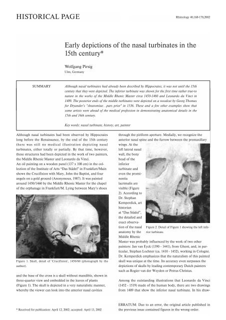
Figure 1. Skull, detail <strong>of</strong> 'Crucifixion', 1450/60 (photograph by <strong>the</strong><br />
author).<br />
and <strong>the</strong> base <strong>of</strong> <strong>the</strong> cross is a skull without mandible, shown <strong>in</strong><br />
three-quarter view and embedded <strong>in</strong> <strong>the</strong> leaves <strong>of</strong> plants<br />
(Figure 1). The skull is depicted <strong>in</strong> a very naturalistic manner,<br />
whereby <strong>the</strong> viewer can look <strong>in</strong>to <strong>the</strong> anterior <strong>nasal</strong> cavities<br />
through <strong>the</strong> piriform aperture. Medially, we recognize <strong>the</strong><br />
anterior <strong>nasal</strong> sp<strong>in</strong>e and <strong>the</strong> furrow between <strong>the</strong> premaxillary<br />
w<strong>in</strong>gs. At <strong>the</strong><br />
left lateral <strong>nasal</strong><br />
wall, <strong>the</strong> bony<br />
head <strong>of</strong> <strong>the</strong><br />
<strong>in</strong>ferior<br />
turb<strong>in</strong>ate and<br />
even <strong>the</strong> prom<strong>in</strong>entia<br />
lacrimalis are<br />
visible (Figure<br />
2). Accord<strong>in</strong>g to<br />
Dr. Stephan<br />
Kemperdick, art<br />
historian<br />
at “Das Städel”,<br />
<strong>the</strong> detailed and<br />
exact observation<br />
<strong>of</strong> <strong>the</strong> <strong>nasal</strong> Figure 2. Detail <strong>of</strong> Figure 1 show<strong>in</strong>g <strong>the</strong> left <strong>in</strong>ferior<br />
turb<strong>in</strong>ate.<br />
anatomy by <strong>the</strong><br />
Middle Rhenic<br />
Master was probably <strong>in</strong>fluenced by <strong>the</strong> work <strong>of</strong> two o<strong>the</strong>r<br />
pa<strong>in</strong>ters: Jan van Eyck (1390 - 1441), from Ghent, and, <strong>in</strong> particular,<br />
Stephan Lochner (ca. 1410 - 1452), work<strong>in</strong>g <strong>in</strong> Cologne.<br />
Dr. Kemperdick emphasizes that <strong>the</strong> naturalism <strong>of</strong> this pa<strong>in</strong>ted<br />
skull was unique at <strong>the</strong> time. Its accuracy even surpasses <strong>the</strong><br />
<strong>depictions</strong> <strong>of</strong> skulls by lead<strong>in</strong>g contemporary Dutch pa<strong>in</strong>ters<br />
such as Rogier van der Weyden or Petrus Christus.<br />
Among <strong>the</strong> outstand<strong>in</strong>g illustrations that Leonardo da V<strong>in</strong>ci<br />
(1452 - 1519) made <strong>of</strong> <strong>the</strong> human body, <strong>the</strong>re are two draw<strong>in</strong>gs<br />
from 1489 that show <strong>the</strong> <strong>in</strong>ferior <strong>nasal</strong> turb<strong>in</strong>ate. In his draw-<br />
* Received for publication: April 12, 2002; accepted: April 13, 2002<br />
ERRATUM: Due to an error, <strong>the</strong> orig<strong>in</strong>al article published <strong>in</strong><br />
<strong>the</strong> previous issue conta<strong>in</strong>ed figures <strong>in</strong> <strong>the</strong> wrong order.
Maxillary pneumos<strong>in</strong>us dilitans 169<br />
<strong>in</strong>g 'Sagittal section <strong>of</strong> skull' (Clark 19057r/FB 40 r), we see <strong>the</strong><br />
right half <strong>of</strong> <strong>the</strong> skull where <strong>the</strong> <strong>nasal</strong> floor, <strong>the</strong> lateral <strong>nasal</strong><br />
wall with <strong>the</strong> <strong>in</strong>ferior turb<strong>in</strong>ate, <strong>the</strong> frontal base, and <strong>the</strong><br />
frontal s<strong>in</strong>us are clearly depicted (Figure 3, bottom).<br />
Leonardo's comment <strong>in</strong> mirror-writ<strong>in</strong>g on <strong>the</strong> left-hand side <strong>of</strong><br />
<strong>the</strong> draw<strong>in</strong>g does not mention <strong>the</strong> <strong>in</strong>ferior turb<strong>in</strong>ate: “Where<br />
<strong>the</strong> l<strong>in</strong>e a m, <strong>in</strong>tersects <strong>the</strong> l<strong>in</strong>e c b, will be <strong>the</strong> confluence <strong>of</strong><br />
all senses; ... understand <strong>the</strong> whole.”<br />
Figure 4. Draw<strong>in</strong>g by da V<strong>in</strong>ci, 1489 (Clark 19058v; from O’Malley, 1952).<br />
translation from O’Malley & Saunders, 1952). On <strong>the</strong> left half<br />
<strong>of</strong> <strong>the</strong> skull, <strong>the</strong> draw<strong>in</strong>g shows, <strong>in</strong> precise detail, <strong>the</strong> supraorbital<br />
and <strong>the</strong> supratrochlear foram<strong>in</strong>a, <strong>the</strong> foram<strong>in</strong>a nutricia <strong>of</strong><br />
Figure 3. Draw<strong>in</strong>g by da V<strong>in</strong>ci, 1489 (Clark 19057r; from O’Malley,<br />
1952).<br />
In his draw<strong>in</strong>g ‘Anterior view <strong>of</strong> skull with frontal and maxillary<br />
s<strong>in</strong>us exposed’ (Clark 19058v/FB 41v), <strong>the</strong> maxillary an<br />
frontal s<strong>in</strong>uses, <strong>the</strong> <strong>nasal</strong> cavity, and <strong>the</strong> roots <strong>of</strong> teeth <strong>in</strong> <strong>the</strong><br />
upper and lower jaw have been exposed on <strong>the</strong> right half <strong>of</strong> <strong>the</strong><br />
specimen (Figure 4). Besides <strong>the</strong> foramen <strong>of</strong> <strong>the</strong> right <strong>in</strong>fraorbital<br />
nerve <strong>in</strong> <strong>the</strong> orbital floor and <strong>the</strong> nasolacrimal duct, <strong>the</strong><br />
draw<strong>in</strong>g also shows part <strong>of</strong> <strong>the</strong> <strong>in</strong>ferior turb<strong>in</strong>ate and <strong>the</strong> right<br />
<strong>in</strong>cisive foramen <strong>in</strong> <strong>the</strong> <strong>nasal</strong> floor. Aga<strong>in</strong>, Leonardo’s comment<br />
does not mention <strong>the</strong>se anatomical details: “The cavity<br />
<strong>of</strong> <strong>the</strong> orbit and <strong>the</strong> cavity <strong>of</strong> <strong>the</strong> bone which supports <strong>the</strong><br />
cheek, and that <strong>of</strong> <strong>the</strong> nose and <strong>of</strong> <strong>the</strong> mouth, are <strong>of</strong> equal<br />
depth and term<strong>in</strong>ate <strong>in</strong> a perpendicular l<strong>in</strong>e below <strong>the</strong> sensus<br />
communis; and each <strong>of</strong> <strong>the</strong>se cavities is as long as <strong>the</strong> third<br />
part <strong>of</strong> a man’s face, that is from <strong>the</strong> ch<strong>in</strong> to <strong>the</strong> hair.” (English<br />
Figure 5. Woodcut by Georg Thomas, 1536 (from Herrl<strong>in</strong>ger, 1981).
170 Juhl et al<br />
<strong>the</strong> <strong>nasal</strong> bone, <strong>the</strong> superior and <strong>in</strong>ferior orbital fissures, as<br />
well as <strong>the</strong> <strong>in</strong>fraorbital and mental foram<strong>in</strong>a!<br />
It was not until 1536 that a woodcut was made – by Georg<br />
Thomas <strong>of</strong> Basel – depict<strong>in</strong>g <strong>the</strong> posterior ends <strong>of</strong> <strong>the</strong> middle<br />
<strong>turb<strong>in</strong>ates</strong> <strong>in</strong> a skull (Figure 5). This pr<strong>in</strong>t appeared <strong>in</strong><br />
Johannes Dryander’s “Anatomiae, hoc est corporis ... pars<br />
prior” <strong>in</strong> 1537. This view <strong>of</strong> <strong>the</strong> base <strong>of</strong> <strong>the</strong> skull allows one to<br />
look <strong>in</strong>to <strong>the</strong> <strong>nasal</strong> choanae. It is very schematic, though, and<br />
<strong>of</strong> poor quality <strong>in</strong> comparison with Leonardo’s true-to-nature<br />
anatomical details. Georg Thomas depicted nei<strong>the</strong>r <strong>the</strong> sutura<br />
palat<strong>in</strong>a mediana nor <strong>the</strong> <strong>in</strong>cisive foramen.<br />
o<strong>the</strong>r. 'Cuculla,' some call <strong>the</strong>m I know not through what comparison,<br />
unless perchance <strong>the</strong>y wish to liken <strong>the</strong> two superior<br />
to a hood which, however, I would ra<strong>the</strong>r compare to <strong>the</strong><br />
Concha Veneris. Hippocrates not <strong>in</strong>aptly calls <strong>the</strong>m sleeves<br />
(manica). Turb<strong>in</strong>es I would call <strong>the</strong>m from <strong>the</strong>ir form and<br />
function, and I always use this term <strong>in</strong> humans.” (This English<br />
translation is taken from Wright, 1914.)<br />
Casserio <strong>the</strong>n describes <strong>the</strong> <strong>turb<strong>in</strong>ates</strong> <strong>in</strong> cattle, <strong>the</strong> horse,<br />
sheep, <strong>the</strong> hare, <strong>the</strong> cat, and <strong>the</strong> dog, referr<strong>in</strong>g to <strong>the</strong>se structures<br />
as bones, not cartilages as o<strong>the</strong>rs had done. He notes that<br />
<strong>the</strong>y orig<strong>in</strong>ate from <strong>the</strong> lateral <strong>nasal</strong> wall, emphasiz<strong>in</strong>g that <strong>the</strong><br />
turb<strong>in</strong>ated bones (turb<strong>in</strong>ata ossa) are always found <strong>in</strong> sets <strong>of</strong><br />
three on both sides. Unlike <strong>the</strong> <strong>in</strong>ferior turb<strong>in</strong>ate, <strong>the</strong> two<br />
o<strong>the</strong>r <strong>turb<strong>in</strong>ates</strong> are hollow. All <strong>of</strong> <strong>the</strong>m are spongy and fragile.<br />
It was not until <strong>the</strong> 18th <strong>century</strong> that a more detailed and more<br />
exact description and depiction was made available by<br />
Albrecht von Haller <strong>in</strong> his ”Icones anatomicae” (1743 – 1756).<br />
Figure 6. ‘Skull <strong>of</strong> a man and dog’ from ”Fabrica”, 1543 (from S<strong>in</strong>ger,<br />
1957).<br />
The figures <strong>of</strong> Vesalius’ “e Humani Corporis Fabrica libri<br />
septem” (Basel 1543) are outstand<strong>in</strong>g <strong>in</strong> <strong>the</strong>ir artistic conception<br />
and much better <strong>in</strong> dist<strong>in</strong>guish<strong>in</strong>g anatomical details <strong>of</strong><br />
<strong>the</strong> skeleton. These draw<strong>in</strong>gs have been attributed with relatively<br />
great certa<strong>in</strong>ty to Jan Stevens <strong>of</strong> Kalkar (1499-1546/50),<br />
who was a student <strong>of</strong> Tiziano Vecellio (1477/1480-1576). With<br />
equal certa<strong>in</strong>ty, however, it is generally accepted that Andreas<br />
Vesalius (1514-1564) himself would have made some <strong>of</strong> <strong>the</strong><br />
draw<strong>in</strong>gs for <strong>the</strong> “Fabrica,” ei<strong>the</strong>r totally or partially (Van Hee,<br />
2000). As for <strong>the</strong> <strong>nasal</strong> <strong>turb<strong>in</strong>ates</strong>, not much can be found <strong>in</strong><br />
<strong>the</strong> illustrations <strong>of</strong> <strong>the</strong> “Fabrica”. The woodcut ‘Skull <strong>of</strong> a man<br />
and dog’ (Fabrica Liber I) clearly depicts <strong>the</strong> anterior <strong>nasal</strong><br />
sp<strong>in</strong>e and <strong>the</strong> premaxillary w<strong>in</strong>gs, while only <strong>the</strong> very anterior<br />
part <strong>of</strong> <strong>the</strong> right <strong>in</strong>ferior turb<strong>in</strong>ate can be recognized (Figure<br />
6). Incidentally, Vesalius used this figure to demonstrate that<br />
<strong>the</strong> human skull has no separate premaxillary bone and that <strong>in</strong><br />
this respect it differs from that <strong>of</strong> <strong>the</strong> dog (S<strong>in</strong>ger, 1957).<br />
At <strong>the</strong> end <strong>of</strong> this historical vignette, we turn to Giulio<br />
Casserio (1561? -1616), <strong>the</strong> anatomist who gave <strong>the</strong> <strong>turb<strong>in</strong>ates</strong><br />
<strong>the</strong>ir present name <strong>in</strong> his work “Pentaes<strong>the</strong>sion h.e. de<br />
qu<strong>in</strong>que sensibus liber” (1610). As Casserio says <strong>in</strong> “de<br />
turb<strong>in</strong>atibus ossibus,” “There are, hidden <strong>in</strong> <strong>the</strong> depths <strong>of</strong> <strong>the</strong><br />
nostrils oblong little bones which may be called spongy, and<br />
seem like <strong>the</strong> steps <strong>of</strong> a ladder, because one is placed over <strong>the</strong><br />
Study<strong>in</strong>g <strong>the</strong> early <strong>depictions</strong> <strong>of</strong> a cleft lip, we could show that<br />
it was late Gothic and Renaissance artists who depicted <strong>the</strong><br />
conspicuous signs <strong>of</strong> surgically treated patients with cleft lip<br />
more than 130 years before <strong>the</strong> surgeons illustrated such cases<br />
(Pirsig et al., 2001). In <strong>the</strong> same period – that is, between<br />
1450/60, when <strong>the</strong> Middle Rhenic Master created <strong>the</strong><br />
‘Crucifixion’, and 1536, when Georg Thomas illustrated<br />
Dryander’s “Anatomiae” – <strong>the</strong>re was also a long stretch <strong>of</strong> time<br />
<strong>in</strong> which some artists were ahead <strong>of</strong> <strong>the</strong> medical pr<strong>of</strong>ession <strong>in</strong><br />
depict<strong>in</strong>g anatomical details <strong>of</strong> <strong>the</strong> <strong>nasal</strong> <strong>turb<strong>in</strong>ates</strong>.<br />
REFERENCES<br />
1. Anonymous (1987) Städelsches Kunst<strong>in</strong>stitut und Städtische<br />
Galerie: Verzeichnis der Gemälde. Frankfurt am Ma<strong>in</strong>.<br />
2. Dryander J (1537) Anatomiae, hoc est corporis....pars prior.<br />
Eucharium Cervicornus, Marpurgi.<br />
3. Herrl<strong>in</strong>ger R (1981) Geschichte der mediz<strong>in</strong>ischen Abbildung I.<br />
Antike bis um 1600. 4th ed. He<strong>in</strong>z Moos Verlag, München.<br />
4. Kemperdick S (2001) Personal communication.<br />
5. O’Malley CD, Saunders JbdeCM (1952) Leonardo da V<strong>in</strong>ci on <strong>the</strong><br />
human body. Henry Schuman, New York.<br />
6. Pirsig W, Haase S, Palm F (2001) Surgically repaired cleft lips<br />
depicted <strong>in</strong> pa<strong>in</strong>t<strong>in</strong>gs <strong>of</strong> <strong>the</strong> late Gothic period and <strong>the</strong><br />
Renaissance. Brit J Oral Maxill<strong>of</strong>ac Surg 39: 127-133.<br />
7. S<strong>in</strong>ger Ch (1957) A short history <strong>of</strong> anatomy & physiology from<br />
<strong>the</strong> Greeks to Harvey (The Evolution <strong>of</strong> Anatomy). Dover<br />
Publications, New York.<br />
8. Van Hee R (2000) Andreas Vesalius and his pupils: The breakthrough<br />
<strong>of</strong> anatomy. In R van Hee (Ed.) Emperor Charles V and<br />
Medic<strong>in</strong>e. Academia Press Scientific Publisher, Ghent, pp.37-62.<br />
9. Wright J (1914) A History <strong>of</strong> Laryngology and Rh<strong>in</strong>ology. 2nd ed.<br />
Lea & Feb<strong>in</strong>ger, Philadelphia and New York.<br />
Wolfgang Pirsig<br />
Mozartstrasse 22/1<br />
D-89075 Ulm<br />
Germany