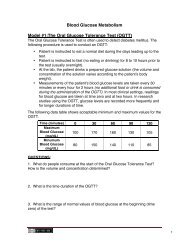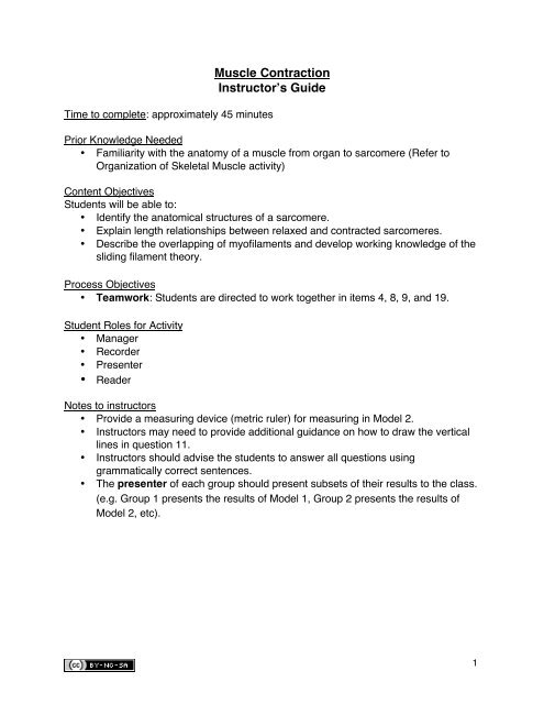
Muscle Contraction Instructor's Guide - Anatomy and Physiology ...
Muscle Contraction Instructor's Guide - Anatomy and Physiology ...
Muscle Contraction Instructor's Guide - Anatomy and Physiology ...

Create successful ePaper yourself
Turn your PDF publications into a flip-book with our unique Google optimized e-Paper software.
<strong>Muscle</strong> <strong>Contraction</strong><br />
Instructor’s <strong>Guide</strong><br />
Time to complete: approximately 45 minutes<br />
Prior Knowledge Needed<br />
• Familiarity with the anatomy of a muscle from organ to sarcomere (Refer to<br />
Organization of Skeletal <strong>Muscle</strong> activity)<br />
Content Objectives<br />
Students will be able to:<br />
• Identify the anatomical structures of a sarcomere.<br />
• Explain length relationships between relaxed <strong>and</strong> contracted sarcomeres.<br />
• Describe the overlapping of myofilaments <strong>and</strong> develop working knowledge of the<br />
sliding filament theory.<br />
Process Objectives<br />
• Teamwork: Students are directed to work together in items 4, 8, 9, <strong>and</strong> 19.<br />
Student Roles for Activity<br />
• Manager<br />
• Recorder<br />
• Presenter<br />
• Reader<br />
Notes to instructors<br />
• Provide a measuring device (metric ruler) for measuring in Model 2.<br />
• Instructors may need to provide additional guidance on how to draw the vertical<br />
lines in question 11.<br />
• Instructors should advise the students to answer all questions using<br />
grammatically correct sentences.<br />
• The presenter of each group should present subsets of their results to the class.<br />
(e.g. Group 1 presents the results of Model 1, Group 2 presents the results of<br />
Model 2, etc).<br />
1
<strong>Muscle</strong> <strong>Contraction</strong><br />
Model 1: <strong>Anatomy</strong> of a Sarcomere<br />
The sarcomere is the functional (contractile) unit of skeletal muscle. It is the region of a<br />
myofibril between two Z discs. Each sarcomere is approximately 2 mm long.<br />
Thick filament<br />
Thin filament<br />
A<br />
I<br />
H<br />
Z<br />
M<br />
Z<br />
QUESTIONS:<br />
1. Label the thick horizontal filament THICK filament.<br />
2. Label the thin horizontal filament THIN filament.<br />
3. How many sarcomeres are shown in the above model?<br />
4. Based on your observations of the location of thick <strong>and</strong> thin filaments, describe each<br />
of the following: (answers may vary among groups).<br />
a) A b<strong>and</strong>- extends the length of the thick filament <strong>and</strong> includes a portion of the<br />
thin filament<br />
b) I b<strong>and</strong>- area between the ends of the thick filaments; contains only thin<br />
filaments<br />
c) H zone- areas between the ends of the thin filaments; contains only a portion<br />
of the thick filaments<br />
d) Z disc- vertical line at each end of the sarcomere located in the middle of the I<br />
b<strong>and</strong><br />
e) M line- vertical line in the middle of the H zone that connects the thick<br />
filaments together<br />
5. Using complete grammatically correct sentences, describe how the H zone differs<br />
from the A b<strong>and</strong>.<br />
The H zone is located in the middle of the A b<strong>and</strong>. The H zone contains only<br />
thick filaments. The A b<strong>and</strong> consists of thick <strong>and</strong> thin filaments. The H zone is a<br />
subset of the A b<strong>and</strong><br />
2
6. How many sarcomeres do you think are in a muscle cell found in your quadriceps?<br />
Many thous<strong>and</strong>s to millions<br />
7. Do you think you would have more or fewer sarcomeres in an eye muscle?<br />
Fewer, eye muscles are smaller<br />
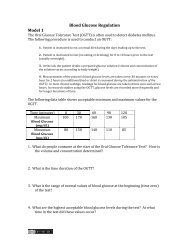
Model 2: Comparing Relaxed <strong>and</strong> Contracted Sarcomeres<br />
Figure 1. Relaxed sarcomeres.<br />
Figure 2. Contracted sarcomeres.<br />
QUESTIONS:<br />
8. In Figures 1 <strong>and</strong> 2 above, label the A-b<strong>and</strong>s, I-b<strong>and</strong>s, <strong>and</strong> H-zones. Measure <strong>and</strong><br />
record the lengths (in mm) of these structures <strong>and</strong> the thick <strong>and</strong> thin filaments in the<br />
chart below:<br />
Structure<br />
Thick filament<br />
Length in Relaxed<br />
Sarcomere (mm)<br />
Length in<br />
Contracted<br />
Sarcomere (mm)<br />
Did the length<br />
change between<br />
Figures 1 <strong>and</strong> 2?<br />
(Y/N)<br />
N<br />
Thin filament<br />
A b<strong>and</strong><br />
I b<strong>and</strong><br />
H zone<br />
Sarcomere<br />
N<br />
N<br />
Y<br />
Y<br />
Y<br />
3
8. Discuss the data from the table in Question 8 with your group <strong>and</strong> describe what<br />
happens to thick <strong>and</strong> thin filaments when muscles contract:<br />
A sarcomere shortens when the thin filaments <strong>and</strong> thick filament overlap to a greater<br />
extent. The filaments do not shorten, but overlap, causing a shortening of the<br />
sarcomere as a whole.<br />
9. As a group, examine the diagram in Model 2. Why is there a limit to the amount of<br />
shortening that can occur in a sarcomere during muscle contraction?<br />
Answers may vary. Possible answer:<br />
Depending upon the length of the thin filaments, there is a limit to the amount of<br />
overlapping that can occur between the thick <strong>and</strong> thin filaments. Also, the Z discs<br />
may run into the ends of the thick filaments <strong>and</strong> not be able to shorten any<br />
further.<br />
4
Model 3: Cross Sections Through a Sarcomere<br />
Model 3 shows cross-sections of a sarcomere that show the filaments at various<br />
locations within a sarcomere.<br />
thin<br />
thick<br />
thin<br />
thick<br />
Fig. A Fig. B Fig. C<br />
QUESTIONS:<br />
10. Label the thick <strong>and</strong> thin filaments in Figs. A, B, <strong>and</strong> C above.<br />
11. There are three sarcomeres shown in the diagram below.<br />
Sarcomere 1 Sarcomere 2 Sarcomere 3<br />
A B C<br />
a) In Sarcomere 1, identify the location within the sarcomere of the cross section<br />
indicated by Figure A in Model 3. Draw a vertical line <strong>and</strong> label it A.<br />
b) In Sarcomere 2, identify the location within the sarcomere of the cross section<br />
indicated by Figure B in Model 3. Draw a vertical line <strong>and</strong> label it B.<br />
c) In Sarcomere 3, identify the location within the sarcomere of the cross section<br />
indicated by Figure C in Model 3. Draw a vertical line <strong>and</strong> label it C.<br />
5
12. Which of the figures (A, B, or C) represents a cross section in the H zone?<br />
B<br />
13. Which of the figures (A, B, or C) represents a cross section in the I b<strong>and</strong>?<br />
A<br />
14. Which of the figures (A, B, or C) represents a cross section in the ends of the A<br />
b<strong>and</strong>?<br />
C<br />
15. On the figure below, shade in the area of the A b<strong>and</strong>. Then identify the location of<br />
the I b<strong>and</strong> <strong>and</strong> label it.<br />
I<br />
16. When viewing skeletal muscle through a microscope, you can easily see the dark<br />
<strong>and</strong> light striations of the muscle fiber. Compare the shading in the diagram in Question<br />
15 an in the photograph of muscle fiber as seen through a microscope below. What<br />
forms the dark <strong>and</strong> light b<strong>and</strong>s?<br />
The dark b<strong>and</strong>s consist of the A b<strong>and</strong> which has the thick filaments <strong>and</strong> portions of the<br />
thin filaments. The light b<strong>and</strong>s consist of the thin filaments of the I b<strong>and</strong>.<br />
A B<strong>and</strong> I B<strong>and</strong> Z disc<br />
Sarcomere<br />
e<br />
Courtesy of: LUMEN - Loyola University Medical Education Network<br />
17. On the photograph above, label the A b<strong>and</strong>, I b<strong>and</strong>, Z disc, <strong>and</strong> a sarcomere.<br />
6
18. The sliding filament theory is used to explain the physiology of skeletal muscle<br />
contraction. On your own, using what you have learned from this activity, write your own<br />
description of what the sliding filament theory states.<br />
Answers will vary.<br />
19. Next, discuss your predictions with your group members <strong>and</strong> develop a definition of<br />
the sliding filament theory with regard to thick <strong>and</strong> thin filaments. (Use grammatically<br />
correct sentences).<br />
Answers will vary. The correct answer should be similar to the following: When a<br />
skeletal muscle contracts, thin filaments slide past the thick filaments. In this process,<br />
the H b<strong>and</strong>s <strong>and</strong> I b<strong>and</strong>s get smaller; the zones of overlap get larger, the Z discs move<br />
closer together, <strong>and</strong> the width of the A b<strong>and</strong> remains constant.<br />
This explanation is known as the sliding filament theory (Martini <strong>and</strong> Ober, 2011).<br />
Source: Martini, F.H. <strong>and</strong> Ober, W.C. 2011. Visual <strong>Anatomy</strong> <strong>and</strong> <strong>Physiology</strong>. Benjamin<br />
Cummings, Boston, pp 287.<br />
7