Faculty of Dental Surgery - NHS Business Services Authority
Faculty of Dental Surgery - NHS Business Services Authority
Faculty of Dental Surgery - NHS Business Services Authority

You also want an ePaper? Increase the reach of your titles
YUMPU automatically turns print PDFs into web optimized ePapers that Google loves.
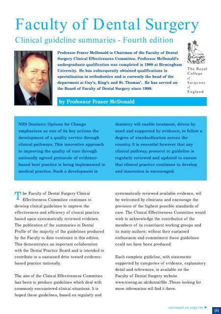
<strong>Faculty</strong> <strong>of</strong> <strong>Dental</strong> <strong>Surgery</strong><br />
Clinical guideline summaries - Fourth edition<br />
Pr<strong>of</strong>essor Fraser McDonald is Chairman <strong>of</strong> the <strong>Faculty</strong> <strong>of</strong> <strong>Dental</strong><br />
<strong>Surgery</strong> Clinical Effectiveness Committee. Pr<strong>of</strong>essor McDonald’s<br />
undergraduate qualification was completed in 1980 at Birmingham<br />
University. He has subsequently obtained qualifications in<br />
specialisation in orthodontics and is currently the head <strong>of</strong> the<br />
department at Guy’s, King’s and St. Thomas’. He has served on<br />
the Board <strong>of</strong> <strong>Faculty</strong> <strong>of</strong> <strong>Dental</strong> <strong>Surgery</strong> since 1998.<br />
by Pr<strong>of</strong>essor Fraser McDonald<br />
<strong>NHS</strong> Dentistry Options for Change<br />
emphasises as one <strong>of</strong> its key actions the<br />
development <strong>of</strong> a quality service through<br />
clinical pathways. This innovative approach<br />
to improving the quality <strong>of</strong> care through<br />
nationally agreed protocols <strong>of</strong> evidencebased<br />
best practice is being implemented in<br />
medical practice. Such a development in<br />
dentistry will enable treatment, driven by<br />
need and supported by evidence, to follow a<br />
degree <strong>of</strong> standardisation across the<br />
country. It is essential however that any<br />
clinical pathway, protocol or guideline is<br />
regularly reviewed and updated to ensure<br />
that clinical practice continues to develop<br />
and innovation is encouraged.<br />
T<br />
he <strong>Faculty</strong> <strong>of</strong> <strong>Dental</strong> <strong>Surgery</strong> Clinical<br />
Effectiveness Committee continues to<br />
develop clinical guidelines to improve the<br />
effectiveness and efficiency <strong>of</strong> clinical practice<br />
based upon systematically reviewed evidence.<br />
The publication <strong>of</strong> the summaries in <strong>Dental</strong><br />
Pr<strong>of</strong>ile <strong>of</strong> the majority <strong>of</strong> the guidelines produced<br />
by the <strong>Faculty</strong> to date continues in this edition.<br />
This demonstrates an important collaboration<br />
with the <strong>Dental</strong> Practice Board and is intended to<br />
contribute to a sustained drive toward evidencebased<br />
practice nationally.<br />
The aim <strong>of</strong> the Clinical Effectiveness Committee<br />
has been to produce guidelines which deal with<br />
commonly encountered clinical situations. It is<br />
hoped these guidelines, based on regularly and<br />
systematically reviewed available evidence, will<br />
be welcomed by clinicians and encourage the<br />
provision <strong>of</strong> the highest possible standards <strong>of</strong><br />
care. The Clinical Effectiveness Committee would<br />
wish to acknowledge the contribution <strong>of</strong> the<br />
members <strong>of</strong> its constituent working groups and<br />
its many authors; without their sustained<br />
enthusiasm and commitment these guidelines<br />
could not have been produced.<br />
Each complete guideline, with statements<br />
supported by categories <strong>of</strong> evidence, explanatory<br />
detail and references, is available on the<br />
<strong>Faculty</strong> <strong>of</strong> <strong>Dental</strong> <strong>Surgery</strong> website<br />
www.rcseng.ac.uk/dental/fds .Those looking for<br />
more information will find it there.<br />
continued on page (ii) ➤<br />
(i)
<strong>Faculty</strong> <strong>of</strong> <strong>Dental</strong> <strong>Surgery</strong> - Clinical guidelines ...continued from page (i)<br />
Treatment <strong>of</strong> intrinsic discolouration in permanent anterior teeth in<br />
children and adolescents: a guideline summary<br />
Principal Authors: Alyson Wray and Richard Welbury<br />
Paediatric Dentistry Working Party<br />
Introduction<br />
Intrinsic discolouration can be defined as discolouration<br />
which is incorporated into the structure <strong>of</strong> either enamel or<br />
dentine and which cannot be removed by prophylaxis with<br />
toothpaste or pumice. Intrinsic tooth discolouration can be a<br />
significant cosmetic, and in some instances, functional,<br />
problem. Loss <strong>of</strong> vitality secondary to trauma or infection<br />
frequently results in tooth discolouration which is not<br />
responsive to conventional endodontic therapy. Similarly<br />
fluorosis, tetracycline staining, localised and chronological<br />
hypoplasia, and both amelogenesis and dentinogenesis<br />
imperfecta can all produce a cosmetically unsatisfactory<br />
dentition and, in the latter two examples, a structurally ‘at<br />
risk’ dentition as well. These Guidelines are designed to<br />
outline the most appropriate options for treating the different<br />
aetiological categories <strong>of</strong> intrinsic discolouration <strong>of</strong> the<br />
anterior permanent dentition in child and adolescent<br />
patients.<br />
1 History<br />
A careful, detailed history is essential for the accurate<br />
diagnosis <strong>of</strong> intrinsic tooth discolouration, as the choice<br />
<strong>of</strong> treatment is greatly influenced by the aetiology.<br />
Specifically: details <strong>of</strong> the mother’s obstetric history<br />
and the delivery; medical history including neo-natal or<br />
early childhood illness and any drugs taken; dental<br />
history including infections relating to primary teeth;<br />
trauma to the primary and permanent teeth; family<br />
history <strong>of</strong> discoloured or abnormal teeth; fluoride<br />
history including supplementation, residence in natural<br />
water fluoridation areas, toothbrushing habits including<br />
the amount <strong>of</strong> paste used, the type <strong>of</strong> paste in<br />
childhood and any admitted swallowing <strong>of</strong> paste.<br />
2 Examination<br />
2.1 Clinical<br />
A standard extra-oral examination and full mouth intraoral<br />
examination should be undertaken, with special<br />
emphasis on the presence and/or absence <strong>of</strong> both<br />
primary and permanent teeth. The distribution <strong>of</strong> any<br />
discolouration or hypoplasia should be clearly<br />
established, specifically whether both dentitions are<br />
affected or not, whether all teeth in one dentition are<br />
equally affected, and whether or not there is a<br />
symmetrical or chronological pattern.The features <strong>of</strong><br />
discolouration may have been evident at tooth eruption,<br />
or may have developed subsequently and become<br />
either more or less severe in the intervening time. If<br />
possible, the extent <strong>of</strong> discolouration should be identified<br />
with respect to the depth <strong>of</strong> affected enamel or dentine.<br />
2.2 Additional investigations<br />
Appropriate radiographs will show abnormalities <strong>of</strong><br />
enamel and dentine structure, abnormal tooth<br />
morphology and the adequacy <strong>of</strong> root canal fillings in<br />
non-vital teeth. Sensibility testing will suggest the<br />
presence or absence <strong>of</strong> a functioning nerve supply,<br />
implying an intact vasculature. Histological sectioning <strong>of</strong><br />
exfoliated or extracted teeth may identify hereditary and<br />
environmental abnormalities.<br />
Management<br />
The treatment <strong>of</strong> choice is dependent on the diagnosis. In<br />
many cases <strong>of</strong> discolouration there is a hierarchy <strong>of</strong><br />
treatment options. These should be pursued in a logical<br />
order until a satisfactory cosmetic outcome is achieved. Pretreatment<br />
photographs, shade taking and sensibility tests<br />
are recommended in all cases.<br />
1 Microabrasion<br />
Microabrasion involves the removal <strong>of</strong> a small amount<br />
<strong>of</strong> surface enamel and classically incorporates both<br />
‘abrasion’ with dental instruments and ‘erosion’ with an<br />
acid mixture. The term ‘abrasion‘ has been used by<br />
some authors. There are two main techniques for<br />
microabrading discoloured or hypoplastic teeth. These<br />
are the hydrochloric acid/pumice technique which<br />
requires very careful isolation <strong>of</strong> the affected teeth, and<br />
the phosphoric acid/pumice technique. These<br />
techniques are simple to perform and the depth <strong>of</strong><br />
enamel removed in 10 applications is approximately<br />
(ii)<br />
DENTAL<br />
pr<strong>of</strong>ile
100 µm. (0.1 mm.). Microabrasion is indicated for<br />
fluorosis, post-orthodontic demineralisation, localised<br />
hypoplasia due to infection or trauma, and idiopathic<br />
hypoplasia where the discolouration is limited to the<br />
outer enamel layer. Analysis <strong>of</strong> the effectiveness <strong>of</strong><br />
microabrasion should be delayed for approximately one<br />
month post-treatment, as the appearance <strong>of</strong> the teeth<br />
will continue to improve during this time.<br />
2 Non-vital bleaching<br />
This technique is indicated for non-vital, endodontically<br />
treated teeth which have become discoloured due to<br />
the deposition <strong>of</strong> blood degradation products in the<br />
dentinal tubules.(19-39) A well-condensed root canal<br />
filling must be present prior to starting non-vital<br />
bleaching. Most techniques utilise hydrogen peroxide or<br />
sodium perborate (Bocasan) either together or<br />
independently. Where a non-vital tooth has an<br />
unsatisfactory root canal filling this should be replaced<br />
with a well-condensed gutta percha restoration prior to<br />
undertaking non-vital bleaching.<br />
alternative and should be used in child and adolescent<br />
patients. Resin can be used by either to camouflage/<br />
replace discrete localised areas <strong>of</strong> abnormal enamel<br />
(localised composites) or to cover the entire enamel<br />
surface (veneer). Composite resin restorations are<br />
indicated in cases <strong>of</strong> hypoplasia caused by moderate to<br />
severe fluorosis, localised hypoplasia not responsive to<br />
microabrasion, chronological hypoplasia, tetracycline<br />
staining, discolouration due to loss <strong>of</strong> vitality not<br />
responsive to non-vital bleaching, amelogenesis and<br />
dentinogenesis imperfecta, and idiopathic hypoplasia.<br />
Composite veneers can be placed directly on to the<br />
tooth surface or fabricated indirectly in the laboratory.<br />
5 Porcelain veneers<br />
Porcelain veneers are indicated for hypoplastic and<br />
discoloured teeth in patients aged 16 years and over,<br />
when techniques such as microabrasion, non-vital<br />
bleaching and composite resins have failed to produce<br />
a satisfactory clinical result.<br />
3 Vital bleaching<br />
3.1 Chairside<br />
This technique involves the external application <strong>of</strong><br />
hydrogen peroxide to the surface <strong>of</strong> the tooth followed<br />
by its activation with a heat source. It is indicated for<br />
mild tetracycline staining without obvious banding, mild<br />
fluorosis, and single teeth with sclerosed pulp<br />
chambers and root canals. The results have been<br />
found to be variable.<br />
3.2 Nightguard application<br />
This technique involves the daily placement <strong>of</strong><br />
carbamide peroxide gel into a custom-fitted tray <strong>of</strong><br />
either the upper or lower arch. It is carried out by the<br />
patient at home and is initially done on a daily basis.<br />
The technique is indicated for mild fluorosis, and<br />
moderate fluorosis as an adjunct to microabrasion.<br />
(Authors’ note regarding Vital Bleaching with products<br />
which release more than 0.1% hydrogen peroxide - At<br />
the time <strong>of</strong> preparation <strong>of</strong> this summary [Dec 2002]<br />
the legal status <strong>of</strong> this technique in the United Kingdom<br />
was under review by the Department <strong>of</strong> Trade and<br />
Industry [DTI].)<br />
4 Composite resin restorations<br />
The large size <strong>of</strong> the immature pulp chamber and pulp<br />
horns, and the immature gingival contour <strong>of</strong> the<br />
adolescent patient contra-indicates the use <strong>of</strong> porcelain<br />
veneers. Composite resin <strong>of</strong>fers a satisfactory<br />
Notes on claiming fees under<br />
the Statement <strong>of</strong> <strong>Dental</strong><br />
Remuneration<br />
Treatment <strong>of</strong> intrinsic discolouration in permanent<br />
teeth in children and adolescents<br />
• Where treatment involving microabrasion or internal<br />
bleaching is proposed, the practitioner will need to apply<br />
for a discretionary fee under item 40 (code 4001).<br />
However, where such treatments are proposed for patients<br />
treated under capitation arrangements attention is drawn<br />
to the provisos to item 41 (capitation) in the SDR;<br />
additional fees shall only be payable for treatment under<br />
item 40, where a laboratory cost is involved (proviso 2), or<br />
where treatment is necessitated by trauma (proviso 3).<br />
• At the present time, the DPB is unable to authorise<br />
discretionary fees for treatment involving external<br />
bleaching <strong>of</strong> vital teeth.<br />
• Treatment under code 1601 is only appropriate for the<br />
provision <strong>of</strong> porcelain veneers at upper incisor and canine<br />
teeth. Where veneers are proposed in any other material,<br />
or at any other tooth notation, a discretionary fee should<br />
be applied for under code 4001.<br />
It is important to note that treatment claimed for under code<br />
4001 requires prior approval.<br />
DENTAL<br />
pr<strong>of</strong>ile<br />
(iii)