MCW X-ray Diffraction Safety Manual - Medical College of Wisconsin
MCW X-ray Diffraction Safety Manual - Medical College of Wisconsin
MCW X-ray Diffraction Safety Manual - Medical College of Wisconsin

Create successful ePaper yourself
Turn your PDF publications into a flip-book with our unique Google optimized e-Paper software.
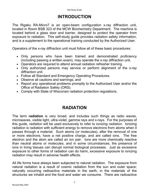
Self Study Guide<br />
INTRODUCTION<br />
The Rigaku RA-Micro7 is an open-beam configuration x-<strong>ray</strong> diffraction unit,<br />
located in Room BSB 323 <strong>of</strong> the <strong>MCW</strong> Biochemistry Department. The machine is<br />
located behind a glass door and barrier, designed to protect the operator from<br />
exposure to radiation. This self-study guide provides radiation safety information,<br />
and is a supplement to the operational training conducted by the Authorized User.<br />
Operators <strong>of</strong> the x-<strong>ray</strong> diffraction unit must follow all <strong>of</strong> these basic procedures:<br />
• Only persons who have been trained and demonstrated pr<strong>of</strong>iciency<br />
(including passing a written exam), may operate the x-<strong>ray</strong> diffraction unit.<br />
• Operators are required to attend annual radiation refresher training.<br />
• Only authorized persons may service or perform alignment <strong>of</strong> the x-<strong>ray</strong><br />
diffraction unit.<br />
• Follow all Standard and Emergency Operating Procedures.<br />
• Observe all cautions and warnings, and<br />
• Report any operational problems promptly to the Authorized User and/or the<br />
Office <strong>of</strong> Radiation <strong>Safety</strong> (ORS).<br />
• Comply with State <strong>of</strong> <strong>Wisconsin</strong> radiation protection regulations.<br />
RADIATION<br />
The term radiation is very broad, and includes such things as radio waves,<br />
microwaves, visible light, ultra-violet, gamma <strong>ray</strong>s and x-<strong>ray</strong>s. For the purposes <strong>of</strong><br />
this guide, radiation will be used exclusively to refer to ionizing radiation. Ionizing<br />
radiation is radiation with sufficient energy to remove electrons from atoms when it<br />
passes through a material. Such atoms (or molecules), after the removal <strong>of</strong> one<br />
or more electrons, have a net positive charge, and are called ions. The free<br />
electron and the atom are called an ion pair. Ions are more chemically reactive<br />
than neutral atoms or molecules, and in some circumstances, the presence <strong>of</strong><br />
ions in living tissues can disrupt normal biological processes. Just as excessive<br />
exposure to other forms <strong>of</strong> radiation can do harm, excessive exposure to ionizing<br />
radiation may result in adverse health effects.<br />
All life forms have always been subjected to natural radiation. The exposure from<br />
natural radiation is a result <strong>of</strong> cosmic radiation from the sun and outer space;<br />
naturally occurring radioactive materials in the earth, in the materials <strong>of</strong> the<br />
structures we inhabit and the food and water we consume. There are radioactive<br />
Revised May 2004<br />
1
Self Study Guide<br />
gases in the air we breathe and our bodies contain some radioactive atoms. The<br />
levels <strong>of</strong> this natural or "background" radiation vary greatly from location to<br />
location. In addition to natural radiation, individuals are also exposed to<br />
technologically developed forms <strong>of</strong> radiation. X-<strong>ray</strong>s and gamma <strong>ray</strong>s used in<br />
medical imaging and therapy are examples <strong>of</strong> this type <strong>of</strong> radiation. Within a<br />
decade after x-<strong>ray</strong>s came into use in the 1890's it became apparent this type <strong>of</strong><br />
radiation could either be beneficial or harmful, depending on its use and control,<br />
and that protective measures were necessary.<br />
REGULATION OF RADIATION<br />
An x-<strong>ray</strong> diffraction unit is a radiation-producing machine, and as such is regulated<br />
by the State <strong>of</strong> <strong>Wisconsin</strong>, Department <strong>of</strong> Health and Family Services, Radiation<br />
Protection Section (hereafter referred to as the State <strong>of</strong> <strong>Wisconsin</strong>). Radiation<br />
protection regulations are found in the <strong>Wisconsin</strong> Administrative Code, HFS 157. 1<br />
A copy <strong>of</strong> the regulations is available to all operators, in print at ORS, or in<br />
electronic form at the State <strong>of</strong> <strong>Wisconsin</strong> website,<br />
http://www.dhfs.state.wi.us/dph_beh/RadiatioP/index.htm. 2<br />
The State <strong>of</strong> <strong>Wisconsin</strong> directed certain rights and responsibilities to persons in<br />
areas where radioactive material or sources <strong>of</strong> radiation are in use or storage.<br />
Refer to Form PPH 45027 “Notice to Employees” 2 in this Study Guide to see what<br />
your general rights and responsibilities are. The responsibility for managing<br />
sources <strong>of</strong> radiation has been delegated to the institution's Radiation <strong>Safety</strong><br />
Committee. The day to day operations involved in the license are overseen by<br />
ORS. The Authorized User and each individual operator <strong>of</strong> the x-<strong>ray</strong> diffraction<br />
unit is responsible for maintaining compliance with the regulations and the terms<br />
<strong>of</strong> usage stipulated by the Radiation <strong>Safety</strong> Committee.<br />
Revised May 2004<br />
2
Self Study Guide<br />
DOSE LIMITS<br />
The State <strong>of</strong> <strong>Wisconsin</strong> in HFS 157.22 1 limits occupational radiation dose (from all<br />
sources <strong>of</strong> radiation at the institution) to individuals as follows:<br />
Type<br />
Whole Body<br />
Extremities<br />
Internal Organs<br />
Skin<br />
Lens <strong>of</strong> the Eye<br />
Minors<br />
Annual Limit<br />
5 rem<br />
50 rem<br />
50 rem<br />
50 rem<br />
15 rem<br />
10% <strong>of</strong> any limit above<br />
DOSIMETERS<br />
Anyone who is likely to exceed 10% <strong>of</strong> any <strong>of</strong> the limits above must be monitored<br />
for exposure to radiation. Even though during normal operation <strong>of</strong> the x-<strong>ray</strong><br />
machine it is unlikely that operators will receive exposure above the limits, the<br />
State requires all operators <strong>of</strong> open-beam x-<strong>ray</strong> diffraction units to wear extremity<br />
monitors when operating the equipment.<br />
AUTHORIZED USERS<br />
Authorization to use x-<strong>ray</strong> diffraction equipment is granted by the Radiation <strong>Safety</strong><br />
Committee (RSC). Investigators authorized by the RSC are responsible for:<br />
• Maintaining the equipment in safe working condition,<br />
• Verifying that only trained and competent staff are allowed to operate the<br />
equipment,<br />
• Ensuring that all State regulations and RSC requirements are followed, and<br />
• Reporting any incidents to ORS.<br />
Revised May 2004<br />
3
Self Study Guide<br />
RADIATION PROTECTION<br />
The major factors in reducing radiation exposure are TIME, DISTANCE and<br />
SHIELDING. The following is a brief explanation <strong>of</strong> each:<br />
TIME<br />
As TI M E<br />
decreases<br />
EXPOSURE<br />
decreases<br />
Radiation exposure is measured as a radiation dose per unit <strong>of</strong> time. The most<br />
common used unit is mR/hr. Radiation dose is directly proportional to time. This<br />
means the less time you spend in a radiation field the less radiation you will<br />
receive.<br />
EXAMPLE: If you spend 10 minutes in a radiation field <strong>of</strong> 60 mR/hr, you<br />
receive a dose <strong>of</strong> 10 mrem.<br />
DISTANCE<br />
As DISTANCE increases<br />
EXPOSURE decreases<br />
Radiation exposure is inversely proportional to the square <strong>of</strong> the distance from the<br />
source. This means as you increase your distance from a source <strong>of</strong> radiation,<br />
your radiation exposure decreases very rapidly. By doubling your distance from a<br />
source <strong>of</strong> radiation, you will receive one-quarter the radiation exposure. It is<br />
important to note, however, if you move to half the distance, you receive four<br />
times the radiation.<br />
EXAMPLE: If you are working 2 feet from a radiation source that is emitting<br />
16 mR/hr, at that distance, and you move 1(one) foot closer, you will be<br />
Revised May 2004<br />
4
Self Study Guide<br />
working in a radiation field <strong>of</strong> 64 mR/hr.<br />
SHIELDING<br />
As SHIELDING increases<br />
EXPOSURE decreases<br />
Radiation can be stopped or attenuated by a barrier or shield. The amount <strong>of</strong><br />
radiation penetrating the barrier depends on the density and thickness <strong>of</strong> the<br />
barrier material and the energy <strong>of</strong> the radiation.<br />
ALARA<br />
The main principle <strong>of</strong> the Radiation <strong>Safety</strong> Program is to maintain radiation<br />
exposures As Low As Reasonably Achievable (ALARA). Simply put, this means<br />
to take appropriate actions to reduce exposure for the procedure being performed.<br />
X-RAY BEAM CHARACTERISTICS<br />
X-<strong>ray</strong>s are generated in the diffraction unit by accelerating electrons from a<br />
filament cathode to a target anode inside the x-<strong>ray</strong> tube. The accelerating force is<br />
created by a voltage difference applied between the cathode and the anode. The<br />
kinetic energy <strong>of</strong> the electrons is proportional to the accelerating voltage, in the<br />
case <strong>of</strong> x-<strong>ray</strong> diffraction units a maximum <strong>of</strong> 40 kilovolts peak potential (kVp).<br />
When the electrons arrive at the anode, they are stopped suddenly in the dense<br />
material. The kinetic energy <strong>of</strong> the electrons is dissipated in the form <strong>of</strong><br />
bremsstrahlung, or braking radiation in the form <strong>of</strong> x-<strong>ray</strong>s. The x-<strong>ray</strong>s are emitted<br />
at a broad range <strong>of</strong> energies, up to a maximum equivalent to the accelerating<br />
voltage, 40 kilo electron-volts (keV). 3 As a practical matter, most <strong>of</strong> the x-<strong>ray</strong>s are<br />
emitted at some lower energy than the maximum, and a “preferred” energy is the<br />
Kα-characteristic energy <strong>of</strong> the copper anode at 7.4 keV. 4 By comparison, a<br />
typical medical x-<strong>ray</strong> radiograph or fluoroscopic image uses x-<strong>ray</strong>s generated by<br />
100 to 125 kVp electrons. The average energy <strong>of</strong> a medical x-<strong>ray</strong> beam is about<br />
50 keV. 5 Consequently, x-<strong>ray</strong>s used in diffraction are less deeply penetrating, and<br />
pose more <strong>of</strong> a skin dose hazard than medical x-<strong>ray</strong>s.<br />
Revised May 2004<br />
5
Self Study Guide<br />
The intensity <strong>of</strong> the x-<strong>ray</strong> beam is proportional to the number <strong>of</strong> electrons striking<br />
the anode, called the beam current, measured in milliamps (mA). The maximum<br />
beam current is 20 mA, 3 or 1.25 ×10 17 electrons per second. Only a fraction <strong>of</strong> the<br />
energy from the electron beam is converted to useful x-<strong>ray</strong>s, however the intensity<br />
<strong>of</strong> the direct beam can be as high as 4 × 10 5 R/minute, and the scattered beam<br />
can be as high as 1.3 R/minute. 6 While not all <strong>of</strong> this energy can contribute to skin<br />
dose, it only takes a fraction <strong>of</strong> a second exposure to the direct beam to reach the<br />
threshold for radiation burns <strong>of</strong> the skin.<br />
BIOLOGICAL EFFECTS OF RADIATION<br />
The biological effects <strong>of</strong> exposure to high doses (hundreds <strong>of</strong> rems) <strong>of</strong> radiation<br />
are well known and documented. The consequences <strong>of</strong> exposure to low doses<br />
(less than the dose limits) <strong>of</strong> radiation are less clear, and are most frequently<br />
expressed as risk factors. The following information is provided for both types <strong>of</strong><br />
radiation exposure.<br />
HIGH-DOSE EXPOSURE<br />
Due to the intensity and penetrating ability <strong>of</strong> x-<strong>ray</strong>s from a diffraction unit, as well<br />
as the physical size <strong>of</strong> the beam and its accessibility within the apparatus, the<br />
most immediate radiation protection concern is exposure <strong>of</strong> the skin <strong>of</strong> the hands<br />
and fingers <strong>of</strong> operators. The following table 7 lists threshold doses and effects<br />
that may be expected within a few days or weeks from an exposure:<br />
Revised May 2004<br />
6
Self Study Guide<br />
Threshold Dose<br />
Description <strong>of</strong> Effect<br />
200 rad Early transient erythema – reddening <strong>of</strong> the skin. At doses <strong>of</strong><br />
600 rad, the peak may occur at 10 to 14 days from the time<br />
<strong>of</strong> exposure.<br />
300 rad Temporary epilation – loss <strong>of</strong> hair to the exposed area.<br />
700 rad Permanent epilation.<br />
1,400 rad Dry desquamation – flaking <strong>of</strong> the skin.<br />
1,800 rad Moist desquamation.<br />
2,400 rad Secondary ulceration.<br />
In addition to the effects listed above, dermal atrophy, telangiectasis (lesions<br />
formed by capillary damage) and delayed necrosis can occur a year after doses<br />
above 1,000-1,200 rads.<br />
The major safety concern with x-<strong>ray</strong> diffraction is accidental exposures to<br />
the direct beam, which can be severe and result in the loss <strong>of</strong> fingers, even<br />
for very short exposures.<br />
LOW-LEVEL EXPOSURE<br />
The effects <strong>of</strong> low-dose radiation exposure are less clearly defined. At doses <strong>of</strong><br />
15-25 rad to the whole body, temporary changes in blood cell ratios in humans<br />
can be observed. 8 Below this level, biological effects are delayed stochastic and<br />
genetic effects that may occur in one individual and not another. For this reason,<br />
low-level radiation exposure effects are expressed in terms <strong>of</strong> risk <strong>of</strong> occurrence in<br />
a population uniformly exposed.<br />
Stochastic effects <strong>of</strong> radiation exposure are effects characterized by the following:<br />
• Occurrence in both exposed and unexposed populations,<br />
• The probability <strong>of</strong> a given effect occurring increases with dose,<br />
• The severity <strong>of</strong> the effect is unrelated to dose, and<br />
• There is no threshold dose below which it can be said with certainty that an<br />
effect will not occur. 5, 8<br />
Revised May 2004<br />
7
Self Study Guide<br />
Risk can be generally defined as the probability (chance) <strong>of</strong> an event (injury,<br />
illness, etc.) from some activity (exposure). Risk estimates are generally<br />
expressed in terms <strong>of</strong> the excess occurrence <strong>of</strong> an effect (the number <strong>of</strong> extra<br />
incidences <strong>of</strong> an effect above the natural occurrence rate) per rem in a group <strong>of</strong><br />
people exposed uniformly.<br />
The main stochastic effects attributed to low-level exposure to radiation are<br />
cancer and genetic effects. The National Council on Radiation Protection and<br />
Measurements (NCRP) has published risk estimates for occupationally exposed<br />
adults, summarized below:<br />
Effect<br />
Excess occurrence per rem<br />
Fatal cancer 0.0004<br />
Nonfatal cancer 0.00008<br />
Severe genetic effects 0.00008<br />
Total 0.00056<br />
According to the estimates above, there will be 4 excess fatal cancers in a<br />
population <strong>of</strong> 10,000 people who each receive a radiation dose <strong>of</strong> 1 rem. It is<br />
important to recognize that these risk estimates are quite low when compared to<br />
the actual number <strong>of</strong> cancer deaths that occur in the U.S. population. According<br />
to the National Cancer Institute, the number <strong>of</strong> cancer deaths per 10,000 is<br />
2,290. 10<br />
Revised May 2004<br />
8
Self Study Guide<br />
TERMS AND DEFINITIONS<br />
ALARA<br />
Alpha particle<br />
"As Low As Reasonably Achievable”. ALARA means making a concerted effort<br />
to maintain exposures to radiation as far below the limits as practical,<br />
consistent with the purpose for which the licensed activity is undertaken, the<br />
economics for improvements in relation to benefits to the public health and<br />
safety and in relation to utilization <strong>of</strong> atomic energy in the public interest.<br />
Charged particle emitted from the nucleus having a mass and charge <strong>of</strong> two<br />
protons and two neutrons<br />
Becquerel (Bq) The SI unit <strong>of</strong> radioactivity, one Becquerel equals one nuclear transformation<br />
per second. 1Bq=2.703E-11Ci.<br />
Beta particle<br />
Curie (Ci)<br />
Gamma <strong>ray</strong><br />
G<strong>ray</strong> (Gy)<br />
Neutron<br />
Proton<br />
Rad<br />
Radiation<br />
Radioactive<br />
Rem<br />
Roentgen (R)<br />
Sievert (Sv)<br />
X-<strong>ray</strong><br />
Negatively charged particle emitted from the nucleus with a mass and charge<br />
<strong>of</strong> the electron.<br />
The unit <strong>of</strong> radioactivity. One curie equals 3.7E10 nuclear transformations per<br />
second. 1Ci=3.7E10Bq<br />
Electromagnetic radiation <strong>of</strong> short wavelength emitted from the nucleus.<br />
The SI unit <strong>of</strong> absorbed dose. 1 Gy = 100 rad.<br />
Neutrally charged particle emitted from the nucleus.<br />
Positively charged particle emitted from the nucleus.<br />
The unit <strong>of</strong> absorbed radiation dose equal to 0.01 J/kg in any medium.<br />
1 rad = 0.01 Gy.<br />
The combined processes <strong>of</strong> emission, transmission and absorption <strong>of</strong> radiant<br />
energy.<br />
Material which spontaneously emits particles or photon radiation.<br />
The unit <strong>of</strong> radiation dose equivalent. The dose equivalent in rems in<br />
numerically equal to the absorbed dose in rads multiplied by the quality factor,<br />
the distribution factor and any other necessary modifying factors.<br />
1 rem = 0.01 Sv.<br />
The unit <strong>of</strong> radiation exposure. One roentgen equals 2.58E-4 coulomb per<br />
kilogram <strong>of</strong> air.<br />
The SI unit <strong>of</strong> dose equivalent. 1 Sv = 100 rem<br />
Electromagnetic radiation emitted usually from the cascade <strong>of</strong> an electron from<br />
a higher orbital to a lower one.<br />
Revised May 2004<br />
9
Self Study Guide<br />
Revised May 2004<br />
10
Self Study Guide<br />
COMPARATIVE RADIATION DOSES<br />
Television<br />
Round trip flight between<br />
London and New York<br />
Self Study Guide<br />
QUESTIONS AND ANSWERS<br />
ABOUT RADIATION SAFETY<br />
1. What is radiation?<br />
Radiation, in basic terms, is simply the movement <strong>of</strong> energy from one place to another by what physicists call an<br />
electromagnetic photon or a particle. Anyone who has ever warmed themselves by a fire is familiar with this<br />
process if the movement <strong>of</strong> energy (from the fire) to another (our "backsides") by photons emitted by the fire. This<br />
common type <strong>of</strong> radiation is only one form <strong>of</strong> electromagnetic radiant energy flow, others are radio, light, ultraviolet,<br />
gamma, cosmic, and x-<strong>ray</strong>. the last three are examples <strong>of</strong> ionizing radiation, the other examples are non-ionizing.<br />
2. What is non-ionizing radiation?<br />
Non-ionizing radiation is a type <strong>of</strong> radiant energy incapable <strong>of</strong> creating ions. Radiant energy, both visible and<br />
ultraviolet, and microwave radiation are examples <strong>of</strong> non-ionizing radiation.<br />
3. What is ionizing radiation?<br />
The type <strong>of</strong> radiant energy which is able to disrupt and atom into separate positively and negatively charged parts.<br />
Gamma and X are among examples <strong>of</strong> electromagnetic ionizing radiation. Alpha and beta particles are examples<br />
<strong>of</strong> particulate radiation.<br />
4. What is electromagnetic radiation?<br />
Energy traveling in a wave motion <strong>of</strong> varying wavelengths. It has no size or mass, but only energy. Since it has no<br />
mass, electromagnetic radiation can be very penetrating. Examples <strong>of</strong> electromagnetic radiation are radio waves,<br />
microwaves, gamma <strong>ray</strong>s and x-<strong>ray</strong>s.<br />
5. What is particulate radiation?<br />
Radiation emitted in the form <strong>of</strong> nuclear particles, having dimension and mass. Since it has mass, particles interact<br />
more readily and therefore do not penetrate very far. Examples <strong>of</strong> particulate radiation are alpha and beta<br />
particles.<br />
6. What are the sources <strong>of</strong> ionizing radiation?<br />
Ionizing radiation is produced by x-<strong>ray</strong> generating equipment, such as a unit that takes dental or chest radiographs.<br />
Ionizing radiation is also released from radioactive materials.<br />
7. What is radioactive material?<br />
Material that spontaneously emits radiation generally as x-<strong>ray</strong>s, gamma <strong>ray</strong>s or other particles.<br />
8. What is radioactive decay?<br />
The decrease in the amount <strong>of</strong> any radioactive material with the passage <strong>of</strong> time, due to spontaneous emission <strong>of</strong><br />
radiation. After the decay event, the atom becomes a nonradioactive atom.<br />
9. What is radioactive contamination?<br />
Deposition <strong>of</strong> radioactive material in any place where it may harm a person or equipment.<br />
10. What is a half-life (T ½ )?<br />
Physical T ½ - the amount <strong>of</strong> time it takes for one-half <strong>of</strong> the radioactive atoms <strong>of</strong> a radioactive material to<br />
disintegrate to another nuclear form. Half-lives vary from millionths <strong>of</strong> a second to billions <strong>of</strong> years.<br />
Biological T ½ - the time required for the body to eliminate half <strong>of</strong> the material taken in by natural biological means.<br />
Effective T ½ - the time required for a radionuclide contained in a biological system, such as human, to reduce its<br />
activity by one-half as a combined result <strong>of</strong> radioactive decay and biological elimination.<br />
11. What is background radiation?<br />
Revised May 2004<br />
12
Self Study Guide<br />
Radiation from cosmic sources, terrestrial and technologically enhanced sources, as well as naturally occurring<br />
radioactive materials in our food, building materials and even in our own bodies. The following table is the<br />
approximate radiation exposure for some common sources <strong>of</strong> radiation:<br />
Chest x-<strong>ray</strong> 20 mrem/exposure<br />
TV set
Self Study Guide<br />
17. If I am exposed to radioactive materials or radiation, do I become radioactive?<br />
No, you do not. In order for you to become radioactive, a radiation source must become incorporated into your<br />
body. This can be done by inhaling a radioactive gas or volatilized particles, ingested by swallowing or absorbed<br />
through the skin.<br />
18. What are the possible health effects <strong>of</strong> radiation exposure?<br />
Health effects from radiation exposure are divided into two types <strong>of</strong> effects. The first is somatic effects, or effects to<br />
your body. Some somatic effects are cataract formation and cancer. The second type <strong>of</strong> effect is genetic. This<br />
affects a descendant, as a result <strong>of</strong> modification <strong>of</strong> genetic material, <strong>of</strong> the exposed individual.<br />
19. What are the risks involved if I become pregnant?<br />
There may be a small risk if you are constantly working in an area where radioactive materials are present. Such<br />
persons work in Nuclear Medicine, Cardiac Catherization Laboratory, or the Angiography section <strong>of</strong> Radiology.<br />
Children are more radiosensitive than adults and fetuses are more radiosensitive than children. Various scientific<br />
organizations and committees have conducted studies to evaluate the risks <strong>of</strong> the unborn when the mother is<br />
occupationally exposed. The recommendations <strong>of</strong> these groups include keeping all the radiation exposure to the<br />
fetus as low as reasonably achievable, but definitely below 500 mrem for the nine month gestation period.<br />
Revised May 2004<br />
14
Self Study Guide<br />
REFERENCES<br />
1. State <strong>of</strong> <strong>Wisconsin</strong>, Department <strong>of</strong> Health and Family Services, Radiation Protection,<br />
<strong>Wisconsin</strong> Administrative Code, Chapter HFS 157<br />
2. State <strong>of</strong> <strong>Wisconsin</strong>, Department <strong>of</strong> Health and Family Services, Radiation Protection<br />
Section, http://www.dhfs.state.wi.us/dph_beh/RadiatioP/index.htm<br />
3. Rigaku Corporation, RA-Micro7 Horizontal RotaFlex Instruction <strong>Manual</strong>, <strong>Manual</strong> No.<br />
ME14042A05.<br />
4. Kocher, D.C., Radioactive Decay Data Tables, A Handbook <strong>of</strong> Decay Data for Application<br />
to Radiation Dosimetry and Radiological Assessments, Technical Information Center,<br />
U.S. Department <strong>of</strong> Energy, 1981.<br />
5. Hendee, W.R., Ritenour, E.R., <strong>Medical</strong> Imaging Physics, 4 th Edition, Wiley-Liss, 2002.<br />
6. Yale University, Office <strong>of</strong> Environmental Health & <strong>Safety</strong>, Radiation <strong>Safety</strong> Procedures<br />
<strong>Manual</strong>, Appendix XI, X-Ray <strong>Diffraction</strong>, http://www.yale.edu/oehs/, 2001.<br />
7. Hall, E.J., Radiobiology for the Radiologist, 5 th Edition, Lippincott Williams & Wilkins,<br />
2000.<br />
8. Cember, H., Introduction to Health Physics, 2 nd Edition, Pergamon Press, 1985.<br />
9. National Council on Radiation Protection and Measurements, Limitation <strong>of</strong> Exposure to<br />
Ionizing Radiation, NCRP Report No. 116, 1993.<br />
10. Ries LAG, Eisner MP, Kosary CL, Hankey BF, Miller BA, Clegg L, Mariotto A, Feuer EJ,<br />
Edwards BK (eds). SEER Cancer Statistics Review, 1975-2001, National Cancer<br />
Institute. Bethesda, MD, http://seer.cancer.gov/csr/1975_2001/, 2004.<br />
11. Stabin, M., RADAR <strong>Medical</strong> Procedure Radiation Dose Calculator, http://www.doseinforadar.com/RADARDoseRiskCalc.html,<br />
2004.<br />
Revised May 2004<br />
15
Self Study Guide<br />
REGULATIONS<br />
HFS 157.87 Radiation <strong>Safety</strong> Requirements for Analytical X-Ray Systems<br />
(2) RADIATION SAFETY REQUIREMENTS FOR ANALYTICAL X–RAY SYSTEMS. The following safety<br />
equipment shall be used with all analytical x–<strong>ray</strong> systems except as otherwise noted:<br />
(a) <strong>Safety</strong> device. An analytical x–<strong>ray</strong> system utilizing an open beam configuration shall incorporate a<br />
safety device that prevents any portion <strong>of</strong> an individual’s body from entering the primary x–<strong>ray</strong> beam path<br />
or that causes the beam to be shut <strong>of</strong>f upon entry into its path. The person in control at the facility may<br />
apply to the department for an exemption from the requirement for a safety device. The application shall<br />
include all the following information:<br />
1. A description <strong>of</strong> the various safety devices that have been evaluated by the person in control.<br />
2. The reason each device evaluated in subd. 1. cannot be used.<br />
3. A description <strong>of</strong> the alternative safety methods available to minimize the possibility <strong>of</strong> an<br />
accidental exposure, including procedures to assure that operators and others in the area will be<br />
informed <strong>of</strong> the absence <strong>of</strong> safety devices. The department shall approve the alternate safety<br />
devices prior to their installation on the system.<br />
(b) Warning devices. Open–beam configurations shall be provided with a readily discernible indication <strong>of</strong><br />
either <strong>of</strong> the following:<br />
1. An indication <strong>of</strong> whether the x–<strong>ray</strong> tube is on or <strong>of</strong>f, if the primary beam is controlled in this<br />
manner.<br />
2. An indication <strong>of</strong> whether the shutter is open or closed, if the primary beam is controlled in this<br />
manner. Warning devices shall be labeled so that their purpose is easily identified.<br />
(c) Ports. Unused ports on radiation source housings shall be secured in the closed position in a manner<br />
that will prevent casual opening.<br />
(d) Labeling. All analytical x–<strong>ray</strong> equipment shall be labeled with a readily discernible sign or signs bearing<br />
the radiation symbol and the words:<br />
1. “CAUTION – HIGH INTENSITY X–RAY BEAM,” or words having a similar intent, on an x–<strong>ray</strong><br />
source housing.<br />
2. “CAUTION RADIATION – THIS EQUIPMENT PRODUCES RADIATION WHEN ENERGIZED,”<br />
or words having a similar intent, near any switch that energizes an x–<strong>ray</strong> tube if the radiation<br />
source is an x–<strong>ray</strong> tube.<br />
(e) Shutters. On open–beam configurations installed after January 1, 1979, each port on the radiation<br />
source housing shall be equipped with a shutter that cannot be opened unless a collimator or a coupling<br />
has been connected to the port.<br />
(f) Warning lights. An easily visible warning light labeled with the words “X–RAY ON,” or words having a<br />
similar intent, shall be located as follows:<br />
1. Near any switch that energizes an x–<strong>ray</strong> tube and illuminates only when the tube is energized.<br />
2. In the case <strong>of</strong> a radioactive source, near any switch that opens a housing shutter and illuminates<br />
only when the shutter is open.<br />
(g) Radiation source housing. An x–<strong>ray</strong> tube housing shall be so constructed that with all shutters closed<br />
the leakage radiation measured at a distance <strong>of</strong> 5 centimeters from its surface is not be capable <strong>of</strong><br />
producing an air kerma in excess <strong>of</strong> 25 uSv (2.5 mrem) in one hour at any specified tube rating.<br />
(h) Generator cabinet. An x–<strong>ray</strong> generator shall be contained within a protective cabinet which limits<br />
leakage radiation measured at a distance <strong>of</strong> 5 centimeters from its surface to no more than 2.5 uSv (2.5<br />
mrem) in one hour.<br />
(3) AREA REQUIREMENTS.<br />
Revised May 2004<br />
16
Self Study Guide<br />
(a) Radiation levels. The local components <strong>of</strong> an analytical x–<strong>ray</strong> system shall be located and arranged and<br />
shall include sufficient shielding or access control so no radiation levels exist in any area surrounding the<br />
local component group that could result in a dose to any individual in excess <strong>of</strong> the dose limits in s. HFS<br />
157.23 (1). For systems utilizing x–<strong>ray</strong> tubes, the permissible radiation levels shall be met at any specified<br />
tube rating.<br />
(b) Surveys. To demonstrate compliance with par. (a), radiation surveys <strong>of</strong> an analytical x–<strong>ray</strong> system shall<br />
be performed according to all the following criteria:<br />
1. Upon installation <strong>of</strong> the equipment.<br />
2. Following any change in the initial arrangement, number or type <strong>of</strong> local components in the<br />
system.<br />
3. Following any maintenance requiring the disassembly or removal <strong>of</strong> a local component in the<br />
system.<br />
4. During the performance <strong>of</strong> maintenance and alignment procedures if the procedures require the<br />
presence <strong>of</strong> a primary x–<strong>ray</strong> beam when any local component in the system is disassembled or<br />
removed.<br />
5. Any time a visual inspection <strong>of</strong> the local components in the system reveals an abnormal<br />
condition.<br />
6. Whenever personnel monitoring devices show an increase <strong>of</strong> 50% over the previous monitoring<br />
period or the readings are approaching the limits <strong>of</strong> sub. (2) (g) or (h). Radiation survey<br />
measurements are not be required if a person in control demonstrates compliance with par. (a) in<br />
some other manner.<br />
(c) Posting. Each area or room containing analytical x–<strong>ray</strong> equipment shall have at least one sign<br />
conspicuously posted bearing the radiation symbol and the words “CAUTION – X–RAY EQUIPMENT” or<br />
words having a similar intent.<br />
(4) OPERATING REQUIREMENTS.<br />
(a) Procedures. Operating procedures shall be written and available to all analytical x–<strong>ray</strong> equipment<br />
workers. No individual may operate analytical x–<strong>ray</strong> equipment in any manner other than that specified in<br />
the procedures unless the individual has obtained written approval <strong>of</strong> the person in control.<br />
(b) Bypassing. No individual may intentionally bypass a safety device unless the individual has obtained<br />
the approval <strong>of</strong> the person in control. When a safety device has been bypassed, a readily discernible sign<br />
bearing the words “SAFETY DEVICE NOT WORKING” or words having a similar intent shall be placed on<br />
the radiation source housing.<br />
(5) PERSONNEL REQUIREMENTS.<br />
(a) Instruction. No individual may operate or maintain analytical x–<strong>ray</strong> equipment unless the individual has<br />
received instruction in and demonstrated competence in all the following:<br />
1. Identification <strong>of</strong> radiation hazards associated with use <strong>of</strong> the equipment.<br />
2. Significance <strong>of</strong> the various radiation warning and safety devices incorporated into the equipment<br />
or the reasons the devices have not been installed on certain pieces <strong>of</strong> equipment and the extra<br />
precautions required in such cases.<br />
3. Proper operating procedures for the equipment.<br />
4. Symptoms <strong>of</strong> an acute localized exposure that may cause a radiation burn.<br />
5. Proper procedures for reporting an actual or suspected exposure.<br />
(b) Personnel monitoring. Finger or wrist dosimetry devices shall be provided to and used by any <strong>of</strong> the<br />
following individuals:<br />
1. An analytical x–<strong>ray</strong> equipment worker using a system having an open–beam configuration and<br />
not equipped with a safety device.<br />
2. Personnel maintaining analytical x–<strong>ray</strong> equipment if the maintenance procedures require the<br />
presence <strong>of</strong> a primary x–<strong>ray</strong> beam when any local component in the analytical x–<strong>ray</strong> system is<br />
disassembled or removed. Reported dose values may not be used for the purpose <strong>of</strong> determining<br />
compliance with s. HFS 157.22 unless the dose values are evaluated by a medical physicist.<br />
Revised May 2004<br />
17