Histological intersex (ovotestis) in the European lobster Homarus ...
Histological intersex (ovotestis) in the European lobster Homarus ...
Histological intersex (ovotestis) in the European lobster Homarus ...

Create successful ePaper yourself
Turn your PDF publications into a flip-book with our unique Google optimized e-Paper software.
Stentiford: Intersex <strong>in</strong> <strong>Homarus</strong> gammarus<br />
187<br />
were cut at a thickness of 3 to 5 μm on a rotary microtome<br />
and were mounted onto glass slides before<br />
sta<strong>in</strong><strong>in</strong>g with haematoxyl<strong>in</strong> and eos<strong>in</strong> (H&E). Sta<strong>in</strong>ed<br />
sections were analysed by light microscopy (Nikon<br />
Eclipse E800) and digital images were taken us<strong>in</strong>g<br />
<strong>the</strong> Lucia Screen Measurement System (Nikon).<br />
RESULTS<br />
External visual exam<strong>in</strong>ation of <strong>European</strong> <strong>lobster</strong>s<br />
sampled from <strong>the</strong> Weymouth Bay site revealed 10<br />
normal male and 10 normal female animals. Dur<strong>in</strong>g<br />
histological exam<strong>in</strong>ation of <strong>the</strong> tissues and organs<br />
from <strong>the</strong>se <strong>lobster</strong>s, one of <strong>the</strong> males, with a carapace<br />
length of 112 mm, displayed abnormal gonadal<br />
development consistent with <strong>the</strong> ‘<strong>ovotestis</strong>’ condition<br />
described <strong>in</strong> numerous species of mar<strong>in</strong>e teloest fish<br />
(e.g. Bateman et al. 2004).<br />
The majority of <strong>the</strong> gonadal tissue mass with<strong>in</strong> <strong>the</strong><br />
<strong><strong>in</strong>tersex</strong> <strong>lobster</strong> consisted of apparently normal sem<strong>in</strong>iferous<br />
tubules conta<strong>in</strong><strong>in</strong>g a progressive l<strong>in</strong>eage of<br />
male reproductive cell types. Spermatogonia and<br />
accessory cells l<strong>in</strong>ed <strong>the</strong> basement membrane of<br />
<strong>in</strong>dividual sem<strong>in</strong>iferous tubules, and apparent spermatocytes<br />
and mature spermatozoa were found <strong>in</strong><br />
<strong>the</strong> lum<strong>in</strong>al region of each tubule (Fig. 1A). Closely<br />
opposed sem<strong>in</strong>iferous tubules at similar stages of<br />
development were present throughout <strong>the</strong> testis.<br />
Sem<strong>in</strong>iferous tubules delivered mature spermatozoa<br />
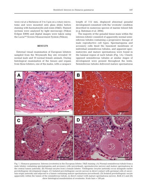
Fig. 1. <strong>Homarus</strong> gammarus. Intersex (ovotestes) <strong>in</strong> <strong>the</strong> <strong>European</strong> <strong>lobster</strong>. H&E sta<strong>in</strong><strong>in</strong>g. (A) Normal sem<strong>in</strong>iferous tubule from a<br />
male <strong>lobster</strong> conta<strong>in</strong><strong>in</strong>g spermatogonia and accessory cells (arrowhead), spermatocytes (arrow) and mature spermatozoa <strong>in</strong><br />
<strong>the</strong> central lumen (asterisk). (B) Normal oocytes from a female <strong>lobster</strong>. Vitellogenic oocytes (asterisk) occur alongside earlier<br />
previtellogenic development stages. (C) Isolated previtellogenic oocyte (arrow) <strong>in</strong> direct contact with germ<strong>in</strong>al cells of uncerta<strong>in</strong><br />
orig<strong>in</strong> (asterisk) and adjacent to a lumen conta<strong>in</strong><strong>in</strong>g mature spermatozoa (arrowhead). (D) Isolated previtellogenic oocyte<br />
apparently with<strong>in</strong> <strong>the</strong> lumen of <strong>the</strong> sem<strong>in</strong>iferous tubule, <strong>in</strong> direct contact with mature spermatozoa (arrow). Both (C) and (D)<br />
show histological manifestation of ovostestes. Scale bars = 100 µm