MCL reconstruction - ISAKOS
MCL reconstruction - ISAKOS
MCL reconstruction - ISAKOS

Create successful ePaper yourself
Turn your PDF publications into a flip-book with our unique Google optimized e-Paper software.
CURRENT CONCEPT<br />
MEDIAL COLLATERAL LIGAMENT RECONSTRUCTION<br />
USING ACHILLES TENDON ALLOGRAFT<br />
Iftach Hetsroni MD<br />
Department of Orthopedic Surgery,<br />
Meir General Hospital, Sackler Faculty of Medicine,<br />
Tel Aviv University, Tel Aviv, Israel<br />
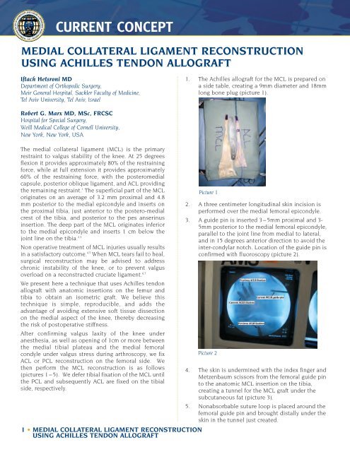
1. The Achilles allograft for the <strong>MCL</strong> is prepared on<br />
a side table, creating a 9mm diameter and 18mm<br />
long bone plug (picture 1).<br />
Robert G. Marx MD, MSc, FRCSC<br />
Hospital for Special Surgery,<br />
Weill Medical College of Cornell University,<br />
New York, New York, USA<br />
The medial collateral ligament (<strong>MCL</strong>) is the primary<br />
restraint to valgus stability of the knee. At 25 degrees<br />
flexion it provides approximately 80% of the restraining<br />
force, while at full extension it provides approximately<br />
60% of the restraining force, with the posteromedial<br />
capsule, posterior oblique ligament, and ACL providing<br />
the remaining restraint. 1 The superficial part of the <strong>MCL</strong><br />
originates on an average of 3.2 mm proximal and 4.8<br />
mm posterior to the medial epicondyle and inserts on<br />
the proximal tibia, just anterior to the postero-medial<br />
crest of the tibia, and posterior to the pes anserinus<br />
insertion. The deep part of the <strong>MCL</strong> originates inferior<br />
to the medial epicondyle and inserts 1 cm below the<br />
joint line on the tibia. 2,3<br />
Non operative treatment of <strong>MCL</strong> injuries usually results<br />
in a satisfactory outcome. 4,5 When <strong>MCL</strong> tears fail to heal,<br />
surgical <strong>reconstruction</strong> may be advised to address<br />
chronic instability of the knee, or to prevent valgus<br />
overload on a reconstructed cruciate ligament. 6,7<br />
We present here a technique that uses Achilles tendon<br />
allograft with anatomic insertions on the femur and<br />
tibia to obtain an isometric graft. We believe this<br />
technique is simple, reproducible, and adds the<br />
advantage of avoiding extensive soft tissue dissection<br />
on the medial aspect of the knee, thereby decreasing<br />
the risk of postoperative stiffness.<br />
After confirming valgus laxity of the knee under<br />
anesthesia, as well as opening of 1cm or more between<br />
the medial tibial plateau and the medial femoral<br />
condyle under valgus stress during arthroscopy, we fix<br />
ACL or PCL <strong>reconstruction</strong> on the femoral side. We<br />
then perform the <strong>MCL</strong> <strong>reconstruction</strong> is as follows<br />
(pictures 1–5). We defer tibial fixation of the <strong>MCL</strong> until<br />
the PCL and subsequently ACL are fixed on the tibial<br />
side, respectively.<br />
Picture 1<br />
2. A three centimeter longitudinal skin incision is<br />
performed over the medial femoral epicondyle.<br />
3. A guide pin is inserted 3–5mm proximal and 3-<br />
5mm posterior to the medial femoral epicondyle,<br />
parallel to the joint line from medial to lateral,<br />
and in 15 degrees anterior direction to avoid the<br />
inter-condylar notch. Location of the guide pin is<br />
confirmed with fluoroscopy (picture 2).<br />
Picture 2<br />
4. The skin is undermined with the index finger and<br />
Metzenbaum scissors from the femoral guide pin<br />
to the anatomic <strong>MCL</strong> insertion on the tibia,<br />
creating a tunnel for the <strong>MCL</strong> graft under the<br />
subcutaneous fat (picture 3).<br />
5. Nonabsorbable suture loop is placed around the<br />
femoral guide pin and brought distally under the<br />
skin in the tunnel just created.<br />
1 • MEDIAL COLLATERAL LIGAMENT RECONSTRUCTION<br />
USING ACHILLES TENDON ALLOGRAFT
CURRENT CONCEPT<br />
14. The reconstructed <strong>MCL</strong> graft is appreciated and<br />
tightness is confirmed (picture 5).<br />
Picture 3<br />
6. The distal tip of the suture loop is firmly held<br />
against the tibia at the estimated anatomic<br />
insertion point, just posterior to the pes anserinus<br />
insertion. Isometricity of the suture loop is checked<br />
through knee motion from 0–90 degrees. In case<br />
isometricity is not obtained, the tibial insertion<br />
point is changed until the loop is isometric.<br />
7. The isometric point is marked on the tibia.<br />
8. The soft tissue around the femur guide pin is<br />
debrided to allow for the future insertion of the<br />
Achilles bone plug.<br />
9. A 9mm reaming is performed over the femur<br />
guide pin to a depth of 20mm.<br />
10. The Achilles allograft bone plug is inserted into<br />
the femoral tunnel and fixed with a 7mm by<br />
20mm interference screw.<br />
11. The Achilles tendon tissue is passed under the<br />
skin and brought to the point of the previously<br />
marked tibial insertion for the <strong>MCL</strong>.<br />
12. The cruciate grafts are tensioned and fixed on<br />
the tibia.<br />
13. The knee is then brought to 20 degrees of flexion<br />
and varus stress is applied. The <strong>MCL</strong> graft is then<br />
tensioned distally and fixed on the tibia with a<br />
spiked screw and washer (picture 4).<br />
Picture 5<br />
15. Subcutaneous tissue and skin are closed. Postoperative<br />
protocol:<br />
If the PCL is reconstructed also, the post-operative<br />
protocol should follow PCL post-operative protocol<br />
guidelines. If the ACL is reconstructed but not the PCL,<br />
then the following post-operative guidelines<br />
are recommended:<br />
• Immediate post-op: toe touch is allowed with a<br />
knee brace locked in extension for 2 weeks.<br />
• At 2 weeks post-op: knee motion in the brace is<br />
allowed from 0 to 60 degrees.<br />
• At 4 weeks post-op: knee motion is expected to<br />
reach 60 degrees flexion. Full weight bearing is<br />
allowed and knee flexion is allowed beyond 60<br />
degrees to reach 90 degrees.<br />
• At 6 weeks: brace removal is allowed and the<br />
patient is progressed to full range of motion.<br />
• Crutches are used until gait is normal.<br />
DISCUSSION<br />
Several procedures have been described in the<br />
literature to reconstruct the <strong>MCL</strong>. Some of these used<br />
semitendinosus autograft with preservation of the tibial<br />
insertion. 8-11 Others used allografts and double bundle<br />
<strong>reconstruction</strong>s to recreate a limb for the posterior<br />
oblique ligament, 12-14 requiring across the joint long<br />
incisions at the medial aspect of the knee. The current<br />
described technique is unique since it is performed with<br />
minimal skin incisions, creates an isometric<br />
<strong>reconstruction</strong>, avoids the need for extensive soft tissue<br />
dissection across the medial aspect of the joint,<br />
relatively simple and reproducible in surgical terms,<br />
and has provided excellent stability in our initial<br />
experience. We are currently in process of reviewing our<br />
Picture 4<br />
results at minimum 2 year follow up.<br />
2 • MEDIAL COLLATERAL LIGAMENT RECONSTRUCTION<br />
USING ACHILLES TENDON ALLOGRAFT
CURRENT CONCEPT<br />
CONCLUSION<br />
The technique described is relatively simple technically<br />
in our opinion, and utilizes the advantage of a wide and<br />
strong allograft tissue with bone to bone healing at the<br />
femoral attachment. Our preliminary results indicate<br />
that this <strong>MCL</strong> <strong>reconstruction</strong> provides good stability,<br />
including cases that involve <strong>MCL</strong> <strong>reconstruction</strong> in<br />
conjunction with revision ACLR. In a small minority of<br />
our cases, an additional medial procedure such as<br />
posteromedial capsular plication may be performed for<br />
cases of extreme laxity, and each case should be<br />
evaluated individually.<br />
REFERENCES<br />
1. Grood ES, Noyes FR, Butler DL, et al. Ligamentous<br />
and capsular restraints preventing straight medial<br />
and lateral laxity in intact human cadaver knees. J<br />
Bone Joint Surg Am 1981;63:1257–1269.<br />
2 La Prade RF, Engebretsen AH, Ly TV, et al. The<br />
anatomy of the medial part of the knee. J Bone<br />
Joint Surg Am 2007;89:2000–2010.<br />
3 Feeley BT, Muller MS, Allen AA, et al. Isometry of<br />
medial collateral ligament <strong>reconstruction</strong>. Knee<br />
Surg Sports Traumatol Arthrosc<br />
2009;17:1078–1082.<br />
4 Kannus P. Long-term results of conservatively<br />
treated medial collateral ligament injuries of the<br />
knee. Clin Orthop Relat Res 1988;226:103–112.<br />
5 Indelicato PA. Non-operative treatment of<br />
complete tears of the medial collateral ligament of<br />
the knee. J Bone Joint Surg Am 1983;65:323–329.<br />
6 Larson RL. Combined instabilities of the knee. Clin<br />
Orthop Relat Res 1980;147:68–75.<br />
7 Robins AJ, Newman AP, Burks RT. Postoperative<br />
return of motion in anterior cruciate ligament and<br />
medial collateral ligament injuries: the effect of<br />
medial collateral ligament rupture locations. Am J<br />
Sports Med 1993;21:20–25.<br />
8 Lind M, Jakobsen BW, Lund B, et al. Anatomical<br />
<strong>reconstruction</strong> of the medial collateral ligament<br />
and posteromedial corner of the knee in patients<br />
with chronic medial collateral ligament instability.<br />
Am J Sports Med 2009;37:1116–1122.<br />
9 Kim SJ, Lee DH, Kim TE, Choi NH. Concomitant<br />
<strong>reconstruction</strong> of the medial collateral and<br />
posterior oblique ligaments for medial instability<br />
of the knee. J Bone Joint Surg Br<br />
2008;90:1323–1327.<br />
10 Azar FM. Evaluation and treatment of chronic<br />
medial collateral ligament injuries of the knee.<br />
Sports Med Arthrosc Rev 2006;14:84–90.<br />
11 Bosworth DM. Transplantation of the<br />
semitendinosus for repair of laceration of medial<br />
collateral ligament of the knee. J Bone Joint Surg<br />
Am 1952;34:196–202.<br />
12 Borden PS, Kantaras AT, Caborn DNM. Medial<br />
collateral ligament <strong>reconstruction</strong> with allograft<br />
using a double-bundle technique. Arthroscopy<br />
2002;18:E19.<br />
13 Fanelli GC, Tomaszewski DJ. Allograft use in the<br />
treatment of the multiple ligament injured knee.<br />
Sports Med Arthrosc Rev 2007;15:139–148.<br />
14 Feeley BT, Muller MS, Allen AA, et al.<br />
Biomechanical comparison of medial collateral<br />
ligament <strong>reconstruction</strong>s using computer-assisted<br />
navigation. Am J Sports Med 2009;37:1123–1130.<br />
3 • MEDIAL COLLATERAL LIGAMENT RECONSTRUCTION<br />
USING ACHILLES TENDON ALLOGRAFT