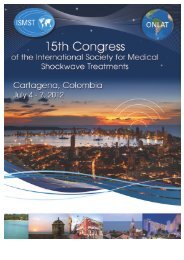
InternatIonal SocIety for MedIcal Shockwave treatMent - ISMST ...
InternatIonal SocIety for MedIcal Shockwave treatMent - ISMST ...
InternatIonal SocIety for MedIcal Shockwave treatMent - ISMST ...
Create successful ePaper yourself
Turn your PDF publications into a flip-book with our unique Google optimized e-Paper software.
<strong>Shockwave</strong><br />
International Society <strong>for</strong> Medical ShOckwave Treatment<br />
JUNE 2012 – VOLUME 8 – ISSUE 1<br />
Early Angiogenic Response to Shock Waves in a Three–Dimensional<br />
Model of Human Microvascular Endothelial Cell (HMEC – 1)<br />
MC. d’Agostino and P. Romeo<br />
Shock Waves Effects on Ca++ Deposition by Human Osteoblasts in Vitro<br />
S. Russo ET AL<br />
Mechanotransduction – Role in tissue adaptation<br />
Frank Suhr and Wilhelm Bloch<br />
Anti-Inflammatory Effects of Extracorporeal <strong>Shockwave</strong> Therapy<br />
Vlado Antonic and Alexander Stojadinovic<br />
Accumulated Total Energy Flux Density an Indicator to Compare<br />
Electrohydraulic and Piezoelectric Devices?<br />
Kerstin Neumann and Hans-Jürgen Duchstein<br />
Extracorporeal Shock Waves modulate osteogenic differentiation of<br />
human mesenchymal stem cells<br />
Roberto Frairia ET AL<br />
Unfocused Extracorporeal Shock Waves Induce Anabolic<br />
Responses in Osteoporotic Bone<br />
OP van der Jagt MD ET AL<br />
Extracorporeal <strong>Shockwave</strong> Therapy and Gene Expressions<br />
Ching-Jen Wang, M. D.
Chief Editor<br />
Paulo Roberto Dias dos Santos,MD<br />
(Brazil) - prds@uol.com.br<br />
Associate Editor<br />
Ching-Jen Wang,MD (Taiwan)<br />
w281211@adm.cgmh.org.tw<br />
EDITORIAL<br />
<strong>Shockwave</strong> Medicine<br />
Past, Present and Future<br />
Editorial Board<br />
Ana Claudia Souza,MD (Brazil)<br />
anaclaudia@cortrel.com.br<br />
Carlos Leal,MD (Columbia)<br />
leal@owc.com.co<br />
Heinz Kuderna,MD (Áustria)<br />
kuderna-unfallchir.wien@aon.ato<br />
John Furia, MD (USA)<br />
jfuria@ptd.net<br />
Leonardo Jaime Guiloff Waissbluth,MD<br />
(Chile) - lguiloff@davila.ch<br />
Ludger Gerdsmeyer,MD (Germany)<br />
gerdesmeyer@aol.com<br />
M. Cristina d’Agostino,MD (Italy)<br />
frogg@tele2.it<br />
Matthias Buch,MD (Germany)<br />
matthiasbuch@aol.com<br />
Paulo Rockett,MD (Brazil)<br />
rockett@terra.com.br<br />
Richard Thiele,MD (Germany)<br />
rithi@t-online.de<br />
Robert Gordon,MD (Canada)<br />
gordon@skockwavedoc.com<br />
Sergio Russo,MD (Italy)<br />
serghiey@hotmail.com<br />
Vinzenz Auersperg,MD (Áustria)<br />
vinzenz.auersperg@gespag.at<br />
Weil Lowel Jr,MD (USA)<br />
lweiljr@weil4feet.com<br />
Wolfgang Schaden,MD (Austria)<br />
med.eswt.schaden@aon.at<br />
Veterinary Committee<br />
Scott Mc Clure,DVM (USA)<br />
mcclures@iastate.edu<br />
Ana Liz Garcia Alves,DVM (Brazil)<br />
anaalves@fmvz.unesp.br<br />
Ana Cristina Bassit,DVM (Brazil)<br />
anafbassit@uol.com.br<br />
Editorial Office<br />
Rua Monte Alegre, 428 - conj. 56<br />
Perdizes - São Paulo - Brazil<br />
CEP 05014-000<br />
Fone: 55-11-2386-4092<br />
E-mail: prds@uol.com.br<br />
<strong>ISMST</strong> office<br />
Ebelsberger Schlossweg 5<br />
A-4030 Linz<br />
Austria - Europe<br />
Tel.: +43 (732) 302373<br />
Fax.: +43 (732) 303375<br />
E-mail: shockwave@ismst.com<br />
website: www.ismst.com<br />
<strong>Shockwave</strong> Medicine. Two words<br />
that mean much more than just<br />
a therapy. The simple concept of<br />
enhancing tissue regeneration through<br />
mechanical controlled stimuli is a<br />
great step ahead in the management of<br />
many low healing diseases. The more<br />
complex effect of neo-angiogenesis and<br />
the generation of capillaries enhancing<br />
blood supply to avascular areas through<br />
mechanotransduction is just fascinating.<br />
The findings of gene expression changes,<br />
the stimulation of cell metabolism<br />
pathways, the secret language of cell<br />
migration and activation has shown us<br />
a rather unknown life line: mechanical<br />
<strong>for</strong>ces. We have been able to translate<br />
part of this language, and create systems<br />
that can be used in reproducible<br />
parameters to obtain constant<br />
repeatable cell and tissue responses.<br />
This is <strong>Shockwave</strong> Medicine. Not an<br />
analgesic extracorporeal device, but a<br />
safe non-invasive tissue regeneration<br />
system.<br />
Two decades of basic and clinical<br />
research are very encouraging. These<br />
years of research and development have<br />
shown solid scientific results. Many of<br />
the most relevant findings in medicine<br />
and therapeutics have started this way:<br />
following a biologically logical pathway,<br />
asking simple research questions<br />
and organizing the results in simple<br />
research answers. Many of our <strong>ISMST</strong><br />
researchers started in <strong>Shockwave</strong><br />
Medicine with the surgeon’s incredulity<br />
<strong>for</strong> a funny therapeutic procedure<br />
coming from urologic lithotripsy.<br />
Prof. Dr. Carlos Leal MD<br />
Universidad el Bosque, Bogotá<br />
President of the <strong>ISMST</strong>, International Society<br />
<strong>for</strong> Medical <strong>Shockwave</strong> Treatments<br />
President of the ONLAT – Federación<br />
Iberoamericana de Sociedades y Asociaciones<br />
de Ondas de Choque en Medicina<br />
Once the results came up, the patients<br />
healed, the pain subsided, there were<br />
more questions than answers. Many<br />
turned their backs. Many continued<br />
against the wind with the clear<br />
commitment of finding why and how<br />
did this work.<br />
Orthopedic sports medicine had<br />
a new tool. <strong>Shockwave</strong> Medicine gave<br />
the opportunity to researchers to<br />
understand the physiopathology of<br />
tendinopathies. Now we know the lack<br />
of regeneration of tenocytes in tennis<br />
elbow, the poor vascularity patterns<br />
of plantar fasceiitis and the limited<br />
capacity <strong>for</strong> healing of the proximal<br />
patellar tendon. We also know that<br />
using extracorporeal stimulation we<br />
can enhance all these conditions,<br />
control pain and recover function at<br />
no risk. Pain is an emotion, so it is very<br />
hard to measure. But after 20 years of<br />
shockwave medicine I only see good<br />
results everywhere. Some bad reports<br />
have been published, but most of them<br />
with a poor design, done by nonexperts<br />
and with unknown protocols. If<br />
<strong>Shockwave</strong> Medicine was a bad thing it<br />
would have disappeared years ago. On<br />
the contrary, it grows day by day.<br />
Bone healing is the most important<br />
challenge <strong>for</strong> the orthopedic surgeon.<br />
The transition from the full stability<br />
treatment protocols from the past<br />
century to the full biology treatment<br />
of our days is quite a change. The<br />
minimally invasive fracture fixations<br />
done today by my residents would<br />
have had me expelled from my hospital<br />
2<br />
shockwave International Society <strong>for</strong> Medical ShOckwave Treatment
in the 80`s. We know now that the<br />
vascularity and biological environment<br />
of the fracture is key to bone healing.<br />
Evidence was always our enemy,<br />
until researchers like Wang, Schaden,<br />
Caccio and Rompe have shown level<br />
one EBM data that proves equivalent<br />
results in bone healing using <strong>Shockwave</strong><br />
Medicine or Surgery. The literature<br />
results, with all the possible variables in<br />
power and evidence, can only show one<br />
thing: we have at least the same results<br />
as surgery, with no complications and at<br />
a lower cost.<br />
I always remember when Prof.<br />
Wolfgang Schaden came to my<br />
hospital`s orthopedic grand round<br />
a few years ago. He showed the<br />
evidence, the data, the cases. He<br />
brilliantly answered all questions in<br />
the most scientific manner. In the end,<br />
a young orthopedic surgeon said she<br />
will never recommend <strong>Shockwave</strong><br />
Medicine in the hospital because of a<br />
conflict of interests with the providers.<br />
I must confess I was really angry with<br />
this answer. Wolfgang was smiling.<br />
I asked why. He said: When there is<br />
nothing else to argue in a medical<br />
procedure but a commercial issue, is<br />
because all the scientific answers are<br />
undisputable.<br />
Plastic surgery faces a huge challenge<br />
with tissue regeneration: complex<br />
wound healing. The poor vascularity<br />
results in delayed coverage and increases<br />
infection rates. It is costly, and the<br />
surgical solutions are usually large and<br />
painful. Bad healing causes functional<br />
and cosmetic problems that require<br />
many other interventions. <strong>Shockwave</strong><br />
Medicine has proved a solution in skin<br />
regeneration. Mechanotransduction<br />
has been studied thoroughly in skin<br />
fibroblasts with great results. The work<br />
of researchers like Stodjadinovic has<br />
shown amazing results in both in vitro<br />
and in vivo series. The clinical use<br />
of <strong>Shockwave</strong> Medicine <strong>for</strong> wound<br />
healing is now widespread, and<br />
patients with these difficult conditions<br />
are healing all over the world. The<br />
protocols <strong>for</strong> diabetic foot and burns<br />
are showing excellent results. I believe<br />
skin applications will soon be the most<br />
popular and well accepted protocol of<br />
<strong>Shockwave</strong> Medicine.<br />
Research in myocardium tissue<br />
regeneration has also been very active<br />
in the past five years. The use of<br />
extracorporeal systems <strong>for</strong> angina has<br />
evolved to the next step: intra-corporeal<br />
<strong>Shockwave</strong> stimulation of ischemic<br />
heart during open coronary by-pass<br />
surgery. The logic is there: changing<br />
the heart`s pipeline sometimes is not<br />
enough. Some kind of tissue stimulation<br />
must be used. Many researchers<br />
have found the use of intra-cardiac<br />
injected growth factors can enhance<br />
muscle regeneration. The results are<br />
encouraging but variable. The use of<br />
intra-corporeal <strong>Shockwave</strong> stimulation<br />
has proved more reliable results both in<br />
animal and human trials. We are excited<br />
to see the results that will come up in<br />
the next years.<br />
Many other applications of a<br />
non-invasive system that creates<br />
angiogenesis, tissue regeneration<br />
and pain control are being used and<br />
under investigation. The development<br />
of pressure and radial wave systems<br />
have proved a great therapeutic tool<br />
in superficial conditions such as<br />
insertional tendinopatias, trigger points<br />
and bursitis. These conditions are by<br />
far the most common consults at the<br />
<strong>Shockwave</strong> Medicine units all over<br />
the world. Providing a non-expensive,<br />
easy to use system that controls pain<br />
and regenerates local tissue is very<br />
attractive, and that is the reason <strong>for</strong> this<br />
growing field.<br />
I am very encouraged to see the<br />
results in spasticity and rehabilitation<br />
of muscle complications of strokes<br />
and cerebral palsy in the near future.<br />
Some of our <strong>ISMST</strong> researchers are<br />
moving <strong>for</strong>ward on this field. The<br />
local stimulation of bone turnover in<br />
osteoporotic patients may be the long<br />
seeked solution <strong>for</strong> prophylaxis in the<br />
contralateral hip in elder femoral neck<br />
fractures. The use of shockwaves in<br />
subchondral stimulation of articular<br />
cartilage lesions has proved some<br />
preliminary results. We hope to show<br />
some more numbers in the near future.<br />
The main goal of a scientific society<br />
like the <strong>ISMST</strong> is to provide the<br />
direction of all ef<strong>for</strong>ts from our corner<br />
of science to improve health. We are<br />
the world experts in tissue regeneration<br />
by <strong>Shockwave</strong> Medicine, and we are<br />
responsible to keep the technology<br />
in the perfect balance between<br />
safety and efficacy. This is the place<br />
where scientists, clinicians, surgeons,<br />
researchers, industry, governments and<br />
insurance companies find a common<br />
ground to put all ef<strong>for</strong>ts together. We<br />
work on this goal day by day.<br />
Every year we get together and<br />
tell each other what we are doing in<br />
<strong>Shockwave</strong> Medicine. Every year the<br />
<strong>ISMST</strong> gives the world an update of this<br />
fascinating and growing technology.<br />
This year we have a beautiful congress<br />
that brings together the world leaders<br />
to discuss in 14 keynote lectures,<br />
51 scientific papers and one fullday<br />
Instructional Course the best<br />
knowledge update in <strong>Shockwave</strong><br />
Medicine.<br />
I have been privileged not only<br />
to direct the ef<strong>for</strong>ts of the <strong>ISMST</strong> in<br />
the past year, but also to organize the<br />
XV world congress in my country,<br />
Colombia. The city of Cartagena is<br />
ready to welcome all the <strong>ISMST</strong> family<br />
in the best Caribbean atmosphere.<br />
We will be able to discuss science and<br />
to meet our friends from all over the<br />
world. This is also the first congress<br />
of ONLAT, the Ibero-American<br />
<strong>Shockwave</strong> Medicine Society as a<br />
regional intercontinental federation.<br />
We expect to have the best congress<br />
ever, and we are committed to it.<br />
There is a lot of past, present and<br />
future of <strong>Shockwave</strong> Medicine. It is<br />
the mission of the <strong>ISMST</strong> to keep it<br />
growing.<br />
Prof. Dr. Carlos Leal MD<br />
Bosque University - Bogota<br />
<strong>ISMST</strong> & ONLAT President<br />
JUNE 2012 – VOLUME 8 – ISSUE 1<br />
3
EDITORIAL<br />
Richard Thiele,MD<br />
Member of the <strong>ISMST</strong><br />
Supervisory Board<br />
Inside this Issue<br />
Editorial ··················································· 2<br />
Early Angiogenic Response to<br />
Shock Waves in a Three–Dimensional<br />
Model of Human Microvascular<br />
Endothelial Cell (HMEC – 1) ·················· 5<br />
Dear <strong>ISMST</strong> Colleagues and Friends<br />
around the world,<br />
As you can see from our current<br />
8th issue of the newsletter its look<br />
and layout have received a complete<br />
makeover. We have embarked on an<br />
update and hope the redesign meets with<br />
your approval. Alongside the annual<br />
<strong>ISMST</strong> Conference, we will, as usual, be<br />
publishing research works of interest on<br />
musculoskeletal shockwave therapy.<br />
Acceptance of this therapy continues<br />
to gain ground. Ever more people the<br />
world over are gaining access to this<br />
exciting therapy. The Austrian National<br />
Institute of Health, <strong>for</strong> instance, has<br />
designated the treatment as the “therapy<br />
of choice” in the care of pseudarthrosis.<br />
Consequently, if a physician in Austria<br />
operates on a patient without first<br />
proposing shockwave therapy, this<br />
practitioner may become criminally liable.<br />
It is encouraging to see that ESWT<br />
is increasingly being covered by<br />
insurances and medical plans. A survey<br />
on the current state of affairs regarding<br />
international acceptance, application<br />
and medical coverage will be presented<br />
at the end of the conference. <strong>Shockwave</strong><br />
therapy is nowadays finding application<br />
in urology, orthopedics, surgery,<br />
dermatology, plastic surgery, dental and<br />
veterinarian medicine.<br />
There are hundreds of valid studies<br />
worldwide, and all of which would<br />
not have been possible without basic<br />
research. Only through basic research<br />
work has it been possible to discover<br />
ESWT’s multifaceted mechanisms of<br />
action. It was believed 30 years ago<br />
that shockwaves could effectively break<br />
up kidney stones, calcification, e.g. in<br />
the shoulder, and thereby generate a<br />
healing effect. Today we know that the<br />
mechanical impact is a mere secondary<br />
effect, that these waves can be healing<br />
and pain relieving. They have a positive<br />
impact on the metabolism of living cells<br />
and can moreover, as these studies have<br />
demonstrated, have a beneficial effect on<br />
mesenchymal stem cells.<br />
This has led to the broadening of<br />
ESWT’s spectrum of mechanism of<br />
action on bones, tendons, skin, nerves<br />
and the heart muscle.<br />
Because basic research is so<br />
vitally important, also <strong>for</strong> the further<br />
development of this therapy, a gathering<br />
of many of the world’s leading scientists<br />
in Innsbruck (Austria) was held a second<br />
time this January <strong>for</strong> the purpose of<br />
presenting and exchanging the latest<br />
findings in basic research. Meetings of<br />
this kind should continue to take place<br />
<strong>for</strong> the coordination of scientific research<br />
around the world and the exchange of<br />
knowledge at the highest levels.<br />
So that these exciting discoveries<br />
can be made available to the public, we<br />
are publishing the presentations of the<br />
most important events in this issue of<br />
the newsletter. For this reason, we would<br />
like to express our heartfelt gratitude to<br />
the authors Prof. Wang (Taiwan), Prof.<br />
Frairia (Italy), Prof. Bloch (Germany),<br />
Dr. dÀgostini (Italy), Dr. Antonic (USA),<br />
Dr. Neumann (Germany) and Dr. van<br />
der Jagd (Netherlands).<br />
Un<strong>for</strong>tunately a few of the<br />
presentations on research findings on the<br />
heart and skin are not yet available, but<br />
which we hope to publish later on.<br />
I wish all of you great enjoyment in<br />
the reading of these important works and<br />
most of all lots of fun and new discoveries<br />
at the 15th International Congress of the<br />
<strong>ISMST</strong> in Cartagena led by our President<br />
Professor Carlos Leal.<br />
Yours sincerely,<br />
Dr. Richard Thiele<br />
Shock Waves Effects on Ca ++<br />
Deposition by Human Osteoblasts<br />
in Vitro ···················································· 9<br />
Mechanotransduction – Role in<br />
tissue adaptation ······························· 14<br />
Anti-Inflammatory Effects<br />
of Extracorporeal <strong>Shockwave</strong><br />
Therapy ·················································· 16<br />
Accumulated Total Energy Flux<br />
Density an Indicator to Compare<br />
Electrohydraulic and<br />
Piezoelectric Devices? ······················· 18<br />
Extracorporeal Shock Waves<br />
modulate osteogenic<br />
differentiation of human<br />
mesenchymal stem cells ················· 20<br />
Unfocused Extracorporeal<br />
Shock Waves Induce Anabolic<br />
Responses in Osteoporotic<br />
Bone ······················································· 22<br />
Extracorporeal <strong>Shockwave</strong><br />
Therapy and Gene Expressions ······· 23<br />
4<br />
shockwave International Society <strong>for</strong> Medical ShOckwave Treatment
Early Angiogenic Response to Shock Waves<br />
in a Three–Dimensional Model of Human<br />
Microvascular Endothelial Cell (HMEC – 1)<br />
1<br />
MC. d’Agostino 1 and P. Romeo 2<br />
1 Rehabilitation Department, IRCCS Istituto Clinico Humanitas<br />
2 Orthopaedic Department, Università degli Studi di Milano,<br />
IRCCS Istituto Ortopedico Galeazzi (Milan, Italy)<br />
In the last few decades the field<br />
of Shock Waves (SW) has been<br />
characterized by some great progresses:<br />
their clinical applications have widened<br />
from the urological field (lithotripsy)<br />
to the treatment of both inflammatory<br />
and degenerative orthopaedic diseases<br />
(tendons, ligaments, soft tissues and bone<br />
pathology) (Valchanou and Michailov<br />
1995, Schaden et al. 2001, Wang et al.<br />
2005, C. d’Agostino et al. 2011).<br />
Moreover, in recent years, they have<br />
been applied also in the field of wound<br />
care management and “scar pathologies”,<br />
thank to their tissue trophic effect,<br />
generated by the dissipation of the high<br />
pressure focused acoustic waves into low<br />
energy unfocused shock waves (uSW)<br />
(Schaden et al. 2007, Romeo et al. 2011).<br />
As a general concept: SW in<br />
orthopaedics are not used to<br />
disintegrate tissues, rather to induce<br />
neovascularization, improve blood<br />
supply, and tissue regeneration<br />
(Wang CJ, 2003). This is possible<br />
thanks to mechanotransduction, that is<br />
the possibility to convert a mechanical<br />
stimulation (SW) into a series of<br />
biological reactions at the tissue level;<br />
this mechanism is active in all cells that<br />
are responsive to physical stimulations.<br />
Experimental data suggest that<br />
one of the main biological effects<br />
induced by mechanical stimulation of<br />
SW is the production of Nitric Oxide<br />
(NO) (Mariotto et al. 2005), which is<br />
well known to promote angiogenesis<br />
(<strong>for</strong>mation of new blood vessels from<br />
pre-existing capillaries). Angiogenesis is<br />
itself one of the first step in tissue healing<br />
and it is induced by a variety of growth<br />
and angiogenic factors (Cooke and<br />
Losordo 2002).<br />
Some other studies suggest that<br />
mechanical stimulation produced by<br />
SW can have also a direct effect on the<br />
ExtraCellular Matrix (ECM), which<br />
further triggers cytoplasmatic and<br />
nuclear reactions, varying according to<br />
the experimental model, the energy level,<br />
the number of impulses and the cell type<br />
(Speed 2004). As already demonstrated<br />
<strong>for</strong> some other specific biomechanical<br />
stimuli, SW could induce some<br />
biochemical reactions in responsive cells,<br />
thus affecting growth, development,<br />
differentiation, apoptosis regulation and<br />
gene expression via signal transduction<br />
pathways (Tarbell et al. 2005).<br />
In particular, from the angiogenetic<br />
point of view, in vivo and in vitro models<br />
demonstrated the pro - angiogenic<br />
activity of extracorporeal SWs, via an<br />
up - regulation of mRNA levels <strong>for</strong><br />
VEGF proliferation and differentiation<br />
of VEGFR-2 positive EC, and NO<br />
production (Wang CJ 2003, Mariotto S et<br />
al. 2009).<br />
As already expressed, cell<br />
responsiveness to exogenous stimulation<br />
is correlated to a series of metabolic<br />
activities comparable, as a result, to<br />
the effects of Growth Factors (GF) on<br />
cellular transcription; in particular,<br />
endothelial cells (EC) are particular<br />
mechano - sensitivite cells.<br />
It has been described that definite<br />
hemodynamic characteristics of the<br />
laminar flow induce, from 1 to 6 hours<br />
after stimulation, a regulatory effect<br />
on eNOS (endothelial NO – Synthase)<br />
and ICAM-1 (IntraCellular Adhesion<br />
Molecule) activation; both of them,<br />
in some way, can be considered as<br />
intracellular signalling events (Stolz et al.<br />
2002, Davies et al. 1997).<br />
JUNE 2012 – VOLUME 8 – ISSUE 1 5
Early Angiogenic Response to Shock Waves in a Three–Dimensional Model of Human Microvascular Endothelial Cell (HMEC – 1)<br />
Mechanical stimuli (as shear<br />
stresses) are described to induce<br />
hemodynamic responses of Endothelial<br />
Cells (EC), into different phases and<br />
times:<br />
• EARLY RESPONSE (within some<br />
minutes /seconds, characterized by the<br />
activation of ion channels ad second<br />
messengers, as NO);<br />
• INTERMEDIATE RESPONSE<br />
(within minutes/hours, characterized by<br />
endocytosis, cell replication, gene up/<br />
down - regulation);<br />
• LATE RESPONSE (within hours<br />
/days, during which you can observe<br />
endothelial cells adaptation; structural<br />
and funtional adaptation are NO –<br />
mediated).<br />
(Resnick et al. 2002).<br />
More in details, after mechanical<br />
stimulation as shear stresses, the<br />
angiogenetic and antiapoptotic effects<br />
would interest some particular cellular<br />
compartments, engaged with selective<br />
sites of eNOS (Dimmeler S et al. 1999,<br />
Balligand et al. 2009).<br />
FOR SUMMARIZING<br />
• on UNSTIMULATED CELLS:<br />
eNOS is maintained in inactive state,<br />
through its association with Cav-1<br />
(Caveoline);<br />
• after MECHANICAL<br />
STIMULATION (shear stresses), it<br />
is possible to observe the following<br />
reactions (if there is an adeguate<br />
availability of substrate (Calmoduline)<br />
and Arginine Transporter CAT-1)<br />
(Traub O et al. 1998):<br />
- eNOS - Cav1 dissociation<br />
- eNOS activation<br />
How to explain in detail angiogenesis<br />
and which effects on human endothelial<br />
cell lines, soon after uSW stimulation ?<br />
On the basis of what above described,<br />
it is reasonable some questions arise:<br />
can SW, under definite experimental<br />
conditions, simulate the effects of<br />
shear stresses and stimulate endothelial<br />
cells? What does it happen in the earlier<br />
stages of EC stimulation ?<br />
Aim of our study was to verify the<br />
capability of uSW to induce new vessels<br />
proliferation (neoangiogenesis) in vitro<br />
and, at the same time, to investigate the<br />
initial response of endothelial cells to this<br />
type of acoustic stimulation. For this<br />
purpose we employed an in vitro system,<br />
consisting of a matrix support, seeded<br />
with microvascular endothelial cells<br />
which resembles, as closely as possible,<br />
the structure of the natural tissues<br />
(Sansone V et al, 2012). By reproducing<br />
the architecture of a mechano-sensitive<br />
structure, such as the capillary network,<br />
one may provide a valid model <strong>for</strong><br />
analyzing the behaviour of EC (HMEC<br />
- 1), when subjected to an acoustic signal<br />
comparable to shear stress.<br />
HMEC-1 is the first immortalized<br />
human microvascular endothelial cell<br />
line, that retains the morphologic,<br />
phenotypic, and functional<br />
characteristics of normal human<br />
microvascular endothelial cells.<br />
When endothelial cells are plated on<br />
BD Matrigel, they can <strong>for</strong>m a three<br />
dimensional network of capillary<br />
tubes comparable to the final step of<br />
the angiogenic cascade (Ades EW<br />
et al. 1992).<br />
In our experimental experience, cell<br />
cultures were stimulated with uSW,<br />
according to various protocols (different<br />
energies and number of shots).<br />
For angiogenesis experiments, cells<br />
were grown in 24-well plates on Matrigel<br />
matrix and vessels-like structures were<br />
quantified by counting the capillary<br />
connections under an inverted<br />
microscope.<br />
The most responsive group<br />
in terms of numbers of capillary<br />
connections underwent gene expression<br />
analysis using the Super Array kit-<br />
Signal Transduction Pathway Finder<br />
(SABiosciences, Qiagen), able to profile<br />
84 key genes representative of 18<br />
different signal transduction pathways.<br />
After 12 hours, the treated cells<br />
showed a substantial increase in the<br />
number of new vessel-like structures<br />
if compared to untreated cells. This<br />
morphological differentiation was more<br />
remarkable in the samples that were<br />
exposed to low energies and limited<br />
numbers of shots (fig. 1 and 2).<br />
As reported by other authors (Steinbach<br />
et al. 1993), we observed a disaggregation<br />
of the Matrigel scaffold and a negative<br />
effect on the <strong>for</strong>mation of capillary<br />
connections at higher energies (data<br />
not published).<br />
In samples showing the most<br />
marked increase in number of capillary<br />
connections, we observed a decreased<br />
gene expression 3 hours after uSW<br />
treatment. Indeed, cells showed a strong<br />
down-regulation of genes involved in the<br />
apoptotic process (BAX, anti-apoptotic<br />
BCL2LI, GADD45A, PRKCA), also<br />
in the cell cycle (CDKN2C, CEBPB,<br />
HK2, IRF1, PRKCA), oncogenes (JUN,<br />
WNT1), cell adhesion (ICAM-1), and<br />
proteolytic systems (CTSD, KLK2,<br />
MMP10).<br />
However, we did not observe any<br />
increased expression of receptors <strong>for</strong><br />
angiogenic agents like endothelial nitric<br />
oxide synthase (eNOS) or vascular<br />
endothelial growth factor (VEGF).<br />
• These preliminary results seem to<br />
indicate that endothelial cells in vitro<br />
quickly respond to SW, by proliferating<br />
and <strong>for</strong>ming vessel-like structures,<br />
depending on the energy level<br />
employed and the number of shocks<br />
released.<br />
In the first 3 hours after SW<br />
stimulation we did not observe either<br />
VEGF or eNOS modulation, whereas<br />
other authors were able to demonstrate<br />
the production of VEGF and the<br />
increased expression of the specific<br />
angiogenesis pathway after 6 hours<br />
(Stojadinovic A, et al. 2008, Wang FS et<br />
al. 2004).<br />
• Nevertheless, the osberved<br />
downregulation of the genes involved<br />
in cell cycle and cell adhesion, could be<br />
interpreted as the preparatory signal<br />
correlated to an upcoming detachment<br />
of endothelial junctions, in other<br />
words: the “first reactive response” of<br />
the endothelial cells to the external<br />
stimuli and the prelude to the events<br />
characterizing the neo-angiogenic<br />
sequence.<br />
Comments<br />
As already mentioned, Endothelial<br />
cells (ECs) are mechano-sensitive cells<br />
which physiologically react to flow<br />
shear stress. Particular regions of the<br />
cell membrane seem to be involved in<br />
the recognition of the different features<br />
of the laminar flow (Fleming and Busse<br />
1999; Ziegler et al. 1998; Barakat et al.<br />
2006) which are subsequently transferred<br />
6<br />
shockwave International Society <strong>for</strong> Medical ShOckwave Treatment
MC. d’Agostino and P. Romeo<br />
to the cytoskeleton (Corson et al. 1996;<br />
Davies et al. 1997). Those cellular<br />
compartments engaged with selective<br />
sites of eNOS are thought to mediate the<br />
angiogenic (Baum et al. 2004) and antiapoptotic<br />
effect (Dimmeler et al. 1999) of<br />
the shear stress.<br />
In clinical practice, several vascular<br />
pathologies are characterized by<br />
an intrinsic ECs dysfunction and a<br />
diminished production of growth factors<br />
(GF) (Madeddu 2005). Hence, new<br />
therapeutic options attempt to correct<br />
this sort of “biologic imbalance” by<br />
inducing neovascularisation, a process<br />
which can be achieved by supplementing<br />
the VEGF either via gene therapy or<br />
transplanting endothelial progenitor cells<br />
(Madeddu 2005; Chenggang et al. 2006).<br />
On the other hand, SW stimulation<br />
represents an alternative, and innovative,<br />
therapeutic approach in those<br />
conditions where a strong angiogenic<br />
impulse is required – <strong>for</strong> example in<br />
severe skin wounds (Schaden et al.<br />
2007) or in myocardial ischemic lesions<br />
(Fukumoto et al. 2006). Moreover,<br />
recent experimental studies suggest<br />
that the treatment of ischemic tissue<br />
with low energy SWs improves the<br />
recruitment of circulating endothelial<br />
progenitor cells (EPCs) due to enhanced<br />
expression of specific chemo-attractant<br />
factors (Aicher et al. 2006).<br />
But, if the late neoangiogenic<br />
response has been documented<br />
adequately, much less attention has been<br />
given to the very early changes induced<br />
by mechanical stimulus. Our study was<br />
established to investigate the early effects<br />
of unfocused shock waves on HMEC-1<br />
cultured in a three-dimensional Matrigel<br />
model, where cells were stimulated<br />
using a source of low energy unfocused<br />
shock waves with a pulsation frequency<br />
of 3 Hz per second. The experimental<br />
Matrigel model, as with any in vitro<br />
model, is obviously not a perfect replica<br />
of the biological tissues. However,<br />
by reproducing the architecture of a<br />
mechano-sensitive structure such as<br />
the capillary network, it is possible to<br />
provide a valid model <strong>for</strong> analyzing the<br />
behaviour of ECs when submitted to<br />
an acoustic signal comparable to shear<br />
stress, since with defocused waves the<br />
flow of the acoustic pulse is close to<br />
being laminar.<br />
HMEC–1 are different from the<br />
lining EC of large vessels, and are<br />
thought to be involved in angiogenesis<br />
and in wound healing. We demonstrated<br />
that unfocused shock waves induced<br />
a quick morphological response (12<br />
hours), characterized by a significant<br />
increase of vessel-like structures<br />
<strong>for</strong>mation (Sansone V et al. 2012).<br />
As described above, the proangiogenic<br />
effects of SW are most<br />
likely mediated by VEGF and NO. In<br />
our study, we did not observe either<br />
VEGF or eNOS modulation 3 hours<br />
after SW stimulation; we demonstrated<br />
a significant down-regulation of genes<br />
involved in the apoptotic process, in cell<br />
cycle and adhesion, and in proteolytic<br />
systems. However, this SW-induced<br />
modulation, which is more significant<br />
<strong>for</strong> the antiapoptotic genes and could<br />
represent the “early reactive response”<br />
of HMEC-1 to the physical impulse<br />
induced by the uSW. These observations<br />
compare favorably with Kim and Von<br />
Recum (2008) who remarked that<br />
mechanical stimulus induced by shear<br />
stress could improve the differentiation<br />
EPCs in ECs, regulating the expression<br />
of several genes involved in apoptosis<br />
and inducing EPCs to <strong>for</strong>m capillary–<br />
like networks in 3D cultures.<br />
In conclusion, our results seem to<br />
confirm that some aspects of the early<br />
gene response of ECs to uSW stimulation<br />
are comparable to those of the laminar<br />
shear stress flow, mainly characterized by<br />
an anti-apoptotic effect.<br />
Further experimental studies are<br />
necessary to validate these hypotheses<br />
and to investigate if the biological<br />
response of EC to SW stimulation<br />
involves the same intercellular pathways<br />
and regulatory mechanisms that<br />
characterise other types of biophysical<br />
stimuli. At the same time, new research<br />
into the gene expression could shed<br />
light on the triggering of the angiogenic<br />
process when acoustic stimulation is<br />
applied.<br />
FIGURES<br />
Fig. 1: Count of capillary<br />
connections (bifurcations) in<br />
HMEC-1 seeded at different cell<br />
concentrations, 12 hours after<br />
low energy uSW treatment (E1)<br />
with different number of shots<br />
(200 shots, light grey bars; 800<br />
shots, dark grey bars). Untreated<br />
HMEC-1 are shown as negative<br />
control (white bars). Under E1<br />
condition the behaviour of EC<br />
was related to the number of<br />
shots applied; indeed at all<br />
densities, cells receiving 200 shots<br />
showed a significant increase in<br />
the number of bifurcations in<br />
comparison to untreated cells. On<br />
the other hand, E1 uSW treatment<br />
with 800 shots was able to<br />
induce a significant increase in<br />
the number of bifurcations just<br />
in HMEC-1 cells plated at high<br />
density (10000 cells/well) in<br />
comparison to untreated cells<br />
(Sansone V et al, 2012).<br />
Fig. 2: Detail of strict capillary connections and<br />
cellular organization in HMEC-1 culture treated<br />
with uSW (E1, 200 shots) (Sansone V et al, 2012).<br />
JUNE 2012 – VOLUME 8 – ISSUE 1<br />
7
Early Angiogenic Response to Shock Waves in a Three–Dimensional Model of Human Microvascular Endothelial Cell (HMEC – 1)<br />
MC. d’Agostino and P. Romeo<br />
REFERENCES<br />
Aicher A, Heeshen C, Sasaki K, Urbich C, Zehier A,<br />
Dimmeler S. Low Energy Shock Waves <strong>for</strong> Enhancing<br />
Recruitment of Endothelial Progenitor Cells. A new<br />
modality to increase efficiency of cell therapy in<br />
chronic hind limb ischemia. Circulation 114 (2006),<br />
pp. 2823-2830.<br />
Ades EW, Candal FJ, Swerlick RA et all. HMEC-<br />
1: establishment of an immortalized human<br />
microvascular endothelial cell line. J Invest Dermatol.<br />
99(6), (dec. 1992), pp. 683-90.<br />
Balligand et al. eNOS Activation by Physical Forces:<br />
From Short-Term Regulation of Contraction to<br />
Chronic Remodeling of Cardiovascular Tissues.<br />
Physiol Rev 89(2) (2009), pp 481-534.<br />
Barakat AI, Lieu DK, Gojova A. Secrets of the code: do<br />
vascular endothelial cells use ion channels to decipher<br />
complex flow signals? Biomaterials 27 (2006), pp.<br />
671-678.<br />
Baum O, Da Silva- Azevedo L, Willerding G, Wockel<br />
A, Planitzer G, Gossrau R, Pries AR, Zakrzewicz<br />
A. Endothelial NOS is main mediator <strong>for</strong> shear<br />
stress–dependent angiogenesis in skeletal muscle<br />
after prazosin administration. Am J Physiol Heart Circ<br />
Physiol. 287 (2004), pp. H2300–H2308.<br />
Chenggang Y, Wei X, Yan Z, Lingxi Z, Maoguo S, Jie<br />
L, Yan H, Shuzhong G. Transplantation of Endothelial<br />
Progenitors Cells Transferred By Vascular Endothelial<br />
Growth Factor Gene For Vascular Regeneration of<br />
Ischemic Flaps. J Surg Res. 135 (2006), pp. 100-106.<br />
Cooke JP, Losordo DW. Nitric Oxide and<br />
Angiogenesis. Circulation 105 (2002), pp. 2133-2135.<br />
Corson MA, James NL, Latta SE, Nerem RM, Berck<br />
BC, Harrison DG. Phos<strong>for</strong>ylation of Endothelial Nitric<br />
Oxide Syntetase in response to fluid shear stress. Circ<br />
Res 79 (1996), pp. 984-991.<br />
d’Agostino C, Romeo P, Amelio E, Sansone V.<br />
Effectiveness of ESWT in the treatment of Kienbock’s<br />
disease. Ultrasound in Med & Biol. Vol. 37 (n° 9)<br />
(2011), pp. 1452 – 1457.<br />
Davies PF, Barbee KA, Volin MV, Robotewskyj A,<br />
Chen J, Lorenz J, Griem ML, Wernick MN, Jacobs<br />
E, Polaceck DC, De Paola N, Barakat A. Spatial<br />
relationships in early signals events of flow mediated<br />
endothelial mechanotransduction. Annu Rev Physiol.<br />
59 (1997), pp. 527-49.<br />
Dimmeler S, Hermann C, Galle J, Zehier AM.<br />
Upregulation of superoxide dismutase and nitric oxide<br />
synthase mediates the apoptosis suppressive effect of<br />
shear stress on endothelial cells. Arterioscler Thromb<br />
Vasc Biol. 19 (1999), pp. 656-664.<br />
Fleming I, Busse R. Signal transduction of eNOS<br />
activation. Cardiovasc Res. 43 (1999), pp. 532-541.<br />
Fukumoto Y, Ito A, Uwatoku T, Matoba T, Kishi T,<br />
Tanaka H, Takeshita A, Sunagawa K, Shimokawa H.<br />
Extracorporeal cardiac shock wave therapy ameliorates<br />
myocardial ischemia in patients with severe coronary<br />
artery disease. Coron. Artery Dis. 17 (2006), pp.63-70.<br />
Kim S, Von Recum H. Endothelial stem cells and<br />
precursors <strong>for</strong> tissue engineering: cell source,<br />
differentiation, selection, and application. Tissue Eng<br />
Part B Rev. 14 (2008), pp. 133-47.<br />
Fukumoto Y, Ito A, Uwatoku T, Matoba T, Kishi T,<br />
Tanaka H, Takeshita A, Sunagawa K, Shimokawa H.<br />
Extracorporeal cardiac shock wave therapy ameliorates<br />
myocardial ischemia in patients with severe coronary<br />
artery disease. Coron. Artery Dis. 17 (2006), pp.63-70.<br />
Madeddu P. Therapeutic Angiogenesis and<br />
Vasculogenesis <strong>for</strong> tissue regeneration. Exp. Physiol. 90<br />
(2005), pp. 315-326.<br />
Mariotto S, Cavalieri E, Amelio E, Ciampa AR,<br />
De Prati AC, Marlinghaus E, Russo S, Suzuki H.<br />
Extracorporeal Shock Waves: from lithotripsy to antiinflammatory<br />
action by NO production. Nitric Oxide<br />
12 (2005), pp. 89-96.<br />
Mariotto S, Carcereri de Prati A, Cavalieri E, Amelio<br />
E, Marlinghaus E, Suzuki H. Extracorporeal shock<br />
wave therapy in inflammatory diseases. Molecular<br />
mechanisms that trigger anti-inflammatory action.<br />
Curr. Med. Chem. 16 (2009), pp. 2366 – 2372.<br />
Resnick N and Gimbrone MA Jr. Hemodynamic<br />
<strong>for</strong>ces are complex regulators of endothelial cell gene<br />
expression. The FASEB Journal, Vol 9 (2002), pp. 874<br />
– 882.<br />
Romeo P, d’Agostino MC, Lazzerini A, Sansone V.<br />
Extracorporeal Shock Waves Therapy in Pillar Pain<br />
after carpal tunnel release: a preliminary study.<br />
Ultrasound in Med & Biol. Vol 37 (n° 10), (2011), pp.<br />
1603 – 1608.<br />
Sansone V, d’Agostino MC, Bonora C, Sizzano F, Di<br />
Girolamo L, Romeo P. Early angiogenic response to<br />
shockwaves in a three – dimensional model of Human<br />
Microvascular Endothelial Cell (HMEC – 1). Journal<br />
of Biological Regulators and Homeostatic Agents, Vol 26<br />
(n° 1), (2012), pp 29 – 37.<br />
Schaden W, Fischer A, Seiler A. Extracorporeal shock<br />
wave therapy of non–union or delayed osseous union.<br />
Clin Orthop 387 (2001), pp. 90-94<br />
Schaden W, Thiele R, Kölpl C, Pusch M, Nissan A,<br />
Attinger CE, Maniscalco-Theberge ME, Peoples GE,<br />
Elster EA, Stojadinovic A. Shock wave therapy <strong>for</strong><br />
acute and chronic soft tissue wounds: a feasibility<br />
study. J Surg Res. 143 (2007), pp. 1-12.<br />
Speed CA. Extracorporeal Shock Wave Therapy in<br />
Management of Chronic Soft-tissue Conditions. J Bone<br />
Joint Surg Br 86B (2004), pp. 165-71.<br />
Steinbach P, Hofstaedter F, Nicolai H, Roessler W,<br />
Wieland W. Determination of the energy-dependent<br />
extent of vascular damage caused by high-energy<br />
shock waves in an umbilical cord model. Urol Res. 21<br />
(1993), pp. 279-282.<br />
Stojadinovic A, Elster AE, Khairul A, Douglas T,<br />
Mihret A, Zins S, Thomas AD. Angiogenic response to<br />
extracorporeal shock wave treatment in murine skin<br />
isografts. Angiogenesis. 11 (2008), pp. 369–380.<br />
Stoltz JF, Wang X. From biomechanics to<br />
mechanobiology . Biorehology 39 (2002), pp. 5 - 10.<br />
Tarbell JM, Weinbaum S, Kamm RD. Cellular Fluid<br />
Mechanics and Mechanotransduction. Ann Biomed<br />
Eng 12 (2005), pp. 1719-1723.<br />
Traub O, Berck CB, Laminar Shear Stress :<br />
Mechanism by Which Endothelial Cells Transduce an<br />
Atheroprotective Force. Arterioscler Thromb Vasc Biol.<br />
18 (1998), pp. 677-685.<br />
Valchanou VD, Michailov P. High energy shock<br />
waves in the treatment of delayed and non-union of<br />
fractures. Int. Orthop. 15 (1991), pp. 181-184.<br />
Wang CJ, Wang FS, Yang, KD, Weng LH , Hsu CC,<br />
Huang CS, Yang LC. Shock wave therapy induces<br />
neovascularisation at the tendon-bone junction. A<br />
study in rabbits. J Orthop Res 6 (2003), pp. 984-989.<br />
Wang, CJ. An overview of shock wave therapy in<br />
musculoskeletal disorders. Chang Gung Med. 26<br />
(2003), pp. 220 - 232.<br />
Wang CJ, Wang FS, Huang CC, Yang KD, Weng LH,<br />
Huang HY. Treatment <strong>for</strong> osteonecrosis of the femoral<br />
head: comparison of extracorporeal shock waves with<br />
core decompression and bone-grafting. J Bone Joint<br />
Surg Am. 87 (2005), pp. 2380-2387.<br />
Wang FS, Wang CJ, Huang HJ. RAS induction of<br />
superoxide activates ERK-dependent angiogenic<br />
transcription factor HIF-1 α and VEGF-A expressions<br />
in shock wave-stimulated osteoblasts. J Biol Chem. 279<br />
(2004), pp. 10331-10337.<br />
Ziegler T, Silacci P, Harrison VJ, Hayoz D. Nitric<br />
oxide synthase expression in endothelial cells exposed<br />
to mechanical <strong>for</strong>ces . Hypertension. 32 (1998), pp.<br />
351-355.<br />
Zimpfer D, Seyedhossein A, Holfeld J, Thomas A,<br />
Dumfarth J, Rosenhek R, Czerny M, Schaden W,<br />
Gmeiner M, Volner E, Grimm M. Direct epicardial<br />
Shock Wave Therapy improves ventricular function<br />
and induces angiogenesis in ischemic heart failure. J<br />
Thorac Cardiovasc Surg. 137 (2009), pp. 963–970.<br />
8<br />
shockwave International Society <strong>for</strong> Medical ShOckwave Treatment
Shock Waves Effects on Ca ++ Deposition<br />
by Human Osteoblasts in Vitro<br />
1*<br />
S. Russo 1* , L. Vallefuoco 2 , M. D’Anna 2 , , H. Suzuki 3 , A. Ciampa 3 ,<br />
E. Marlinghaus 4 , E. M. Corrado 1 , S. Montagnani 2<br />
1 Orthopaedics Clinic Dept. of Surgical and Orthopaedic Sciences<br />
2 Dept. of Biomorphological and Functional Sciences, Faculty of Medicine,<br />
“Federico II” University, Naples<br />
3 Dept. of Morphological and Biomedical Sciences, University of Verona<br />
4 Applied Research Center, Storz Medical AG, Kreuzlingen, Switzerland<br />
Correspondent<br />
Sergio Russo<br />
Orthopedics Clinic Dept.<br />
of Surgical and Orthopedic<br />
Sciences, Faculty of Medicine,<br />
“Federico II”University,<br />
Via Pansini 5, I - 80131 Naples<br />
e-mail: serghiey@hotmail.com<br />
Background<br />
In order to contribute to clarify the biological effects of Shock Waves (SW) treatment,<br />
we per<strong>for</strong>med it on in vitro cultures of human osteoblasts. Here we evaluate the effects<br />
of SW on cell proliferation, Ca++ deposition and ALP and NOS activities.<br />
Methods<br />
In vitro cultured human osteoblasts obtained from surgical fragments were submitted<br />
to three SW applications. Ca ++ deposition, ALP and NOS activities and cell growth<br />
rate and differentiation were evaluated and data related with control group of cells.<br />
Results<br />
SW treatment can inhibit both growth rate both Ca ++ deposition. In the same time SW<br />
modifies the NOS activity in human osteoblasts, so presumably affecting ALP activity.<br />
Interpretation<br />
The low rate of Ca ++ deposition induced by SW treatment may be due to a decrease<br />
in enzymatic activity linked to bone mineralization, such as ALP activity, as well as to<br />
change in Ca ++ intra and extracellular flow.<br />
The use of Shock Wave (SW) in<br />
orthopaedic disease was reviewed with<br />
special regard to the clinical application<br />
(Haupt G,1997). Our interest was appointed<br />
on some diseases of bone and tendons,<br />
as abnormalities in intratendineous<br />
calcification, which are restored to normal<br />
conditions after SW treatment.<br />
A SW is a transient pressure<br />
disturbance that propagates rapidly in<br />
three-dimensional space; it is associated<br />
with a sudden rise from ambient pressure<br />
to its maximum pressure and with a<br />
cavitation due to the negative phase of<br />
the wave propagation (Ogden JA et al,<br />
2001). Various authors have studied the<br />
effects of SW on normal and pathological<br />
cells (Smits GA et al, 1991), and recently<br />
this physical stimulation has been shown<br />
to cause a transient increase in the<br />
permeability of the cell membrane. In<br />
fact it causes the <strong>for</strong>mation of dimples on<br />
cell membrane and membrane potential<br />
hyperpolarization (Martini L et al. 2005).<br />
In vivo and in vitro studies have<br />
shown that mechanical stimulation is<br />
associated with physiological changes<br />
in bone cells (Wang FS et al. 2003). The<br />
response to mechanical stimuli in bone<br />
cells is associated with an increase in<br />
ion-channel opening, an alteration in<br />
membrane potential, in cell proliferation<br />
and in the synthesis activity of proteins<br />
related to the regulation of bone<br />
<strong>for</strong>mation and bone turnover (Harter LV<br />
et al. 1995).<br />
As biological effects of these<br />
applications on Ca ++ cellular metabolism<br />
are nevertheless quite unknown, our<br />
study was focused on the morphology,<br />
the proliferation and the enzymatic<br />
activities related to mineralization<br />
like Alkaline Phosphatase (ALP) of<br />
in vitro cultured human osteoblasts.<br />
In vivo conditions of SW treatment<br />
are obviously very different from our<br />
experimental system, as biological<br />
structures like muscle, articular and<br />
JUNE 2012 – VOLUME 8 – ISSUE 1 9
SHOCK WAVES EFFECTS ON Ca ++ DEPOSITION BY HUMAN OSTEOBLASTS IN VITRO<br />
tendon tissues and so on create a<br />
biological filter among the SW source<br />
and the cells that are the final objective<br />
of the treatment. This doesn’t happen<br />
in vitro, where SW act directly on cell<br />
populations.<br />
As vasodilation is probably<br />
important in SW in vivo effects, we<br />
decided to evaluate also NOS activity<br />
and NO production in our cultures after<br />
SW treatment.<br />
Recently, it was demonstrated that<br />
extracorporeal SW, at a low energy<br />
density value, quickly increase neuronal<br />
nitric oxide synthase (nNOS) activity<br />
and basal nitric oxide (NO) production<br />
in the rat glioma cell line C6 (Ciampa<br />
A, 2005). Nitric oxide (NO) is a highly<br />
versatile signaling molecule which is<br />
produced in different cell types by at least<br />
three iso<strong>for</strong>ms of NO synthase (NOS)<br />
through the conversion of L-arginine and<br />
oxygen into L-citrulline. Two enzymes,<br />
neuronal NOS (nNOS) and endothelial<br />
NOS (eNOS), are constitutively<br />
expressed and their enzymatic activity<br />
is Ca ++ /calmodulin-dependent. These<br />
constitutive NOS (cNOS) are responsible<br />
<strong>for</strong> the production of physiological<br />
levels of NO involved in events such<br />
as vasodilation, angiogenesis, and<br />
neurotransmission (Ciampa A et al.<br />
2005). The third enzyme is an inducible<br />
and Ca ++ -independent iso<strong>for</strong>m of NOS<br />
(iNOS), virtually expressed in all cell<br />
types and increasing after stimulation<br />
with different cytokines.<br />
MATERIALS AND METHODS<br />
Cell cultures<br />
Surgical specimens of bone tissue<br />
were obtained from young patients<br />
(8 males 18-24 years range) under<br />
traumatic accident. They were washed<br />
with antibiotic-added physiological<br />
solution, submitted to microdissection<br />
both mechanically under the microscope<br />
both enzymatically by Collagenase<br />
(Collagenase IV-S8 Sigma, St. Louis,<br />
Missouri USA) <strong>for</strong> 40’ at 37°C, and<br />
finally seeded in culture dishes and<br />
cultured in MEM α-medium with 10%<br />
FBS, L-glutammine 20mM, P/S 1%,<br />
Vitamin C 50μg/ml and fungyzone 0,2%.<br />
Cells were cultured in a humidified<br />
95% air /5% CO2 incubator, at 37°C.<br />
When cells migrated from the tissue<br />
microfragments into culture dishes after<br />
3-4 weeks, we added 1-2ng/ml of basic<br />
FGF (FGF b, Sigma, St. Louis, Missouri<br />
USA) and Desamethasone 10 -8 M and<br />
β-glycerol-phosphate 2mg/ml (Sigma,<br />
St. Louis, Missouri USA) to the medium<br />
to induce osteoblasts differentiation and<br />
bone matrix-like mineralization.<br />
Treatment with shock waves<br />
We per<strong>for</strong>med the treatment with<br />
SW on our primary cultures of human<br />
osteoblasts as summarized in tab. 1;<br />
between every application there was<br />
a 48h break. The SW generator we<br />
utilized <strong>for</strong> our in vitro experiments<br />
is a electromagnetic device especially<br />
designed <strong>for</strong> clinical use in orthopaedics<br />
and traumatology. The lithotripter<br />
MODULITH SLK was kindly provided<br />
by STORZ Medical AG (Kreuzlingen,<br />
Switzerland). Cell cultured in 30 mm<br />
Petri dishes with 2 ml medium were<br />
treated with SW directly focusing the<br />
centre of the plate. The SW unit was<br />
kept in contact with the cell containing<br />
culture dish by means of a water-filled<br />
cushion. Common ultrasound gel was<br />
used as a contact medium between the<br />
cushion and the culture dish.<br />
Tab. 1<br />
Group<br />
A<br />
B<br />
C<br />
D<br />
E<br />
Control<br />
Impulses<br />
1000<br />
1000<br />
250<br />
500<br />
1000<br />
–<br />
mJ/mm 2<br />
0,1<br />
0,030<br />
0,006<br />
0,006<br />
0,006<br />
–<br />
Cell proliferation<br />
Cells were detached with 0,25%<br />
Trypsin in EDTA and cellular<br />
proliferation was evaluated with<br />
Neubauer haemocytometer at 2, 4<br />
and 7 days of culture. Cell growth rate<br />
was studied during the treatment, i.e.<br />
between an application and the following<br />
one, and then at the end of the treatment.<br />
Cytochemistry<br />
For cytochemical analysis of Ca ++<br />
deposition, treated and untreated cells<br />
were plated on glass coverslips and<br />
stained with the Von Kossa method<br />
modified <strong>for</strong> cell cultures (Postiglione<br />
et al. 2003). Briefly, cells were fixed <strong>for</strong><br />
3 min with 3% <strong>for</strong>maldehyde in PBS,<br />
washed twice in PBS, covered with a<br />
0.5% aqueous solution of silver nitrate<br />
and exposed to UV light (Philips, 30W)<br />
<strong>for</strong> 30 min at 25°C. Coverslips were then<br />
washed with distilled water and treated<br />
with 5% Na 2<br />
SO 4<br />
. After washing in tap<br />
water, coverslips were covered with 1%<br />
neutral red to stain nuclei and then<br />
submitted to the routinary passages in<br />
alcohol, xylene and mounting medium.<br />
The stain was per<strong>for</strong>med 7 and 12 days<br />
after the treatment.<br />
Enzymatic activity<br />
ALP activity was observed after<br />
the staining by an enzymatic kit and<br />
quantified by an automatic analyser<br />
immediately after each application.<br />
We measured the eNOS and the iNOS<br />
activities using both DAF fluorescent<br />
staining both biochemical method as<br />
described, at the end of the treatment.<br />
1) Alkaline-Phosphatase activity<br />
According to the protocol suggested<br />
by Sigma Aldrich ( Sigma Diagnostics,<br />
St. Louis, Missouri, USA) fixed cells were<br />
incubated at room temperature<br />
of 18-26°C in a solution containing<br />
Naphthol AS-BI-Phosphate and<br />
Nº f of<br />
applications<br />
3<br />
3<br />
3<br />
3<br />
3<br />
–<br />
rieshly prepared Fast Red Violet<br />
LB salt or Fast Blue BB salt<br />
buffered at pH 9.5 with<br />
2-Amino-2-methyl-1,3-<br />
Propanediol (AMPD). Sites of<br />
activity are either red or<br />
blue, depending upon the<br />
choice of Diazonium salts.<br />
2) Alkaline phosphatase<br />
measurements<br />
Alkaline phosphatase<br />
(ALP) was determined using<br />
p-nitrophenylphosphate as a substrate<br />
(Sigma). Cells were scraped into<br />
500 µl ice-cold harvest buffer (10mM<br />
Tris HCI, pH 7,4, 0,2% NP-40 and<br />
2mM phenylmethylsulfonyl fluoride,<br />
PMSF,) (Sigma). Enzimatic activity was<br />
measured by an automatic analyser<br />
(Hitachi 747, Boheringer Mannheim,<br />
Indianapolis, IN, USA). The results<br />
were expressed as UI/(enzyme<br />
activity)/10 4 cells.<br />
10<br />
shockwave International Society <strong>for</strong> Medical ShOckwave Treatment
S. RUSSO ET AL.<br />
3) nNOS assay<br />
nNOS activity was estimated by measuring the conversion of<br />
L-2,3,4,5-[3H]arginine to L-2,3-[3H]citrulline, according to the method<br />
described by Colasanti M. et al. (1999).<br />
The production of NO was assayed using the DAF-2DA detection<br />
system, as previously described (Mariotto S et al. 2005). Briefly, 10 lM<br />
4,5-diamino.fluoresceindiacetate (DAF-2DA; Alexis-Corp., San Diego,<br />
CA, USA) was added to the cells cultured in serum free medium and<br />
incubated at 37° C <strong>for</strong> 10 min. After washings with PBS plus 1.2 mM<br />
CaCl2, the cells were fixed with 3% w/v para<strong>for</strong>maldehyde plus 4% w/v<br />
sucrose, and cellular fluorescence was observed using a fluorescence<br />
microscope (Axioplan 2, LSM 510, Carl Zeiss, Gottingen, Germany)<br />
equipped <strong>for</strong> image acquisition.<br />
Statistical analysis<br />
For statistical evaluation of our data on cell proliferation we used<br />
the t-Student test <strong>for</strong> unpaired data; probability value of p < 0,05 was<br />
accepted as statistically significant.<br />
Tab. 2<br />
Osteoblast proliferation during<br />
the treatment with SW<br />
RESULTS<br />
Cell proliferation<br />
Cell proliferation at 2, 4 and 7 days after cell seeding and during the<br />
treatment is represented in tab. 2, were statistically significant steps are<br />
evidentiated. Cellular growth decreased in group A, which was submitted<br />
to the stronger treatment, while the decrease in the proliferation was<br />
lighter <strong>for</strong> lighter treatment, as growth rate in the group C indicates.<br />
Tab. 3<br />
ALP activity in human osteoblasts<br />
during SW treatment<br />
Enzymatic activity<br />
1) Alkaline-Phosphatase activity<br />
ALP reaction staining was evident after 4 days in vitro in our<br />
cultures. This stain was per<strong>for</strong>med both in basal condition to confirm<br />
that our cells were differentiated osteoblasts both after the treatment.<br />
The enzymatic activity is well evident in all cell dishes when examined<br />
by the light microscope, but the morphology of cells, as well as their<br />
number, are really different. In particular, the treatment with SW is<br />
able to modify cell shape, which appears more flat, and slightly<br />
decreases the ALP staining in all treated groups (Fig. 1).<br />
2) Alkaline phosphatase measurements<br />
ALP measured by an automatic analyser and expressed as UI/<br />
(enzyme activity)/10 4 cells demonstrated a decrease in ALP activity<br />
in SW treated cells beginning from the first application (Tab.3).<br />
Enzymatic activity quantification is related to the number of cells,<br />
<strong>for</strong> control as well as <strong>for</strong> treated cells; so the decreased number of<br />
osteoblasts after SW cells is not responsible <strong>for</strong> the decreased ALP<br />
activity we measured. There is a restore in ALP levels in the 24h<br />
following the first and the second application of SW, but this does<br />
not happen after the third application.<br />
Tab. 4<br />
Effect of SW treatment on<br />
NOS activity<br />
3) NOS assay<br />
The SW induced variations in NOS activity are represented in<br />
tab. 4. There is a dramatic increase in nNOS activity in cell group B,<br />
when compared with control group, while the value of this type of<br />
NOS decreased in group A. The iNOS increases in apparent relation<br />
with the increasing power of the treatment. The endothelial <strong>for</strong>m, on the<br />
JUNE 2012 – VOLUME 8 – ISSUE 1<br />
11
SHOCK WAVES EFFECTS ON Ca ++ DEPOSITION BY HUMAN OSTEOBLASTS IN VITRO<br />
contrary, is less expressed as compared<br />
with control group <strong>for</strong> low treatment and<br />
more expressed after strong treatment.<br />
To verify whether enhancement of nNOS<br />
activity after SW resulted in an increase<br />
of the NO synthesis, the intracellular<br />
NO production was measured using<br />
the DAF-2DA detection system. When<br />
the cells were treated with SW, DAF-2T<br />
fluorescence was significantly enhanced<br />
above the background level (data not<br />
shown). As expected, this fluorescence<br />
response was prevented in cells treated<br />
with 1 mM N-nitro-L-arginine methyl<br />
ester (L-NAME) <strong>for</strong> 30 min be<strong>for</strong>e<br />
SW treatment, thus indicating that the<br />
increase of DAF-2T fluorescence was<br />
consequent to the activation of the<br />
L-arginine-NO pathway.<br />
Cytochemistry<br />
We used Ca ++ deposition, showed<br />
by Ca ++ brownish precipitates in the<br />
mineralized matrix, as an evidence of cell<br />
differentiation. The stain was per<strong>for</strong>med<br />
after 7 and 12 days after the treatment<br />
with SW. The Ca ++ brownish precipitates<br />
of mineralized matrix are always more<br />
early evident in control cells; mineralized<br />
ECM appeared in cell cultures treated<br />
with SW only after 7 days and also then<br />
the amount of Ca ++ precipitates was less<br />
evident than in untreated cells.<br />
The stain per<strong>for</strong>med at 12 days after<br />
the treatment demonstrated strong<br />
evidence of mineralization <strong>for</strong> all cell<br />
groups, but confirmed the decreased<br />
level of Ca ++ deposition in cells<br />
submitted to SW treatment (Fig.2).<br />
DISCUSSION<br />
Our interest was appointed on<br />
some diseases of bone and tendons,<br />
as abnormalities in intratendineous<br />
calcification, which are usually<br />
restored to normal conditions after SW<br />
treatment. We investigated the biological<br />
effects of SW treatment by using a<br />
simply experimental model consisting<br />
of human osteoblasts cultured in vitro<br />
in a medium which is permissive <strong>for</strong><br />
their differentiation and <strong>for</strong> mineralized<br />
matrix deposition. This method permits<br />
the evaluation of some morphological<br />
and physiological aspects of cells,<br />
such as growth rate, NOS activity and<br />
the expression of markers of bone<br />
differentiation like ALP activity and Ca ++<br />
precipitation.<br />
Cell cultures were submitted to the<br />
same treatment used <strong>for</strong> patients, and<br />
our data indicate that cell proliferation<br />
decreases after SW treatment at all the<br />
used intensity while cell morphology<br />
is early enlarged, poligonal-shaped and<br />
“adhesive” in treated cells. The “adhesive”<br />
phenotype is usually associated with<br />
the decrease in cell proliferation in vitro<br />
and it is not surprising, but at the same<br />
time we expected an increase of cell<br />
differentiation and Ca ++ deposition. On<br />
the contrary, Ca ++ deposition is clearly<br />
more slow in SW treated cells when<br />
compared with control ones.<br />
We investigated NOS activity in our<br />
experimental system because we think<br />
that our data are probably related to NO<br />
anti-inflammatory action after SW. It is<br />
well known that SW are implied in NO<br />
increase in treated tissue (Ciampa A et al.<br />
2005). We observed that iNOS activity is<br />
increased by SW in a energy-dependent<br />
way. On the contrary, nNOS activity is<br />
dramatically increased by low levels of<br />
energy and inhibited by higher levels<br />
while eNOS is influenced in an opposite<br />
way. It is interesting that both their<br />
enzymatic activities are responsible <strong>for</strong> the<br />
production of physiological levels of NO<br />
involved in vasodilation and angiogenesis<br />
and are Ca++/calmodulin-dependent,<br />
and it is well accepted that SW influence<br />
ionic and Ca++ transmembrane currents.<br />
Recently it was demonstrated that SW<br />
lead to an increase in eNOS activity and<br />
NO <strong>for</strong>mation in other experimental<br />
systems (Mariotto S et al. 2005), while the<br />
presence of nNOS in human osteoblasts is<br />
not yet commonly accepted (MacPherson<br />
H et al. 1999). In effect, the nNOS level<br />
is very low in human osteoblasts when<br />
cultured in vitro, but it rapidly increases al<br />
lower levels of energy in our experimental<br />
system. Our results confirm that SW<br />
rapidly increase NO production by<br />
enhancing catalytic activity of nNOS with<br />
the maximum value (about 10 folds over<br />
the control value) being reached at the<br />
medium level of energy densities.<br />
On the other hand, the observed<br />
decrease in eNOS could contribute to<br />
inhibit the Alkaline Phosphatase activity<br />
and the Ca++ deposition in mineralizing<br />
matrix. Recently, it was demonstrated<br />
a marked retardation both in postnatal<br />
bone <strong>for</strong>mation both in osteoblasts<br />
maturation in eNOS knockout mice,<br />
resulting in reduced bone volume<br />
(Aguirre J et al. 2001).<br />
All together, these results contribute<br />
to clarify why SW treatment is a useful<br />
tool <strong>for</strong> decreasing Ca ++ deposition, as<br />
they act retarding the mineralization of<br />
bone Extra Cellular Matrix in in vitro<br />
cultured osteoblasts as well as they do<br />
in athypical localizations of calcified<br />
tissue in vivo. As regards the different<br />
energy levels we used, our opinion is<br />
that medium-low levels are to prefer<br />
<strong>for</strong> studies on biological effects of SW;<br />
stronger levels (as group A) might<br />
cause cellular damage while too low<br />
applications (group C) might be virtually<br />
undistinguishable from control cultures.<br />
So we decided to show mainly our data<br />
on group B and group E, as indicate.<br />
It is interesting that some evidences<br />
indicate that SW at higher energy levels<br />
induced increase in NO production<br />
and could exert a positive effect on the<br />
differentiation of mesenchimal cells<br />
toward osteoprogenitors (Wang FS et<br />
al. 2002). This is not inconsistent with<br />
our data, as it is well known that SW<br />
are useful in repairing processes as<br />
pseudoarthrosis, in vivo: they could act<br />
inducing differentiation of resident or<br />
circulating mesenchimal cells. On the<br />
contrary, we utilized lower energies and<br />
only well differentiated osteoblasts; the<br />
differences we observed in SW effects<br />
could indicate that we can modulate<br />
their effects by varying their energy level.<br />
Our data indicate that cell<br />
proliferation is always decreased by<br />
SW but do not clarify if there is a<br />
proportional relation between SW<br />
energy level and growth rate. It could<br />
be useful to per<strong>for</strong>m more experiments<br />
on the cell cycle profile of human<br />
osteoblasts to evaluate the influence of<br />
SW on the growth rate as well as on the<br />
production of various components of<br />
ECM. Other studies in progress will help<br />
to clarify the mechanism of action of SW<br />
on Ca ++ transport and deposition.<br />
The Authors thank STORZ Medical<br />
Italia <strong>for</strong> the use of the Lithotripter<br />
Modulith SLK. No competing interest<br />
declared.<br />
12<br />
shockwave International Society <strong>for</strong> Medical ShOckwave Treatment
S. RUSSO ET AL.<br />
REFERENCES<br />
Aguirre J, Buttery L, O’Shaughnessy M, Afzal F, Fernandez<br />
de Marticorena I, Huang M, Huang P, MacIntyre I, Polak J.<br />
Endothelial nitric oxide syntase gene-deficient mice demonstrate<br />
marked retardation in postnatal bone <strong>for</strong>mation, reduced bone<br />
volume, and defects in osteoblast maturation and activity.<br />
American Journal of Pathology 2001; 158 (n.1): 247-257.<br />
Ciampa AR, Carcereri de Pratia A, Amelio A, Cavalieria E,<br />
Persichini T, Colasanti M, Musci G, Marlinghaus E, Suzuki H,<br />
Mariotto S. Nitric oxide mediates anti-inflammatory action of<br />
extracorporeal shock waves. FEBS letters 2005; 579: 6839-6845.<br />
Colasanti M, Persichini T, Cavalieri E, Fabrizi C, Mariotto S,<br />
Menegazzi M, Lauro GM and Suzuki H. Rapid inactivation of<br />
NOS-I by lipopolysaccharide plus interferongamma- induced<br />
tyrosine phosphorylation. J. Biol. Chem. 1999; 274: 9915–9917.<br />
Harter LV, Kruska KA, Duncan RL. Human osteoblast like<br />
bone cells respond to mechanical strain with increased bone<br />
matrix protein production independent of hormonal regulation.<br />
Endocrinology 1995; 136: 528-535.<br />
Haupt G. Use of extracorporeal shock waves in the treatment of<br />
pseudoarthrosis, tendopathy and other orthopaedic diseases. J Urol<br />
1997; 158: 4-11.<br />
MacPherson H, Noble BS, Ralston SH. Expression and functional<br />
role of nitric oxide synthase iso<strong>for</strong>ms in human osteoblast-like<br />
cells. Bone 1999; 24 (n.3): 179-185.<br />
Mariotto S, Cavalieri E, Amelio E, Ciampa A, R de Prati, Carcereri<br />
A and Marlinghaus E et al. Extracorporeal shock waves: from<br />
lithotripsy to anti-inflammatory action by NO production. Nitric<br />
Oxide 2005; 12: 89–96.<br />
Martini L, Giavaresi G, Fini M, Torricelli P, Borsari V, Giardino<br />
R, De Pretto M, Remondini D, Castellani G.C. Shock wave<br />
therapy a san innovative technology in skeletal disorders: study<br />
on transmembrane current in stimulated osteoblast-like cells. The<br />
International Journal of Artificial Organs 2005; Vol. 28 n.8: 841-847.<br />
Meghji S, Lillicrap M, Maguire M, Tabona P, Gaston JSH, Poole<br />
S and Henderson B. Human chaperonin 60 (Hsp60) stimulates<br />
bone resorption: structure/function relationships. Bone 2003; 33:<br />
419-425.<br />
Ogden JA, Alvarez RG, Lewitt R, Marlow M. Shock wave therapy<br />
(orthotripsy) in musculoskeletal disorders. Clin Orthop 2001; 387:<br />
22-40.<br />
Ogden JA, Toth-Kischkat A, Schultheiss R. Principles of shock<br />
wave therapy. Clin Orthop 2001; 387: 8-17.<br />
Pockley A.G. Heat shock proteins as regulators of the immune<br />
response. The Lancet 2003; 362: 469-476.<br />
Postiglione L, Di Domenico G, Montagnani S, Di Spigna G,<br />
Salzano S, Castaldo C, Ramaglia L, Sbordone L, Rossi G.<br />
Granulocyte-Macrophage Colony-Stimulating Factor (GM-<br />
CSF) induces the osteoblastic differentiation of the human<br />
osteosarcoma cell line SaOS-2. Calcified Tissue International 2003;<br />
72: 85-97.<br />
Smits GA, Oosterof GO de Ruyter Ae Chalken JA, Debryne FM.<br />
Cytotoxic effects of high energy shock in different in vitro models:<br />
influence of the experimental set-up. J Urol 1991; 145: 171-175.<br />
Thiel M. Application of shock waves in medicine. Clin Orthop<br />
Relat Res 2001 Jun; 387: 18-21.<br />
Wang FS, Yang KD, Kuo YR, Wang CJ, Sheen-Chen SM, Huang<br />
HC and Chen YJ. Temporal and spatial expression of bone<br />
morphogenetic proteins in extracorporeal shock wave-promoted<br />
healing of segmental defect. Bone 2003; 32: 387-396.<br />
Wang FS, Wang CJ, Sheen-Chen SM, Kuol YR, Chen RF, Yang KD.<br />
Superoxide mediates shock wave induction of ERK-dependent<br />
osteogenic transcription factor (CBFA1) and mesenchymal cell<br />
differentiation toward osteoprogenitors. The Journal of biological<br />
chemistry 2002; 29: 10931-10937.<br />
FIGURES<br />
Fig. 1<br />
ALP staining in<br />
in vitro cultured<br />
human osteoblasts.<br />
SW treatment<br />
modifies cell<br />
morphology and<br />
decreases ALP<br />
enzymatic activity<br />
(A) Control group<br />
(B) Group E<br />
Magnification 130X<br />
Fig. 2<br />
A<br />
B<br />
A<br />
C<br />
The deposition of Ca ++ in vitro is decreased by SW treatment also in the presence of<br />
Dexamethasone and β-glycerophosphate. On the left are imagines from control groups, on<br />
the right from SW treated group B. Data are referred to 7 (A-B) and 12(C-D) days of culture<br />
after SW treatment.<br />
Von Kossa, Magnification 450X<br />
B<br />
D<br />
JUNE 2012 – VOLUME 8 – ISSUE 1 13
Mechanotransduction – Role in tissue<br />
adaptation<br />
1<br />
Frank Suhr and Wilhelm Bloch 1<br />
Institute of Cardiovascular Research and Sport Medicine, Department of<br />
Molecular and Cellular Sport Medicine, German Sport University Cologne,<br />
Cologne, Germany<br />
Correspondent<br />
Prof. Dr. Wilhelm Bloch<br />
Institute of Cardiovascular<br />
Research and Sport Medicine<br />
Department of Molecular and<br />
Cellular Sport Medicine<br />
German Sport University Cologne<br />
Am Sportpark Müngersdorf 6<br />
50933 Cologne<br />
Germany<br />
Telephone: +49 (0) 221 4982 5390<br />
Facsimile: +49 (0) 221 4982 8370<br />
E-mail: w.bloch@dshs-koeln.de<br />
Introduction<br />
Biological tissues, such as tendons,<br />
cartilage, skeletal muscle, connective<br />
tissue, endothelium or epithelia possess<br />
the ability to sense a variety of different<br />
kinds of stress modes. The mentioned<br />
biological structures are regulated,<br />
processed, and maintained by diverse<br />
stimuli that directly or indirectly<br />
fulfill their specific tasks in all kinds of<br />
biological structures. Among others<br />
the most important stimuli and the<br />
best characterized ones are hormonal<br />
stimuli, inflammatory stimuli, metabolic<br />
stimuli, and mechanical stimuli. In<br />
this review the focus will be put on<br />
the influences of mechanical stimuli<br />
on the described biological materials.<br />
Furthermore, the review will highlight<br />
new aspects, such as 1) is there a link<br />
between mechanical and redox signaling<br />
and 2) is mechanotransduction linked to<br />
epigenetic regulation.<br />
What does<br />
mechanotransduction mean?<br />
Mechanotransduction describes the<br />
sensing and transmission of externally<br />
induced mechanical <strong>for</strong>ces into a<br />
cellular system. In a recent review,<br />
Jaalouk and Lammerding (2009) define<br />
mechanotransduction as follows:<br />
“Mechanotransduction describes<br />
the cellular processes that translate<br />
mechanical stimuli into biochemical<br />
signals, thus enabling cells to adapt to<br />
their physical surroundings.”<br />
According to this definition<br />
mechanical stimuli induce two<br />
rebuttals that have to be temporally<br />
discriminated. On the one hand,<br />
mechanical stimulations generate<br />
acute functional responses in<br />
the affected tissue/cells leading<br />
to rapid cellular shifts, like<br />
con<strong>for</strong>mational changes of proteins<br />
or posttranslational modifications,<br />
including phosphorylation, acetylation<br />
or methylation. On the other hand,<br />
mechanical <strong>for</strong>ces applied <strong>for</strong> a longer<br />
period of time will remodel affected<br />
tissues/cells in the way that the tissues’/<br />
cells’ are longtime modulated leading to<br />
structural and functional adaptations of<br />
the tissues and organs.<br />
The process of cellular<br />
mechanotransduction follows several<br />
steps including different phases finally<br />
resulting in cellular responses and<br />
adaptations. Wu et al. (2009) defined<br />
three phases of mechanotransduction.<br />
The first phase is called signal<br />
transduction phase. This phase is a<br />
highly complex phenomenon as diverse<br />
cellular mechanisms are switched on to<br />
lead to the second phase called signal<br />
propagation phase. This phase is crucial<br />
in the way that specific transcription<br />
factors are activated to enter the cell<br />
nucleus to induce and to regulate<br />
specific gene transcriptions. The<br />
primary level of this complex cellular<br />
interplay is the <strong>for</strong>ce transmission<br />
into the tissue where it can be sensed<br />
mechanically. This mechanical signal<br />
has to be transduced within the cell<br />
into a biochemical signal to activate<br />
the downstream connected signal<br />
transmission, including the regulation<br />
of calcium-dependent pathways,<br />
mitogen-activated protein kinases,<br />
second messenger systems, etc. The<br />
signal propagation phase will finally<br />
lead to the cellular response phase.<br />
In the following part, we will focus<br />
on a possible link between mechanical<br />
<strong>for</strong>ce sensing and direct redox<br />
signaling as well as on implications of<br />
mechanotransduction in epigenetic<br />
regulations.<br />
14<br />
shockwave International Society <strong>for</strong> Medical ShOckwave Treatment
Is there a link between<br />
mechanosensing and redox<br />
signaling?<br />
Redox signaling is significantly<br />
involved in cellular regulation and<br />
homeostasis, as almost all cellular<br />
substructures are regulated by redox<br />
systems (Bedard & Krause, 2007).<br />
Important protein complexes involved<br />
in these processes are NAD(P)H oxidase<br />
(Nox) complexes (Bedard & Krause,<br />
2007). These Nox complexes crucially<br />
involve the translocation of p67 phox<br />
subunits to the complex. Otherwise Nox<br />
complexes cannot exert their redox<br />
signaling potential, leading to different<br />
pathologies (Bedard & Krause, 2007).<br />
The translocation of p67 phox to the Nox<br />
complex is mediated by its interaction with<br />
the small GTPase Rac1. Rac1 have been<br />
attributed significant roles in cytoskeletal<br />
arrangements and cellular migration<br />
regulation (Sepulveda & Wu, 2006).<br />
There<strong>for</strong>e, it seems to be highly interesting<br />
to explore the possible link between Rac1<br />
dependent mechanotransduction and<br />
redox signaling.<br />
Mechanical impacts are sensed by<br />
extracellular matrix (ECM)/basement<br />
membrane (BM) structures that travel<br />
the mechanical impact downstream<br />
to intracellular structures via the<br />
connection to focal adhesions (FAs)<br />
(Legate et al., 2006). A central player<br />
of FAs is the integrin-linked kinase<br />
(Ilk) that assembles different regulatory<br />
proteins to the FA site (Legate et al.,<br />
2006; Lange et al., 2009). β-parvin<br />
(Parvb) is one of these regulatory<br />
proteins interacting with Ilk at FA sites<br />
to transduce mechanical stimuli in<br />
cellular processes. Importantly, Parvb<br />
has been shown to interact with Rac1<br />
and thereby stabilizing Rac1 at the<br />
plasma membrane (Sepulveda & Wu,<br />
2006). We thus have investigated the role<br />
of Parvb in redox signaling events in<br />
more detail. We observed an important<br />
role of Parb <strong>for</strong> tissue adaptations<br />
towards physical training. This cardiac<br />
adaptation was associated with impaired<br />
redox signaling (Thievessen, Suhr et al.,<br />
unpublished data), because physiological<br />
redox signaling has been shown <strong>for</strong><br />
physiological tissue adaptations (Zhang<br />
et al., 2010).<br />
These preliminary data demonstrate<br />
that a direct between mechanosensing<br />
structures and physiological redox<br />
signaling exists at least at the plasma<br />
membrane. These findings are of very<br />
high significance as they demonstrate<br />
that physiological loading can result in<br />
maladaptive tissue adaptations due to<br />
disturbed interactions of mechanical and<br />
redox signaling components.<br />
Mechanotransduction and<br />
epigenetics<br />
Epigenetics is a growing field<br />
of research investigating gene<br />
manipulations by external factors rather<br />
than by classical inheritance processes.<br />
A diversity of external factors has been<br />
described in epigenetic gene control and<br />
regulation (McGee & Hargreaves, 2011).<br />
Among these factors, recent evidence<br />
arose attributing mechanical stress a<br />
central role in these epigenetic control<br />
mechanisms. Skeletal muscle tissue is<br />
a classically stimulated by mechanical<br />
impacts, either by external impacts or<br />
by internal <strong>for</strong>ces or by a combination of<br />
both. It was demonstrated that exercising<br />
conditions result in transcriptional<br />
activation in skeletal muscle by<br />
increasing acetylations of histones 3<br />
Fig. 1<br />
and 4 (McGee & Hargreaves, 2011).<br />
These data highlight the importance<br />
of mechanical stimulations on skeletal<br />
muscle plasticity related to epigenetic<br />
modifications and gene regulation. We<br />
used extracorporeal shock waves as<br />
a model of defined mechanical stress<br />
application. Murine immortalized<br />
myoblasts, C2C12, were subjected<br />
to extracorporeal shock waves to<br />
investigate the influence of mechanical<br />
<strong>for</strong>ces on histone modifications, such<br />
as methylations, because methylations<br />
are known to have central roles in gene<br />
transcriptional suppression (McGee &<br />
Hargreaves, 2011). The application of<br />
focused extracorporeal shock waves<br />
resulted in time-dependent regulation<br />
of methylations of histone H3 at lysine<br />
residue 4 (K4) (Fig.1). We have first<br />
evidences that methylation pattern of<br />
shock wave treated cells is decreased<br />
during the first hour post treatment and<br />
turned to an increase of methylation in<br />
the following hours(Willkomm, Suhr,<br />
Bloch, unpublished data). These data<br />
show <strong>for</strong> the first time the importance<br />
of mechanical stimulations as exerted<br />
by extracorporeal shock waves on the<br />
transcriptional regulation pathways,<br />
because histone structures are directly<br />
affected by mechanical impacts.<br />
Epigenetic control by mechanical stimuli<br />
JUNE 2012 – VOLUME 8 – ISSUE 1 15
Mechanotransduction – Role in tissue adaptation<br />
Frank Suhr and Wilhelm Bloch<br />
Conclusion<br />
Mechanical stimuli possess great<br />
potentials in regulating cellular<br />
responses and adaptation. A multitude<br />
of mechanosensitive structures<br />
and molecules are involved in<br />
transmission of the mechanical stimuli<br />
in biological response by different<br />
signal pathways. As highlighted in this<br />
review, new and important aspects<br />
of mechanotransduction pathways<br />
should be taken into account regarding<br />
mechanical regulation of tissue<br />
plasticity and disease manifestations.<br />
Interactions between mechanical<br />
and redox signaling components<br />
seem to be of very high importance.<br />
Furthermore, the emergence of genetic<br />
control by epigenetic pathways, such<br />
as direct histone modifications by<br />
mechanical impacts highlights the<br />
significance <strong>for</strong> defined investigations<br />
exploring the potential of mechanical<br />
impacts in disease development and<br />
gene transcriptional control. In the<br />
future, these two central aspects will<br />
be crucial <strong>for</strong> the understanding of<br />
cellular processes involved in disease<br />
development and manifestations.<br />
However, mechanical stimulations seem<br />
to offer a promising tool to successfully<br />
treat different diseases by inducing a<br />
myriad a signaling pathway positively<br />
influence cellular homeostasis.<br />
REFERENCES<br />
Bedard K & Krause KH (2007). The NOX family of<br />
ROS-generating NADPH oxidases: physiology and<br />
pathophysiology. Physiol Rev 87, 245-313.<br />
Jaalouk DE & Lammerding J (2009).<br />
Mechanotransduction gone awry. Nat Rev Mol Cell<br />
Biol 10, 63-73.<br />
Lange A, Wickstrom SA, Jakobson M, Zent R, Sainio<br />
K, & Fassler R (2009). Integrin-linked kinase is<br />
an adaptor with essential functions during mouse<br />
development. Nature 461, 1002-1006.<br />
Legate KR, Montanez E, Kudlacek O, & Fassler R<br />
(2006). ILK, PINCH and parvin: the tIPP of integrin<br />
signalling. Nat Rev Mol Cell Biol 7, 20-31.<br />
McGee SL & Hargreaves M (2011). Histone<br />
modifications and exercise adaptations. J Appl Physiol<br />
110, 258-263.<br />
Sepulveda JL & Wu C (2006). The parvins. Cell Mol<br />
Life Sci 63, 25-35.<br />
Wu M, Fannin J, Rice KM, Wang B, &<br />
Blough ER (2009). Effect of aging on cellular<br />
mechanotransduction. Ageing Res Rev.<br />
Zhang M, Brewer AC, Schroder K, Santos CX, Grieve<br />
DJ, Wang M, Anilkumar N, Yu B, Dong X, Walker SJ,<br />
Brandes RP, & Shah AM (2010). NADPH oxidase-4<br />
mediates protection against chronic load-induced<br />
stress in mouse hearts by enhancing angiogenesis.<br />
Proc Natl Acad Sci U S A 107, 18121-18126.<br />
Anti-Inflammatory Effects of<br />
Extracorporeal <strong>Shockwave</strong> Therapy<br />
1<br />
Vlado Antonic 1 and Alexander Stojadinovic 2<br />
1 Henry M Jackson Foundation <strong>for</strong> the Advancement in Military Medicine,<br />
Diagnostic and Translational Research Center, Gaithersburg MD, USA<br />
2 Combat Wound Initiative Program, Bethesda, MD, USA<br />
Abstract<br />
Extracorporeal shockwave therapy<br />
has been in use <strong>for</strong> over two decades as<br />
treatment <strong>for</strong> disintegration of kidney<br />
stones and more recently <strong>for</strong> orthopaedic<br />
indications. Experimental observations<br />
that soft tissues enveloping surrounding<br />
bones of interest healed faster after<br />
application of ESWT, established a<br />
new therapeutic direction <strong>for</strong> ESWT,<br />
that of soft tissue pathology such as<br />
tendinopathies. With this finding and<br />
further scientific advances, the list of<br />
indications <strong>for</strong> ESWT was ones more<br />
expanded, this time to include difficult<br />
to heal and non-healing wounds. Even<br />
though these pioneering steps toward<br />
soft tissue applications are yet to be fully<br />
supported by experimental and welldesigned<br />
prospective clinical trials with<br />
large cohorts of patients, preliminary<br />
data and studies thus far demonstrate<br />
efficacy and safety of this promising<br />
technology. Inflammation is a crucial<br />
process in wound healing that has<br />
an established role not only in killing<br />
pathogens and debridement of damaged<br />
tissue, but it continuation of the healing<br />
cascade and ultimately restoration<br />
of tissue structure and function. The<br />
specific aim of this review is to provide<br />
a brief overview of the current peer<br />
reviewed literature on the effects of<br />
ESWT on the inflammatory response<br />
and components of this important<br />
biological process.<br />
16<br />
shockwave International Society <strong>for</strong> Medical ShOckwave Treatment
Anti-Inflammatory Effects of Extracorporeal <strong>Shockwave</strong> Therapy<br />
Vlado Antonic and Alexander Stojadinovic<br />
Inflammation<br />
When describing inflammation it is<br />
imperative to distinguish between two<br />
very different types of inflammation:<br />
acute (a healthy response by the<br />
body to a harmful condition with the<br />
purpose of serving as the body’s first<br />
line of defense); and, chronic (harmful,<br />
uncontrolled inflammatory response).<br />
The ultimate goal of acute inflammation<br />
is to neutralize/destroy injurious<br />
pathogens, create a barrier in order to<br />
limit the nature and extent of injury, to<br />
set in place cells and factors required <strong>for</strong><br />
healing, and to alert the host to injury.<br />
There are two major components<br />
of the inflammatory response: first, the<br />
vascular response, which includes all<br />
the changes of the vasculature within<br />
the inflamed area; and, second, the<br />
cellular response which represents<br />
changes of cells involved in the<br />
initiation, propagation and resolution<br />
of the inflammatory response. Because<br />
of the potential damaging effects of<br />
inflammation to healthy tissues, an active<br />
process of resolution of inflammation is<br />
required. Failure to resolution results in<br />
chronic inflammation, and concomitant<br />
cellular/tissue damage and destruction.<br />
Effects of EWST on components of<br />
inflammatory response cascade.<br />
This overview centers on research<br />
that at least partially explains the<br />
mechanism of the observed antiinflammatory<br />
effects of ESWT. These<br />
studies were conducted in vitro and in<br />
vivo, e.g. ischemic flap animal model,<br />
ischemic muscle animal model, skin<br />
isograft mouse model, composite tissue<br />
allografts in rats, and animal model of<br />
severe burn wounds.<br />
Vascular response to ESWT<br />
Numerous studies have showed<br />
increased tissue perfusion and<br />
oxygenation after ESWT. We showed<br />
that not only tissue perfusion but<br />
also tissue vasculature permeability is<br />
affected by shockwaves. In a standard<br />
rat ischemic flap model, we showed<br />
an increase of topical perfusion in all<br />
zones and decreased edema <strong>for</strong>mation<br />
(Mittermayr, Hartinger et al. 2011)<br />
following ESWT. In random patterns<br />
skin flaps (Yan, Zeng et al. 2008) and<br />
even in skin flaps with underlying comorbidity<br />
(chemically caused diabetes<br />
in rats) a significant increase in blood<br />
perfusion was reported (Kuo, Wang<br />
et al. 2009). The induction of nitric<br />
oxide, a small ubiquitous molecule, is<br />
reproducibly correlated with ESWT,<br />
and its release from endothelial cells is<br />
one of the proposed mechanisms of the<br />
observed short term effects of ESWT on<br />
blood perfusion (Kuo, Wang et al. 2009).<br />
Comparing pre-ischemic treatment<br />
vs. ischemia in the model of ischemic<br />
cremaster muscle, (Krokowicz 2008)<br />
showed a decrease in rolling and sticking<br />
of leukocytes as well as down-regulation<br />
of proteins involved in these two<br />
processes, specifically, ELAM-1, ICAM-1<br />
and VCAM-1. An increased velocity of<br />
red blood cells was also observed in this<br />
model.<br />
In 2011 the same group showed that<br />
shockwave pretreatment was associated<br />
with increases in red blood cell velocity<br />
in the arterioles of up to 40%, relative<br />
to ischemic controls (p
Anti-Inflammatory Effects of Extracorporeal <strong>Shockwave</strong> Therapy<br />
Vlado Antonic and Alexander Stojadinovic<br />
suggesting that it may be mediated by<br />
shockwave-induced increases in NO<br />
production. In vivo, pre-ischeminc<br />
treatment causes increases of iNOS,<br />
CXCL-5 and CCL-2 expression while<br />
post ischemic shock wave treatment<br />
decreases iNOS (Krokowicz, Klimczak et<br />
al. 2012)<br />
Some authors have suggested that<br />
ESWT produces systemic effects. Kuo<br />
et al. 2009. showed that hydrogen<br />
peroxide (H 2<br />
O 2<br />
) levels in circulation<br />
were significantly decreased after ESWT,<br />
indicating potential systemic effect of<br />
ESWT on circulatory cells. Mittermayr<br />
et al. 2011. showed remote effects of<br />
ESWT in a transgenic mouse model<br />
<strong>for</strong> luciferase labeled VEGF receptor,<br />
finding increased expression of this<br />
protein in the contralateral untreated<br />
hind limb. Importantly, preconditioning<br />
of the tissues with shockwaves appears<br />
to have protective effects in ischemia/<br />
reperfusion injury; this occurs through<br />
the down-regulation of numerous proinflammatory<br />
proteins, which has been<br />
repeatedly shown to positively influence<br />
the healing process. Our team is<br />
currently working on evaluating ESWT<br />
pre-conditioning in two animal models:<br />
one of peripheral tissue trauma induced<br />
gastrointestinal dysmotility, another<br />
of postoperative inflammation and<br />
ileus. We are evaluating serum levels of<br />
various pro-inflammatory cytokines and<br />
chemokines after ESWT. Preliminary<br />
data indicates systemic effects of ESWT.<br />
Conclusion<br />
A growing body of peer reviewed<br />
literature supports ESWT effects on<br />
biological systems. Further laboratory<br />
studies and controlled clinical trials are<br />
indicated to better define the mechanism<br />
and role of ESWT, taking into account<br />
that this promising and seemingly<br />
effective therapy is currently approved<br />
<strong>for</strong> only limited a number of indications.<br />
REFERENCES<br />
Aicher, A., C. Heeschen, et al. (2006). “Low-energy<br />
shock wave <strong>for</strong> enhancing recruitment of endothelial<br />
progenitor cells: a new modality to increase efficacy<br />
of cell therapy in chronic hind limb ischemia.”<br />
Circulation 114(25): 2823-2830.<br />
Berta, L., A. Fazzari, et al. (2009). “Extracorporeal<br />
shock waves enhance normal fibroblast proliferation<br />
in vitro and activate mRNA expression <strong>for</strong> TGF-beta1<br />
and <strong>for</strong> collagen types I and III.” Acta Orthop 80(5):<br />
612-617.<br />
Chen, Y. J., T. Wurtz, et al. (2004). “Recruitment of<br />
mesenchymal stem cells and expression of TGF-beta<br />
1 and VEGF in the early stage of shock wavepromoted<br />
bone regeneration of segmental defect in<br />
rats.” J Orthop Res 22(3): 526-534.<br />
Davis, T. A., A. Stojadinovic, et al. (2009).<br />
“Extracorporeal shock wave therapy suppresses the<br />
early proinflammatory immune response to a severe<br />
cutaneous burn injury.” Int Wound J 6(1): 11-21.<br />
Krokowicz, K., Mielniczuk, Grykien,<br />
Siemionow (2008). “Pulsed Acoustic Cellular<br />
Technology Protecting Microcirculation due to<br />
Neovascularization and Wound Healing in Ischemic<br />
Muscle Flap Model.” 11th <strong>ISMST</strong> congress.<br />
Krokowicz, L., A. Klimczak, et al. (2012). “Pulsed<br />
acoustic cellular expression as a protective therapy<br />
against I/R injury in a cremaster muscle flap model.”<br />
Microvasc Res 83(2): 213-222.<br />
Kuo, Y. R., C. T. Wang, et al. (2009). “Extracorporeal<br />
shock-wave therapy enhanced wound healing<br />
via increasing topical blood perfusion and tissue<br />
regeneration in a rat model of STZ-induced diabetes.”<br />
Wound Repair Regen 17(4): 522-530.<br />
Kuo, Y. R., C. T. Wang, et al. (2009). “Extracorporeal<br />
shock wave treatment modulates skin fibroblast<br />
recruitment and leukocyte infiltration <strong>for</strong> enhancing<br />
extended skin-flap survival.” Wound Repair Regen<br />
17(1): 80-87.<br />
Mittermayr, R., J. Hartinger, et al. (2011).<br />
“Extracorporeal shock wave therapy (ESWT)<br />
minimizes ischemic tissue necrosis irrespective<br />
of application time and promotes tissue<br />
revascularization by stimulating angiogenesis.” Ann<br />
Surg 253(5): 1024-1032.<br />
Radu, C. A., J. Kiefer, et al. (2011). “Shock wave<br />
treatment in composite tissue allotransplantation.”<br />
Eplasty 11: e37.<br />
Stojadinovic, A., E. A. Elster, et al. (2008).<br />
“Angiogenic response to extracorporeal shock wave<br />
treatment in murine skin isografts.” Angiogenesis<br />
11(4): 369-380.<br />
Yan, X., B. Zeng, et al. (2008). “Improvement of<br />
blood flow, expression of nitric oxide, and vascular<br />
endothelial growth factor by low-energy shockwave<br />
therapy in random-pattern skin flap model.” Ann<br />
Plast Surg 61(6): 646-653.<br />
Accumulated Total Energy Flux Density an Indicator to<br />
Compare Electrohydraulic and Piezoelectric Devices?<br />
Kerstin Neumann and Hans-Jürgen Duchstein<br />
Institute <strong>for</strong> Pharmacy; University of Hamburg; Hamburg, Germany<br />
Introduction<br />
Piezoelectric and electrohydraulic<br />
devices are commonly used <strong>for</strong> shockwave<br />
therapy. So far there is no way to<br />
compare the application parameters and<br />
energy flux densities of both devices.<br />
Generated shock-waves have different<br />
wave characteristics, which follow a<br />
slight curve in case of electrohydraulic<br />
and a cone shape in case of piezoelectric<br />
devices. Furthermore the load voltage of<br />
the generation principles varies whereby<br />
the emitted energy is different. The<br />
energy flux densities in the defined zones<br />
(-6 dB, 5 MPa, 5 mm) of a pressure-area<br />
curve of a single wave (Figure Pressure<br />
zones) can be measured and help to<br />
compare both machines.<br />
Materials and Methods<br />
Normal human dermal fibroblasts<br />
were treated using the IVSWT Water<br />
Bath. Shock-wave machines used were<br />
Orthowave 180c CP-155, MTS Europe<br />
GmbH or PiezoWave F7G3, Richard<br />
Wolf GmbH. Seven Days after treatment<br />
proliferation results compared to control<br />
were measured.<br />
18<br />
shockwave International Society <strong>for</strong> Medical ShOckwave Treatment
Accumulated Total Energy Flux Density an Indicator to Compare Electrohydraulic and Piezoelectric Devices?<br />
Kerstin Neumann and Hans-Jürgen Duchstein<br />
Several distances between applicator<br />
and cells were tested, as well as different<br />
energy levels and number of pulses.<br />
With the theorem of intersecting<br />
lines the treated diameter was<br />
calculated (Figure Treated diameter).<br />
Considering the number of applied<br />
pulses, the emitted energy in the<br />
observed zone and the treated area, an<br />
accumulated energy flux density <strong>for</strong><br />
every zone was calculated.<br />
Results<br />
First, the measured emitted energies,<br />
which imply the level of intensity, were<br />
plotted against the load voltage and<br />
compared in every energy zone (Figure<br />
Regression curves). As a result it could<br />
be seen that only the 5 MPa zone was<br />
useful <strong>for</strong> analysis. The emitted energy<br />
of the piezoelectric device is in the -6 dB<br />
zone nearly stable <strong>for</strong> every level of<br />
intensity. This follows from an increase<br />
in energy flux density but decrease of the<br />
focus zone. In the 5 mm zone the area<br />
of the focus from the electrohydraulic<br />
device is not covered. Loss of effective<br />
energy compared to the piezoelectric<br />
device follows from that.<br />
Proliferation results <strong>for</strong> both<br />
principles of shock wave generation<br />
compared to control were plotted<br />
against the accumulated energy flux<br />
density (Figure Graphs proliferation<br />
and energy). Both devices show<br />
comparable results, which can be seen<br />
in the first graph. The curve, created<br />
from the results of the PiezoWave and<br />
dermagold, follows a skew distribution<br />
with steep increase to lower and slight<br />
decrease to higher accumulated energy<br />
flux densities. Statistically significant<br />
enhancement of proliferation is shown<br />
between an accumulated energy flux<br />
density of 1-4 mJ/mm 2 in both cases<br />
(n≥3; Mean±SD; *p
Extracorporeal Shock Waves modulate<br />
osteogenic differentiation of human<br />
mesenchymal stem cells<br />
1<br />
Roberto Frairia 1 , Maria Graziella Catalano,<br />
Francesca Marano, Laura Berta<br />
Department of Clinical Pathophysiology, University of Torino, Torino, Italy<br />
Human mesenchymal stem cells<br />
(hMSCs) are a promising candidate cell<br />
type <strong>for</strong> regenerative medicine and tissue<br />
engineering applications due to their<br />
capacity of self-renewal and multipotent<br />
differentiation. In particular, bone<br />
tissue engineering using hMSCs has the<br />
purpose to treat patients with trauma,<br />
spinal fusion and large bone defects. The<br />
adipose tissue holds MSCs which share<br />
characteristics with MSCs derived from<br />
bone marrow, such as high proliferative<br />
capacity, and the ability to differentiate<br />
into diverse mesenchymal cell lines, after<br />
the addition of peculiar growth factors<br />
(1). To date, little is known about the<br />
effects of physical stimulation on the<br />
differentiation of these cells.<br />
Extracorporeal shock waves (ESWs)<br />
are acoustic waves that can induce a<br />
mechanical wave that passes through<br />
the cell compartment with cavitational<br />
effect; the cell response is proportional<br />
to the energy used. Bone and tendon<br />
regeneration enhanced by ESW<br />
treatment suggests that ESW may induce<br />
some signals <strong>for</strong> growth and maturation<br />
of the mesenchymal progenitors.<br />
Aim of the present study was to<br />
evaluate the modulation of ESWs on<br />
the osteogenic differentiation of hMSCs<br />
induced by osteogenic medium.<br />
Methods<br />
Human adipose-derived stem cells<br />
(LPA cells, provided by dr. Laura de<br />
Girolamo, IRCCS Galeazzi Orthopaedic<br />
Institute, Milan, Italy) - obtained from<br />
subcutaneous fat of healthy donors<br />
undergoing plastic surgery by elective<br />
lipoaspiration - were assessed <strong>for</strong> specific<br />
mesenchimal stem cells markers by<br />
cytofluorimetric (FACS) analysis. Cells<br />
were routinely maintained in control<br />
medium (DMEM/F12 plus FCS 10%).<br />
Control cells and cells to be submitted to<br />
ESW treatment (1 ml of cell suspension<br />
[1x10⁶ cell/ml]) were placed into 20<br />
mm polypropylene tubes completely<br />
filled with either control or osteogenic<br />
medium. Osteogenic medium consisted<br />
of control medium supplemented with<br />
10 mM glicerol-2-phosphate, 10 nM<br />
dexamethasone, 150 μM l-ascorbic<br />
acid-2-phosphate and 10 nM<br />
cholecalciferol. Focused ESW treatment:<br />
1000 shots, EFD: 0.32 mJ/mm 2 (Piezoson<br />
100, Richard Wolf, Knittlingen,<br />
Germany).<br />
After combined treatment with<br />
osteogenic medium and ESWs, we<br />
determined: cell viability by trypan<br />
blue exclusion; cell proliferation by<br />
WST-1 colorimetric assay; alkaline<br />
phosphatase (ALP), osteocalcin<br />
(BGLAP) and the two factors Runx2<br />
and Ets-1 (transcription factor<br />
<strong>for</strong> the differentiation of MSCs to<br />
osteoprogenitors), Collagen type I and<br />
CD105 (co-receptor <strong>for</strong> Trans<strong>for</strong>ming<br />
Growth Factor-β1 [TGF-β1], involved<br />
in TGF pathway) by RT-real time PCR<br />
and, respectively, phenotypic profile by<br />
FACS analysis <strong>for</strong> cell markers: CD13,<br />
CD14, CD34, CD44, CD45, CD90,<br />
CD105. The nonspecific fluorescence<br />
was assessed by incubating cells with<br />
monoclonal anti-Human IgG. To detect<br />
and quantify the mineralization process,<br />
calcium deposits were shown up by<br />
staining the cells with Alizarin red S.<br />
Results<br />
The combined treatment (osteogenic<br />
medium [OST] + ESW, 1000 shots)<br />
determined a higher increase of ALP<br />
20<br />
shockwave International Society <strong>for</strong> Medical ShOckwave Treatment
and Runx-2 expression (72 hours after<br />
treating cells) with respect to treatment<br />
with osteogenic medium alone, as<br />
shown in Figure 1.<br />
In addition, ESW treatment<br />
determined a reduction of CD105<br />
expression in cells maintained either<br />
in DMEM or in osteogenic medium<br />
(OST), both on day 5 and day 7<br />
following treatment (Figure 2).<br />
CD105 has been shown to function<br />
as a regulator of TGF-β/TGF-β<br />
receptors signaling. In cells with lower<br />
CD105 expression, an enhanced human<br />
adipose-derived stromal cell<br />
osteogenesis has been reported (2).<br />
Lastly, 21 days after maintaining<br />
cells in osteogenic medium (OST),<br />
histochemical analysis showed that<br />
calcium deposition was more evident in<br />
ESW treated cells than in control cells<br />
that did not receive ESWs (Figure 3).<br />
FIGURES<br />
Figure 1<br />
Figure 2<br />
Conclusions<br />
These data allow to regard ESW<br />
treatment as a new tool to accelerate<br />
osteogenic differentiation of human<br />
mesenchymal stem cells. Our previous<br />
observation on ESW induced activity<br />
of osteoblast-like cells in bioactive<br />
scaffolds (3) suggests to address future<br />
investigations to evaluate the role of<br />
dynamic cell seeding onto scaffolds.<br />
Seeding Mesenchymal Stem Cells<br />
exposed to ESW onto appropriate<br />
scaffolds will allow to verify the effect of<br />
ESW treatment on cell migration within<br />
the scaffold and on bone production.<br />
References<br />
1) de Girolamo L, Arrigoni E, Stanco D, Lopa S, Di<br />
Giancamillo A, Addis A, Borgonovo S, Dellavia C,<br />
Domeneghini C, Brini AT. Role of autologous rabbit<br />
adipose-derived stem cells in the early phases of the<br />
repairing process of critical bone defects. J Orthop<br />
Res 2011; 29: 100-108.<br />
2) Levi B, Wan DC, Glotzbach JP, Hyun J, Januszyk<br />
M, Montoro D, Sorkin M, James AW, Nelson ER,<br />
Li S, Quarto N, Lee M, Gurtner GC, Longaker MT.<br />
CD105 protein depletion enhances human adiposederived<br />
stromal cell osteogenesis through reduction<br />
of trans<strong>for</strong>ming growth factor β1 (TGF-β1) signaling.<br />
J Biol Chem 2011; 286: 39497-39509.<br />
3) Muzio G, Vernè E, Canuto RA, Martinasso G,<br />
Saracino S, Baino F, Miola M, Berta L, Frairia R,<br />
Vitale-Brovarone C. Shock waves induce activity of<br />
human osteoblast-like cells in bioactive scaffolds.<br />
J Trauma 2010; 68: 1439-1444.<br />
Figure 3<br />
JUNE 2012 – VOLUME 8 – ISSUE 1 21
Unfocused Extracorporeal Shock Waves<br />
Induce Anabolic Responses in<br />
Osteoporotic Bone<br />
1*<br />
OP van der Jagt MD 1* ; JH Waarsing PhD 1 , N. Kops BSc 1 ,<br />
W. Schaden MD 2 , H Jahr PhD 1 , JAN Verhaar MD, PhD 1 ,<br />
H Weinans PhD 1<br />
1 Erasmus MC, University Medical Center, Rotterdam, The Netherlands<br />
2 AUVA, Traumacenter Meidling, Vienna, Austria<br />
Introduction<br />
Current therapy <strong>for</strong> osteoporosis<br />
aims at reducing further bone<br />
loss using bisphosphonates. It<br />
was previously shown that nonosteoporotic<br />
rats treated with<br />
unfocused extracorporeal shock waves<br />
(UESW) had higher cortical and<br />
cancellous bone volumes and improved<br />
mechanical properties (Van der Jagt et<br />
al. JBJS 2011;93:38-48) In the current<br />
study we examined the effects of<br />
unfocused ESW in osteoporotic rats.<br />
To explore the clinical value of ESW<br />
<strong>for</strong> patients that do or do not receive<br />
anti-resorptives, rats were treated with<br />
or without a bisphosphonate.<br />
Methods<br />
Female Wistar rats received an<br />
ovariectomy (OVX). Two weeks<br />
after OVX one group received saline<br />
(n=9) and another group received<br />
alendronate (n=9). At 0 weeks 1000<br />
ESW were applied to one hind leg, the<br />
other was not treated and served as<br />
control. At 0,2,4, and 10 weeks after<br />
ESW in vivo microCT-scans were<br />
made. Cancellous and cortical bone<br />
changes were analyzed. Furthermore<br />
mechanical testing and histological<br />
analysis were per<strong>for</strong>med. Paired t-tests<br />
were used <strong>for</strong> statistical analyses.<br />
Results<br />
In saline treated rats ESW resulted<br />
in higher cancellous bone volume at<br />
2 weeks (p=0.003), but not at 4 and<br />
10 weeks (Fig.1a). ESW resulted in<br />
higher cortical volume at 2, 4 and 10<br />
weeks with respectively 3.2, 5.5, 5.5 %<br />
more than the untreated control side<br />
(Fig.2a).<br />
In rats receiving alendronate ESW<br />
resulted in higher cancellous bone<br />
volume at 2, 4 and 10 weeks (p=0.002;<br />
p=0.001; p=0.001, respectively)<br />
(Fig.1b). ESW resulted in higher<br />
cortical volume at 2,4 and 10 weeks<br />
with respectively 7, 10.5, 12 % more<br />
than the untreated control side<br />
(Fig.2b).<br />
In both groups ESW treated legs<br />
showed significant higher maximal<br />
<strong>for</strong>ce at failure. Large areas of direct<br />
bone <strong>for</strong>mation were observed a the<br />
cortex and around de novo bone niches<br />
in the marrow of ESW treated legs.<br />
Intramedullary soft tissue damage, but<br />
no periosteal or bone micro damage<br />
was observed.<br />
Conclusions<br />
Unfocused ESW drastically<br />
increase cancellous and cortical bone<br />
volume and improve biomechanical<br />
properties. When shock wave<br />
treatment is combined with an antiresorptive<br />
treatment these beneficial<br />
effects are enhanced and retained.<br />
This study shows promising results<br />
<strong>for</strong> the use of UESW in the treatment<br />
of osteoporosis, but more research<br />
is needed to further investigate the<br />
biological responses and the safety <strong>for</strong><br />
human therapy.<br />
22<br />
shockwave International Society <strong>for</strong> Medical ShOckwave Treatment
FIGURES<br />
Fig. 1 Cancellous bone volume.<br />
Fig. 2 Cortical bone volume<br />
Extracorporeal <strong>Shockwave</strong> Therapy and<br />
Gene Expressions<br />
Ching-Jen Wang, M. D.<br />
Department of Orthopedic Surgery<br />
Chang Gung University College of Medicine<br />
Kaohsiung Chang Gung Memorial Hospital, Taiwan<br />
Abstract<br />
The recent advance in microarray<br />
technology has made it possible to<br />
investigate the tissue specific patterns of<br />
gene expression and their relationship<br />
with the specific<br />
tissue lineages. Conservatively<br />
expressed genes or gene sets define<br />
common functions in a tissue group<br />
and are related to tissue specific disease.<br />
Differentially expressed genes contribute<br />
to the functional divergence of tissues.<br />
The quantification of gene expression<br />
by real-time polymerase chain reaction<br />
(PCR) has revolutionized the field of<br />
gene expression analysis. Due to its<br />
sensitivity and flexibility, it is becoming<br />
the method of choice <strong>for</strong> many<br />
investigators.<br />
The commonly utilized gene<br />
expressions in shockwave research<br />
include angiogenesis (vWF, eNOS,<br />
VEGF and CD31). Osteogenesis (BMP-<br />
2.osteocalcin, alkaline phosphatase,<br />
RUNX-2, DKK1 and Wnt /ß catenin),<br />
proliferation (PCNA, EGF,TGF-ß,BRDU<br />
and MMP13) and inflammatory (IL1-ß,<br />
IL 1& 6,TNF-α and TUNEL). The<br />
presentations illustrate the changes of<br />
gene expressions in chronic diabetic foot<br />
ulcers (DFU) and osteoarthritis of the<br />
knee.<br />
In DFU, the hypothesis suggests<br />
that the effects of ESWT in DFU are<br />
linked to the improvement in blood flow<br />
perfusion and tissue regeneration. The<br />
blood flood perfusion scan confirmed the<br />
improved blood flood perfusion rate after<br />
ESWT that correlated with the healing<br />
of DFU. The analyses of gene expressions<br />
including vWF, eNOS, VEGF, PCNA,<br />
EGF and TUNEL confirmed the initial<br />
hypothesis.<br />
In osteoarthritis (OA) of the knee,<br />
the initial hypothesis speculates that<br />
ESWT may improve subchondral bone<br />
remodeling that in turn prevents the<br />
initiation of OA with damage to the<br />
articular cartilage. X-rays, BMD and<br />
histomorphology (Mankin score and<br />
Safranin O stain) showed more advanced<br />
OA changes after anterior cruciate<br />
ligament transection (ACLT), while the<br />
ESWT-treated knees showed very subtle<br />
degenerative changes of the knee. The<br />
analyses of gene expressions including<br />
DKK-1, Wnt-5a, β-catenin and MMP13<br />
confirmed the findings and concluded<br />
that ESWT shows chondroprotective<br />
effects in<br />
the initiation of ACLT OA of the knee In<br />
rats.<br />
In conclusion, the sensitive,<br />
convenient and flexible microarray<br />
technology has revolutionized in the<br />
investigation of the tissue specific<br />
patterns of gene expression and their<br />
relationship with tissue specific disease.<br />
With proper selection of the specific<br />
gene expressions <strong>for</strong> the specific disease<br />
condition, the researchers are able to<br />
accomplish the results more accurately<br />
and efficiently be<strong>for</strong>e full-blown disease<br />
manifestation.<br />
JUNE 2012 – VOLUME 8 – ISSUE 1 23
shockwave International Society <strong>for</strong> Medical ShOckwave Treatment JUNE 2012 – VOLUME 8 – ISSUE 1<br />
Instructions <strong>for</strong> Authors<br />
<strong>Shockwave</strong> -ISMT is an international,<br />
peer-reviewed journal produced by<br />
International Society <strong>for</strong> Medical <strong>Shockwave</strong><br />
Treatment (<strong>ISMST</strong>) and is issued three times a<br />
year <strong>Shockwave</strong> - ISMT offers the opportunity<br />
to publish original research, clinical studies,<br />
review articles, case reports, clinical lessons,<br />
abstracts, book reviews, conference reports and<br />
communications regarding the scientific or<br />
medical aspects shockwave therapy.<br />
Manuscript Submission<br />
All manuscripts should be sent to the<br />
Editor:<br />
By e-mail: prds@uol.com.br<br />
On disk and mail to:<br />
Dr. Paulo Roberto Dias dos Santos<br />
Rua Monte Alegre, 428 - conj. 56<br />
Perdizes - São Paulo - Brazil<br />
CEP 05014-000<br />
We encourage authors to submit<br />
manuscripts via e-mail. When submitting by<br />
e-mail, print mail address and telephone and fax<br />
numbers also should be included.<br />
Manuscript Categories<br />
All articles should be well-written in<br />
plain English, whereby jargon, acronyms,<br />
abbreviations and complicated data should be<br />
avoided.<br />
Scientific research<br />
Theoretical or experimental (basic or<br />
applied) scientific research or the practical<br />
application of this research. The article should<br />
consist of an abstract, key words, introduction,<br />
methods, results, discussion, and conclusion.<br />
Length: The manuscript should be no longer<br />
than 2,500 words, including title page, abstract,<br />
references, legends and tables.<br />
Review articles<br />
Review articles on topics of general interest<br />
are welcomed. Reviews should include the<br />
specific question or issue that is addressed<br />
and its importance <strong>for</strong> the shockwave therapy<br />
community, and provide an evidence-based,<br />
balanced review on this topic. The article<br />
should include a description of how the relevant<br />
evidence was identified, assessed <strong>for</strong> quality,<br />
and selected <strong>for</strong> inclusion; synthesis of the<br />
available evidence such that the best-quality<br />
evidence should receive the greatest emphasis;<br />
and discussion of controversial aspects and<br />
unresolved issues. Meta-analyses also will<br />
be considered as reviews. Authors interested<br />
in submitting a review manuscript should<br />
contact the editorial office prior to manuscript<br />
preparation and submission.<br />
Length: Approximately 2,000 to 2,500 words<br />
and no more than 40 references.<br />
Case reports<br />
Authors are encouraged to submit<br />
articles with interesting case reports with<br />
relevant in<strong>for</strong>mation regarding diagnosis and<br />
therapy, unique <strong>for</strong> shockwave therapy. The<br />
articles should be short, accurate and easy to<br />
understand, and should consist of the following:<br />
• A summary with the clinical relevance;<br />
• An introduction explaining the clinical<br />
problem;<br />
• A short report of the cases, consisting<br />
of history, physical examination, further<br />
investigation, treatment and follow-up.<br />
• A discussion, whereby the clinical<br />
consequences are described and the most<br />
interesting aspects of the case report.<br />
Length: Approximately 750 to 1,200 words<br />
and a maximum of 15 references.<br />
Clinical lesson<br />
Authors are invited to give a description and<br />
background in<strong>for</strong>mation of developments in<br />
the field of further diagnostics and clinical tests<br />
and methods that are relevant to all aspects of<br />
shockwave therapy, training and rehabilitation.<br />
It is not necessary to include examples of<br />
patients, as in case reports. The articles should<br />
be up-to-date, short, accurate, and easy to<br />
understand and should contain the following:<br />
• A summary with the clinical relevance<br />
(max. 150 words)<br />
• And introduction with the theme of the<br />
article<br />
• A description of the used test method or<br />
diagnostic<br />
• A conclusion with the practical relevance<br />
and practical tips.<br />
Length: Approximately 750 to 1,200 words<br />
and a maximum of 5 references.<br />
National organisation<br />
communications<br />
National organisations are invited to<br />
describe any aspect of medical care or science in<br />
their country, e.g. the function of their medical<br />
committee, medical care of their players,<br />
research that is being conducted etc.<br />
Approximately 300 to 500 words.<br />
Letters to the editor<br />
Letters discussing an article that has<br />
been published in Journal of Extracorporeal<br />
<strong>Shockwave</strong> Therapy have the greatest chance<br />
of acceptance if they are sent in with 2 months<br />
of publication. Letters that are approved will be<br />
<strong>for</strong>warded to the author, who will have 6 weeks<br />
to respond. The original letter and the reply will<br />
be published simultaneously.<br />
Length: Such letters should not exceed 400<br />
words of text and 5 references. Research Letters<br />
reporting original research also are welcome<br />
and should not exceed 600 words of text and 6<br />
references and may include a table or figure.<br />
Review of the Literature<br />
Authors are invited to submit summaries<br />
of published article of particular interest <strong>for</strong> the<br />
shockwave therapy community. The opinion<br />
of the author should be stated following each<br />
summary.<br />
Length: Such a review should be<br />
approximately 500 to 700 words. A review of<br />
three articles simultaneously should be no<br />
longer than 1,000 words.<br />
Conference reports and Abstracts<br />
Authors are invited to submit reports of<br />
conferences they have attended, and to include<br />
one to three photographs taken at the meetings.<br />
Please include the names and highest titles<br />
of the persons that can be identified in the<br />
photographs. Summaries of work presented at<br />
the conference may be submitted <strong>for</strong> publication<br />
as well.<br />
Length: 300 to 500 words per report or<br />
abstract.<br />
Manuscript Preparation<br />
Manuscripts should be prepared in<br />
accordance with the Uni<strong>for</strong>m Requirements <strong>for</strong><br />
Manuscripts Submitted to Biomedical Journals<br />
(Vancouver Style). http://www.nlm.nih.gov/bsd/<br />
uni<strong>for</strong>m_requirements.html<br />
• If submitting by e-mail, text, tables, and<br />
figures should be included in the same file. Do<br />
not submit duplicate copies by mail or fax.<br />
• Articles should be in Microsoft Word<br />
<strong>for</strong>mat.<br />
• Double-space throughout, including<br />
title page, abstract, text, acknowledgements,<br />
references, figure legends, and tables.<br />
• Do not use abbreviations in the title or<br />
abstract and limit their use in the text.<br />
• Please use Times New Roman, size 12.<br />
• On the title page include the full names,<br />
highest academic degrees, and affiliations of all<br />
authors. If an author’s affiliation has changed<br />
since the work was done, list the new affiliation<br />
as well.<br />
• Figures, summary tables and diagrams<br />
should be numbered consecutively throughout<br />
the paper. Photographs should be clearly<br />
labelled.<br />
• References. Number references in the<br />
order they appear in the text; do not alphabetise.<br />
In text, tables, and legends, identify references<br />
with superscript Arabic numerals. When listing<br />
references, follow AMA style and abbreviate<br />
names of journals according to Index Medicus.<br />
List all authors and/or editors up to 6; if more<br />
than 6, list the first 3 followed by et al.<br />
– Journal: Kibler WB. The role of the<br />
scapula in athletic shoulder function. Am J<br />
Sports Med. 1998;26(2):325-337.<br />
– Book: Perry J. Biomechanics of the<br />
shoulder. In: Rowe CR, ed. The shoulder.<br />
London: Churchill Livingstone, 1988:1-15.<br />
• Footnotes should be avoided.<br />
Review process<br />
Contributions will be reviewed by the<br />
editorial board <strong>for</strong> scientific research, review<br />
papers, case reports, clinical lessons, and<br />
abstracts. Manuscripts should meet the<br />
following criteria: material is original; writing<br />
is clear; study methods are appropriate; the<br />
data are valid; conclusions are reasonable and<br />
supported by the data; in<strong>for</strong>mation is important;<br />
and topic has general shockwave therapy<br />
interest.<br />
Manuscripts with insufficient priority or<br />
quality <strong>for</strong> publication are rejected promptly.<br />
Other manuscripts are sent to expert consultants<br />
<strong>for</strong> peer review. Peer reviewer identities are kept<br />
confidential, but author identities are known by<br />
reviewers. The existence of a manuscript under<br />
review is not revealed to anyone other than peer<br />
reviewers and editorial staff.<br />
Intellectual property<br />
• The article must be your own original<br />
work.<br />
• If the article contains any photographs,<br />
figures, diagrams, summary tables, graphs<br />
or other non-textual elements that are not<br />
your own original work, you must ensure that<br />
you have obtained written permission from<br />
the copyright owner to include their work<br />
in your article <strong>for</strong> publication in Journal of<br />
Extracorporeal <strong>Shockwave</strong> Therapy. Permission<br />
letters must be submitted with your article.


