MAKETA 5/3
MAKETA 5/3
MAKETA 5/3

Create successful ePaper yourself
Turn your PDF publications into a flip-book with our unique Google optimized e-Paper software.
24<br />
A C T A M E D I C A M A R T I N I A N A 2 0 0 5 5/3<br />
or invasion into blood vessels into the peritumoral space. The Nottingham prognostic index (NPI)<br />
was set by the following criteria: 0.2 x tumor size (cm) + lymph node stage (1 - 3) + histological<br />
grade (1 - 3) (19). Cases were divided into three groups: 1.) with score ≤ 3.4; 2.) score 3.4-5.4; and<br />
3.) score > 5.4 in accordance with achieved final score. The estrogen (ER) and progesterone receptors<br />
(PR) were examined immunohistochemically in the majority of cases (n ERPR<br />
= 97). Cases with<br />
> 10 % expression of malignant cells have been considered positive. For their determination we<br />
used monoclonal antibodies against ER (clone ER1D5, Immunotech) and against PR (clone<br />
PR10A9, Immunotech) diluted 1:50. The streptavidin-biotin-peroxidase detection system (Biogenex)<br />
and chromogen 3-amino-9-ethylcarbazol - AEC (Sigma) were used for visualization. ER and PR status<br />
was evaluated semi-quantitatively and cases in which more than 10 % of tumor cells (in DCIS<br />
or invasive component) showed adequate antigen expression were considered positive (20,21).<br />
Statistical analysis was performed using SPSS 12.0 ® (SPSS Inc., Chicago, IL) statistical software<br />
for Windows XP Professional. Chi - square test (χ 2 ) was used to compare values in observed<br />
criteria. p value < 0.05 was considered to be statistically significant.<br />
RESULTS<br />
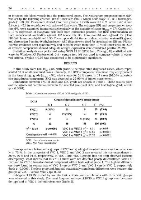
In this study were DIC (n G3<br />
= 55) with grade 3 the most often diagnosed cases, which represented<br />
52 % of all examined cases. Similarly, the DCIS component was most frequently present<br />
in the form of high grade (n VNC3<br />
= 54), what stands for 51 % cases. In 17 cases (16.0 %) an extensive<br />
intraductal component (EIC) was detected in 25-99 % of tumor mass extent.<br />
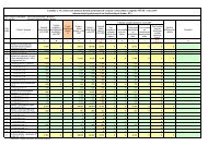
Correlations between VNC of DCIS and DIC grade are shown in Table 1. These results point<br />
out the significant correlation between the selected groups of DCIS and histological grade of DIC<br />
(p < 0.0001).<br />
Table 1 Correlation between VNC of DCIS and grade of DIC<br />
DCIS….ductal carcinoma in situ<br />
VNC….Van Nuys classification<br />
Correspondence between the groups of VNC and grading of invasive breast carcinoma is nearly<br />
in 75 %. In the categories of VNC 1, VNC 2 and VNC 3 was revealed this correspondence in<br />
36 %, 70 % and 94 %, respectively. In VNC 1 and VNC 3 groups has not been observed extreme<br />
discrepancy, what means that in VNC 1 there were not detected poorly differentiated forms of<br />
DIC and in VNC 3 invasive ductal component within histological grade 1. The highest differences<br />
were found in comparison of VNC 1 versus VNC 3 and VNC 2 versus VNC 3, respectively<br />
(both p_ 0.0001). The less profound, but still statistically significant differences were between the<br />
groups of VNC 1 versus VNC 2 (p< 0.05).<br />
Subtypes of DCIS divided by architectonic criteria and correlations with three VNC groups<br />
were observed in this study. The most frequent subtype of DCIS in VNC 3 group was the comedo-type<br />
and in VNC 1 the cribriform one (Table 2).