Pancreatic Hamartoma and SAPHO Syndrome: a ... - ResearchGate
Pancreatic Hamartoma and SAPHO Syndrome: a ... - ResearchGate
Pancreatic Hamartoma and SAPHO Syndrome: a ... - ResearchGate

Create successful ePaper yourself
Turn your PDF publications into a flip-book with our unique Google optimized e-Paper software.
<strong>Pancreatic</strong> <strong>Hamartoma</strong> <strong>and</strong> <strong>SAPHO</strong> <strong>Syndrome</strong>: a Case<br />
Report<br />
Dorel Sampelean 1 , Mircea Adam 1 , Valentin Muntean 2 , Bianca Hanescu 1 , Iacob Domsa 1<br />
1) 4 th Medical Clinic; 2) 4 th Surgical Clinic, University of Medicine <strong>and</strong> Pharmacy Iuliu Hatieganu, Cluj-Napoca,<br />
Romania<br />
Abstract<br />
We report the first case of an association of pancreatic<br />
hamartoma with <strong>SAPHO</strong> syndrome mimicking disseminated<br />
bone metastases. A 46 year old male with intermittent back<br />
pain for 10 years, relieved by NSAIDs <strong>and</strong> desquamation<br />
erythemathous palmo-plantar eruption one year before,<br />
presented with symptoms of duodenal stenosis, a cystic tumor<br />
at the head of the pancreas <strong>and</strong> osteoformative (hyperostosis)<br />
<strong>and</strong> osteodestructive (osteitis) lesions of the clavicle,<br />
m<strong>and</strong>ible, lumbar spine. The bone lesions resembled bone<br />
metastases, but an inflammatory infiltrate <strong>and</strong> fibrosis were<br />
found on the excisional biopsy of left clavicle, compatible<br />
with the <strong>SAPHO</strong> syndrome. The pancreatic tumor grew<br />
rapidly <strong>and</strong> showed a histological aspect of malignancy<br />
at laparoscopy. A cephalic duodenopancreatectomy was<br />
performed, but the histological findings established the<br />
diagnosis of pancreatic hamartoma. Several months later,<br />
the bone Tc99m scintigraphy was normal.<br />
Keywords<br />
<strong>SAPHO</strong> syndrome – pancreatic hamartoma – osteitis<br />
– palmoplantar eruption – hyperostozis.<br />
Case report<br />
A 46-year-old male patient was admitted to the Medical<br />
Clinic IV in June 2006 for: epigastric pain, acid regurgitation<br />
starting a week before, accompanied by 14 kg weight loss<br />
over the past 14 months against a background of appetite<br />
loss, asthenia <strong>and</strong> fatigue. The patient also complained of<br />
intermittent pains in the lumbar spine, for approximately<br />
10 years, which were relieved by NSAIDs, <strong>and</strong> for several<br />
weeks he had had pains at the level of the left clavicle <strong>and</strong><br />
m<strong>and</strong>ible.<br />
Received:18.12.2008 Accepted: 20.01.2009<br />
J Gastrointestin Liver Dis<br />
December 2009 Vol.18 No 4, 483-486<br />
Address for correspondence: Dorel Sampelean<br />
Medical Clinic IV<br />
Cluj-Napoca, Romania<br />
E-mail: steelnox78@yahoo.com<br />
The personal history of the patient included the<br />
appearance of a desquamative erythematous palmoplantar<br />
eruption one year before, which remitted spontaneously.<br />
The family history was insignificant for the current<br />
pathology. The patient was a chronic drinker <strong>and</strong> smoker<br />
(40 cigarettes/day for 10 years).<br />
The clinical examination evidenced: general altered<br />
state, emaciation, a tumor palpable at the level of the left<br />
clavicle, 5-6 cm in size, of hard consistency, immobile <strong>and</strong><br />
painful, tumefaction in the left m<strong>and</strong>ibular angle ~ 2 cm in<br />
size, painful on palpation, left mobile laterocervical lymph<br />
node 0.5 cm in size, pain at the percussion of the lumbosacral<br />
spinous apophyses, pain on palpation in the right upper<br />
abdominal quadrant.<br />
Biological tests on admission showed an inflammatory<br />
syndrome: increased ESR, leukocytosis, thrombocytosis,<br />
hyperfibrinogenemia, increased alpha1 <strong>and</strong> alpha 2<br />
globulins. high serum IgG levels. Renal, <strong>and</strong> liver function<br />
tests were within normal limits, serum <strong>and</strong> urinary calcium<br />
<strong>and</strong> phosphorus within normal limits, normal alkaline<br />
phosphatase, negative rheumatoid factor, ASLO titer < 200<br />
U.<br />
The abdominal ultrasound evidenced a tumor in the<br />
duodenum, involving the mucosa circumferentially,<br />
extending from the bulb to the lower extremity of the<br />
second segment (D2). The other abdominal organs appeared<br />
normal.<br />
Upper digestive endoscopy was performed, which<br />
detected esophageal hyperemia, antropyloric mucosal<br />
hyperemia, <strong>and</strong> an infiltrative process in the duodenal bulb<br />
<strong>and</strong> D2. Biopsies were taken, which found inflammatory<br />
infiltration with eosinophils <strong>and</strong> polymorphonuclears.<br />
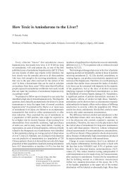
Echoendoscopy showed the head of the pancreas of<br />
normal appearance with the duct of Wirsung 2 mm in<br />
diameter, a thickened up to 12 mm duodenal wall at the apex<br />
of the bulb, several round hypoechogenic adenopathies 8 mm<br />
in size <strong>and</strong> the common bile duct without suspect images<br />
inside. The established diagnosis was stenosing duodenitis<br />
(Fig. 1).<br />
Abdominal CT showed the liver without nodular images,<br />
a homogeneous spleen, <strong>and</strong> a cyst 0.6 cm in size in the body
484<br />
of the pancreas. Increased volume of the head of the pancreas<br />
(Fig. 2), with a 0.9 cm cyst. In the peripancreatic region, in<br />
the head of the pancreas, <strong>and</strong> around the pancreatic duct, a<br />
dense weakly iodophilic muff-shaped mass was visualized.<br />
In D2 <strong>and</strong> the lower jejunum, the wall was significantly<br />
infiltrated. Lumbo-aortic <strong>and</strong> interaortic caval adenopathies<br />
were also described.<br />
The thorax CT evidenced areas of osteolysis <strong>and</strong><br />
sclerosis in the middle third of the left clavicle up to the<br />
sternal extremity level. The radiograph of the lumbosacral<br />
spine showed a homogeneous bone compression of the L4<br />
vertebra by anterior osteophytosis in the middle dorsal <strong>and</strong><br />
lower lumbar region (Fig. 2). As a first bone scintigraphy<br />
suggested bone metastases, the patient was referred to the<br />
Oncology Institute Cluj, where an excisional biopsy of the<br />
left clavicle was performed, revealing a spongy bone with<br />
marked fibrosis of medullary spaces. A second Tc99m bone<br />
scintigraphy detected a pathological hyperfixation at the level<br />
of the L3 <strong>and</strong> L4 vertebrae, left hemi-m<strong>and</strong>ible, left clavicle<br />
over the whole length, with the involvement of the left<br />
sternocostal joint, hyperfixation in the right sternoclavicular<br />
joint, sternal manubrium, left chondro-costal joints VI <strong>and</strong><br />
VII <strong>and</strong> in the right iliac bone.<br />
Based on clinical data (inflammatory osteoarticular<br />
symptomatology relieved by NSAIDs), the biological<br />
inflammatory syndrome, lytic <strong>and</strong> osteocompressive lesions<br />
detected by imaging, scintigraphic changes, clavicular<br />
biopsy, <strong>and</strong> the history of palmoplantar skin lesions, the<br />
diagnosis of <strong>SAPHO</strong> syndrome was established.<br />
After one month, the patient presented a general good<br />
state, remission of epigastric pain, improvement of bone<br />
Sampelean et al<br />
pain, disappearance of m<strong>and</strong>ibular tumefaction <strong>and</strong> minimal<br />
tumefaction of the left clavicle. Biological tests detected<br />
an increased ESR, serum fibrinogen, PCR, with the rest of<br />
biological values normal. The tumor markers CEA <strong>and</strong> CA<br />
19-9 were negative.<br />
The patient was transferred to Surgical Clinic IV for<br />
diagnostic laparoscopy with intraoperative duodenal,<br />
hepatic, <strong>and</strong> (peripancreatic) lymph node biopsy. The<br />
histopathological examination of the duodenal bioptic<br />
sample evidenced medium sized cells with a high nucleocytoplasmic<br />
ratio, moderate nuclear pleomorphism (an<br />
aspect which might indicate malignancy). A clinical <strong>and</strong><br />
biological reassessment was decided after 4 weeks, but the<br />
patient came back only 9 months later, complaining of upper<br />
abdominal pain <strong>and</strong> vomiting, as well as of intermittent pain<br />
in the lumbar spine that was relieved by NSAIDs. Abdominal<br />
ultrasound showed a nodular tumor 80/65 mm in size in<br />
the head of the pancreas, invading the duodenal wall, also<br />
confirmed by echoendoscopy <strong>and</strong> abdominal CT scan, which<br />
excluded secondary hepatic metastases.<br />
Cephalic duodenopancreatectomy was performed in June<br />
2007. The histopathological examination established the<br />
diagnosis of pancreatic hamartoma (Figs. 3, 4).<br />
Fig 3. <strong>Hamartoma</strong> myoepithelial. Exocrine ducts<br />
with acinar compound (H&Ex100 ).<br />
Fig 1. Echoendoscopy showing a thickened duodenal<br />
wall.<br />
Fig 4. <strong>Hamartoma</strong> myoepithelial. <strong>Pancreatic</strong> duct<br />
necrosis (H&Ex100 ).<br />
Discussion<br />
Fig 2. Abdominal CT of the patient.<br />
In 1987, a group of French rheumatologists established<br />
the <strong>SAPHO</strong> acronym for a clinico-radiological entity
<strong>Pancreatic</strong> hamartoma <strong>and</strong> <strong>SAPHO</strong> syndrome 485<br />
combining bone <strong>and</strong> joint symptoms with skin lesions [1].<br />
Its history started much earlier, in 1961, when Windom et<br />
al reported on the association between acne conglobata <strong>and</strong><br />
inflammatory polyarthritis. In 1967 Sasaki et al described the<br />
association between palmoplantar pustulosis <strong>and</strong> clavicular<br />
hyperostosis.<br />
Until 1987 there were more than 50 terms referring to<br />
the clinical picture of <strong>SAPHO</strong> such as pustulotic arthroosteitis<br />
[2] <strong>and</strong> the acquired hyperostosis syndrome, known<br />
to German radiologists [3].<br />
In 1986, Schilling et al described two clinical entities<br />
frequent to the syndrome: spondylarthritis hyperostotica<br />
pustulo-psoriatica (SHPP) <strong>and</strong> chronic recurrent multifocal<br />
osteomyelitis (CRMO).<br />
Due to its clinical heterogenicity the diagnosis is difficult<br />
to establish, therefore in 1994 Kahn et al established the three<br />
diagnostic criteria specific to the <strong>SAPHO</strong> syndrome [4], one<br />
of them being sufficient to establish the diagnosis: multifocal<br />
osteitis with or without skin manifestations; sterile acute or<br />
chronic joint inflammation associated with either pustules<br />
or psoriasis of palms <strong>and</strong> soles; <strong>and</strong> sterile osteitis in the<br />
presence of one of the skin lesions mentioned below.<br />
The prevalence is difficult to establish, <strong>and</strong> many<br />
cases remain undetected because of non-recognition of the<br />
syndrome. It has been described especially in Japan, Western<br />
Europe, particularly in Sc<strong>and</strong>inavian countries, but all ethnic<br />
groups can be affected. This is a disease of the young adult,<br />
the mean age at the time of diagnosis being 38 years, but it<br />
can also be found in children, where it manifests as aseptic<br />
CRMO. Sex distribution seems to be equal.<br />
The etiology of the syndrome is not completely<br />
understood. A recent hypothesis suggests that it might be<br />
caused by an immunological response to an infectious<br />
agent that triggers an inflammatory reaction in bone or<br />
joint tissue, taking advantage of a type of “paresis” of<br />
the immune system, which is favored by a predisposing<br />
genetic background. The infectious agent incriminated by<br />
some authors is Propionibacterium Acnes, a common skin<br />
saprophyte isolated from some open bone biopsies in one<br />
study [5], which might explain the therapeutic effect of<br />
antibiotics; but this gram-negative bacterium grows under<br />
anaerobic conditions <strong>and</strong> can be difficult to culture [6].<br />
On the other h<strong>and</strong>, the elements supporting the<br />
connection between <strong>SAPHO</strong> syndrome <strong>and</strong> seronegative<br />
spondylarthropathies should not be neglected: association<br />
with psoriasis, involvement of the spine, of the sacroiliacs,<br />
association with inflammatory bowel disease (Crohn‘s<br />
disease, ulcerative colitis), presence of the HLA B27<br />
antigen.<br />
Osteoarticular disorder may be clinically acute or<br />
chronic, having the appearance of inflammatory osteitis.<br />
The main site of the inflammatory process is the<br />
anterior thoracic wall, with the inflammation of the sternum<br />
<strong>and</strong> sternocostoclavicular joint. If thoracic involvement<br />
facilitates diagnosis, the involvement of the spine, of long<br />
bones in adults, especially the tibia <strong>and</strong> the femur, of cranial<br />
bones raise differential diagnosis problems with a tumor or<br />
infectious lesion. Sterile osteitis of the m<strong>and</strong>ible represents<br />
10% of bone lesions. This should be taken into consideration,<br />
because tooth ablation or long duration antimicrobial therapy<br />
have no results [6]. Peripheral joints are equally affected,<br />
sometimes having a pseudoseptic appearance.<br />
Skin involvement is not a requirement for making the<br />
diagnosis. It is most frequently preceded by 1 to 20 years<br />
osteoarticular involvement in both children <strong>and</strong> adults.<br />
Palmoplantar pustulosis, conglobate acne, acne fulminans,<br />
suppurative hydradenitis or different types of psoriasis may<br />
occur. Our patient also reported a history of palmoplantar<br />
skin disease, most probably psoriatic.<br />
There are no biological stigmata specific for the<br />
syndrome, only an increase in inflammatory parameters<br />
(ESR, PCR), in immunoglobulin levels or in the complement<br />
fractions C3, C4.<br />
The radiological aspect is frequently suggestive:<br />
hypertrophy <strong>and</strong> compression or even osteolysis. CT shows<br />
lytic lesions surrounded by sclerotic areas, changes also<br />
found in our patient.<br />
Bone scintigraphy with Tc 99m shows hyperfixation <strong>and</strong><br />
is extremely useful for the detection of osteoproliferative<br />
(hyperostosis) <strong>and</strong> osteodestructive lesions (osteitis), left<br />
undetected by imaging. In our patient, the bone lesions<br />
were first interpreted as metastases, but histopathological<br />
examination <strong>and</strong> the second scintigraphy refuted this<br />
diagnosis. Histopathological examination supported<br />
the diagnosis of the disorder by early lesions, in which<br />
polymorphonuclear cells were dominant, later the<br />
inflammatory infiltrate mainly consisted of monocytes <strong>and</strong><br />
fibroblasts, <strong>and</strong> bone remodeling <strong>and</strong> bone marrow fibrosis<br />
occurred [7].<br />
Patients diagnosed with <strong>SAPHO</strong> syndrome require<br />
long-term monitoring in order to prevent complications:<br />
retroperitoneal fibrosis, mediastinal fibrosis, compressive<br />
venous thrombosis, intercostal neuralgia.<br />
The literature reports no case of association of this<br />
syndrome with pancreatic hamartoma or other digestive<br />
disease, except for enterocolopathies (Crohn‘s disease,<br />
ulcerative colitis).<br />
The long-term prognosis is favorable, the evolution of<br />
the disease being characterized by prolonged remissions<br />
followed by relapses.<br />
The treatment of the disease is symptomatic. It consists of<br />
NSAIDs or sulfasalazine [6]. The dosage is adapted to each<br />
individual case, taking the side effects into consideration.<br />
Corticoids are prescribed quite rarely <strong>and</strong> are indicated<br />
for emergency cases in limited time periods. Some trials<br />
have studied the effect of immunomodulatory treatment<br />
represented by anti TNF-alpha [8]. TNF-alpha inhibitors<br />
such as Infliximab or Etanercept have been successfully<br />
used due to the high TNF-alpha concentrations found<br />
during bone biopsy [9]. In some cases, methotrexate has<br />
been used. Antibiotics have not proved to be useful, except<br />
for azithromycin, which has both an anti-inflammatory <strong>and</strong><br />
immunomodulatory effect [10]. Some authors recommend<br />
long-term treatment with azithromycin (or clarithromycin) as
486<br />
first line treatment in CRMO, especially when the presence<br />
of Propionibacterium Acnes is confirmed [9].<br />
At the same time, the usefulness of a hormone<br />
treatment for this disease was discovered. Calcitonin has<br />
both an osteotrophic <strong>and</strong> anti-inflammatory effect. Over<br />
the past years, due to a number of cases resistant to this<br />
treatment, calcitonin has been replaced by bisphosphonates<br />
(pamidronate, zoledronic acid), which have an antiosteoclastic,<br />
antiinflammatory action, with the suppression<br />
of IL6, IL1 or TNF-alpha. All pharmacological options<br />
require careful interdisciplinary monitoring, as well as the<br />
participation of a rheumatologist <strong>and</strong> dermatologist in<br />
decision.<br />
In conclusion, we presented the first case of an<br />
association of pancreatic hamartoma with <strong>SAPHO</strong> syndrome<br />
mimicking disseminated bone metastases.<br />
References<br />
1. Chamont AM, Benhamou CL,Kahn MF, Beraneck I, Kaplan G,<br />
Prost A. Le syndrome acne pustulose hyperostose osteite (<strong>SAPHO</strong>):<br />
resultats d’une enquete nationale. 85 observations. Rev Rhum Mal<br />
Osteoartic 1987; 54: 187-196.<br />
Sampelean et al<br />
2. Sonozaki H, Mitsui H, Miyanaga Y, et al. Clinical features of 53 cases<br />
with pustulotic arthro-osteitis. Ann Rheum Dis 1981; 40: 547-553.<br />
3. Dihlmann W, Dihlmann SW, Hering L. Acquired hyperostosis<br />
syndrome –AYHS - (sternocostoclavicular hyperostosis, pustulotic<br />
arthro-osteitis, <strong>SAPHO</strong>-syndrome): bone scintigraphy of the anterior<br />
chest wall. Clin Rheumatol 1997; 16: 13-24.<br />
4. Kahn MF, Kahn MA.The <strong>SAPHO</strong>-<strong>Syndrome</strong>. In: Wright <strong>and</strong><br />
Helliwell (Eds): Psoriatic Arthritis. Bailliere‘s Clin Rheumatol 1994;<br />
8: 333-362.<br />
5. Edlund E, Johnsson U, Lidgren L, et al. Palmoplantar pustulosis<br />
<strong>and</strong> sternoclavicular arthoro-osteitis. Ann Rheum Dis 1988; 47:<br />
809-815.<br />
6. Hayem G, Bouchaud-Chabot A, Benali K, et al. <strong>SAPHO</strong> <strong>Syndrome</strong>:<br />
A long- term follow-up study of 120 cases. Semin Arthritis Rheum<br />
1999; 29: 159-171.<br />
7. Van Doornum S, Barraclough D, McColl G, Wicks I. <strong>SAPHO</strong>: rare<br />
or just not recognised? Semin Arthritis Rheum 2000; 30: 70-77.<br />
8. Wagner AD, Andresen J, Jendro MC, Hülsemann JL, Zeidler H.<br />
Sustained response to tumor necrosis factor alpha-blocking agents<br />
in two patients with <strong>SAPHO</strong> syndrome. Arthritis Rheum 2002; 46:<br />
1965-1968.<br />
9. Schillling F,Wagner AD. Azythromycin: an anti-inflammatory effect<br />
in chronic recurrent multifocal osteomyelitis? A preliminary report.<br />
Z Rheumatol 2000; 59: 352-353.