Iatrogenic Left Diaphragmatic Hernia as a Complication of Hydatid ...
Iatrogenic Left Diaphragmatic Hernia as a Complication of Hydatid ...
Iatrogenic Left Diaphragmatic Hernia as a Complication of Hydatid ...
Create successful ePaper yourself
Turn your PDF publications into a flip-book with our unique Google optimized e-Paper software.
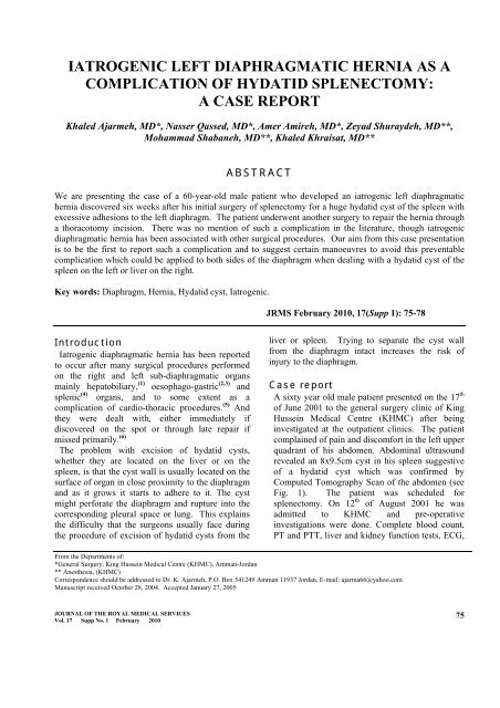
IATROGENIC LEFT DIAPHRAGMATIC HERNIA AS A<br />
COMPLICATION OF HYDATID SPLENECTOMY:<br />
A CASE REPORT<br />
Khaled Ajarmeh, MD*, N<strong>as</strong>ser Q<strong>as</strong>sed, MD*, Amer Amireh, MD*, Zeyad Shuraydeh, MD**,<br />
Mohammad Shabaneh, MD**, Khaled Khraisat, MD**<br />
ABSTRACT<br />
We are presenting the c<strong>as</strong>e <strong>of</strong> a 60-year-old male patient who developed an iatrogenic left diaphragmatic<br />
hernia discovered six weeks after his initial surgery <strong>of</strong> splenectomy for a huge hydatid cyst <strong>of</strong> the spleen with<br />
excessive adhesions to the left diaphragm. The patient underwent another surgery to repair the hernia through<br />
a thoracotomy incision. There w<strong>as</strong> no mention <strong>of</strong> such a complication in the literature, though iatrogenic<br />
diaphragmatic hernia h<strong>as</strong> been <strong>as</strong>sociated with other surgical procedures. Our aim from this c<strong>as</strong>e presentation<br />
is to be the first to report such a complication and to suggest certain manoeuvres to avoid this preventable<br />
complication which could be applied to both sides <strong>of</strong> the diaphragm when dealing with a hydatid cyst <strong>of</strong> the<br />
spleen on the left or liver on the right.<br />
Key words: Diaphragm, <strong>Hernia</strong>, <strong>Hydatid</strong> cyst, <strong>Iatrogenic</strong>.<br />
JRMS February 2010, 17(Supp 1): 75-78<br />
Introduction<br />
<strong>Iatrogenic</strong> diaphragmatic hernia h<strong>as</strong> been reported<br />
to occur after many surgical procedures performed<br />
on the right and left sub-diaphragmatic organs<br />
mainly hepatobiliary, (1) oesophago-g<strong>as</strong>tric (2,3) and<br />
splenic (4) organs, and to some extent <strong>as</strong> a<br />
complication <strong>of</strong> cardio-thoracic procedures. (5) And<br />
they were dealt with, either immediately if<br />
discovered on the spot or through late repair if<br />
missed primarily. (6)<br />
The problem with excision <strong>of</strong> hydatid cysts,<br />
whether they are located on the liver or on the<br />
spleen, is that the cyst wall is usually located on the<br />
surface <strong>of</strong> organ in close proximity to the diaphragm<br />
and <strong>as</strong> it grows it starts to adhere to it. The cyst<br />
might perforate the diaphragm and rupture into the<br />
corresponding pleural space or lung. This explains<br />
the difficulty that the surgeons usually face during<br />
the procedure <strong>of</strong> excision <strong>of</strong> hydatid cysts from the<br />
liver or spleen. Trying to separate the cyst wall<br />
from the diaphragm intact incre<strong>as</strong>es the risk <strong>of</strong><br />
injury to the diaphragm.<br />
C<strong>as</strong>e report<br />
A sixty year old male patient presented on the 17 th<br />
<strong>of</strong> June 2001 to the general surgery clinic <strong>of</strong> King<br />
Hussein Medical Centre (KHMC) after being<br />
investigated at the outpatient clinics. The patient<br />
complained <strong>of</strong> pain and discomfort in the left upper<br />
quadrant <strong>of</strong> his abdomen. Abdominal ultr<strong>as</strong>ound<br />
revealed an 8x9.5cm cyst in his spleen suggestive<br />
<strong>of</strong> a hydatid cyst which w<strong>as</strong> confirmed by<br />
Computed Tomography Scan <strong>of</strong> the abdomen (see<br />
Fig. 1). The patient w<strong>as</strong> scheduled for<br />
splenectomy. On 12 th <strong>of</strong> August 2001 he w<strong>as</strong><br />
admitted to KHMC and pre-operative<br />
investigations were done. Complete blood count,<br />
PT and PTT, liver and kidney function tests, ECG,<br />
From the Departments <strong>of</strong>:<br />
*General Surgery, King Hussein Medical Centre (KHMC), Amman-Jordan<br />
** Anesthesia, (KHMC)<br />
Correspondence should be addressed to Dr. K. Ajarmeh, P.O. Box 541249 Amman 11937 Jordan, E-mail: ajarma66@yahoo.com<br />
Manuscript received October 28, 2004. Accepted January 27, 2005<br />
JOURNAL OF THE ROYAL MEDICAL SERVICES<br />
Vol. 17 Supp No. 1 February 2010<br />
75
Fig. 1. CT scan <strong>of</strong> the abdomen showing the splenic<br />
hydatid cyst with adhesions on the left diaphragm<br />
(posterolaterally)<br />
and chest X-ray were normal (see Fig. 2).<br />
On the 13 th <strong>of</strong> August 2001 the patient underwent<br />
laparotomy through a left sub-costal incision and the<br />
operative findings were a huge cyst arising from the<br />
spleen with excessive adhesions to the left<br />
diaphragm. The liver and the rest <strong>of</strong> the intraperitoneal<br />
organs were inspected and found to be<br />
free <strong>of</strong> any gross pathology.<br />
The surgeon's aim w<strong>as</strong> to resect the spleen with the<br />
cyst without trying to excise or evacuate the cyst<br />
alone, but due to the excessive adhesions faced<br />
during the dissection <strong>of</strong> the cyst wall from the<br />
diaphragm the cyst w<strong>as</strong> opened and it w<strong>as</strong><br />
immediately secured and irrigation with 10%<br />
hypertonic saline <strong>of</strong> the area w<strong>as</strong> performed. The<br />
cyst w<strong>as</strong> completely evacuated and dissection<br />
continued with difficulty, then splenectomy w<strong>as</strong><br />
completed, irrigation with normal saline w<strong>as</strong> done,<br />
haemost<strong>as</strong>is w<strong>as</strong> secured, a rubber open drain w<strong>as</strong><br />
inserted in the left sub-diaphragmatic space, and the<br />
wound w<strong>as</strong> closed.<br />
The patient had an uneventful postoperative<br />
recovery and the drain w<strong>as</strong> removed on the second<br />
postoperative day. He w<strong>as</strong> discharged on the 15 th <strong>of</strong><br />
August 2001. After his discharge the patient w<strong>as</strong><br />
followed up in the general surgery outpatient’s<br />
clinic twice (2 nd & 15 th <strong>of</strong> September) and on both<br />
occ<strong>as</strong>ions he w<strong>as</strong> symptom free apart from a<br />
superficial wound infection managed by daily<br />
dressing for 10 days before it completely healed.<br />
On the 23 rd <strong>of</strong> Sep 2001 he w<strong>as</strong> referred from a<br />
peripheral hospital where he w<strong>as</strong> admitted for five<br />
days for the complaint <strong>of</strong> shortness <strong>of</strong> breath, chest<br />
Fig. 2. Chest X ray <strong>of</strong> the patient before splenectomy<br />
reported <strong>as</strong> normal.<br />
pain, fever and chills and w<strong>as</strong> managed <strong>as</strong> a c<strong>as</strong>e <strong>of</strong><br />
left sided pneumonia by intravenous antibiotics. He<br />
did not improve and w<strong>as</strong> transferred to KHMC <strong>as</strong> a<br />
suspected c<strong>as</strong>e <strong>of</strong> sub-diaphragmatic collection for<br />
further investigations and management.<br />
His chest X-ray showed a raised left hemi<br />
diaphragm suggestive <strong>of</strong> a sub-diaphragmatic<br />
collection with left pleural effusion (see Fig. 3), but<br />
when ultr<strong>as</strong>ound w<strong>as</strong> performed it excluded the<br />
presence <strong>of</strong> a collection. CT scan indicated the<br />
presence <strong>of</strong> an iatrogenic left diaphragmatic hernia<br />
(see Fig. 4a and 4b).<br />
On the 3 rd <strong>of</strong> October 2001 the patient underwent<br />
left thoracotomy, the hernia w<strong>as</strong> found to be through<br />
a lateral defect in the left hemi-diaphragm. It w<strong>as</strong><br />
reduced and the defect repaired using prolene 0/0<br />
sutures in double layers. A chest tube w<strong>as</strong> inserted<br />
and the wound w<strong>as</strong> closed. Chest X-ray on the<br />
fourth post operative day w<strong>as</strong> clear (see Fig.5). The<br />
chest tube w<strong>as</strong> removed on the same day and the<br />
patient w<strong>as</strong> discharged home on the eighth post<br />
operative day. He w<strong>as</strong> followed by both the general<br />
surgery and thoracic surgery clinics till May 2002<br />
where he w<strong>as</strong> discharged from both clinics.<br />
Most <strong>of</strong> the c<strong>as</strong>es <strong>of</strong> iatrogenic diaphragmatic<br />
herni<strong>as</strong> mentioned in the literature are those missed<br />
at the time <strong>of</strong> primary procedures which are<br />
diagnosed later when patients start to be<br />
symptomatic with chest pain, and discomfort<br />
<strong>as</strong>sociated with dyspnea and possible cough. The<br />
diagnosis is confirmed usually after radiological<br />
investigations. (6) JOURNAL OF THE ROYAL MEDICAL SERVICES<br />
76<br />
Vol. 17 Supp No. 1 February 2010
Fig. 3. Chest X ray <strong>of</strong> the patient on the second admission showing a raised left hemi-diaphragm.<br />
Fig. 4a. Scout view <strong>of</strong> the CT scan <strong>of</strong> the lower chest<br />
and upper abdomen showing a left diaphragmatic hernia<br />
containing stomach and splenic flexure <strong>of</strong> the colon.<br />
Fig. 4b. Four cuts <strong>of</strong> the CT scan <strong>of</strong> lower chest and upper<br />
abdomen. Showing the hernial sac containing the stomach<br />
with air-fluid level.<br />
Fig. 5. Chest X-ray <strong>of</strong> the patient after the thoracotomy where the chest tube is still in situ. The left hemi-diaphragm is<br />
back to normal.<br />
Discussion<br />
<strong>Diaphragmatic</strong> herni<strong>as</strong> are cl<strong>as</strong>sified according to<br />
cause into congenital and acquired, where the<br />
acquired type is either traumatic or iatrogenic. (7,8)<br />
Late diagnosis <strong>of</strong> acquired diaphragmatic herni<strong>as</strong><br />
is a frequent occurrence, especially when they are<br />
<strong>as</strong>sociated with small penetrating wounds.<br />
In the absence <strong>of</strong> strangulated or obstructed<br />
JOURNAL OF THE ROYAL MEDICAL SERVICES<br />
Vol. 17 Supp No. 1 February 2010<br />
77
Table I. Common procedures complicated by diaphragmatic hernia<br />
Surgical procedure<br />
Laparoscopic splenectomy (4)<br />
Laparoscopic cholecystectomy (1)<br />
Anti-reflux procedures (2,3)<br />
<strong>Left</strong> nephrectomy (9)<br />
<strong>Left</strong> Hepatic lobectomy (10)<br />
Oesophagectomy (6)<br />
Coronary artery byp<strong>as</strong>s (5)<br />
Miscellaneous (11)<br />
*Occurrence <strong>of</strong> iatrogenic hiatal hernia is also reported.<br />
Site <strong>of</strong> Occurrence<br />
<strong>Left</strong><br />
Right<br />
<strong>Left</strong>*<br />
<strong>Left</strong><br />
Right and <strong>Left</strong><br />
<strong>Left</strong>*<br />
Central<br />
Right and left<br />
viscera, chronic pain with or without mild<br />
respiratory changes may be the only presenting<br />
complaint. (6)<br />
<strong>Iatrogenic</strong> diaphragmatic herni<strong>as</strong> are not <strong>as</strong><br />
common <strong>as</strong> the traumatic type, and have been<br />
reported several times in the literature <strong>as</strong> a<br />
complication <strong>of</strong> different upper abdominal and<br />
cardio-thoracic surgical procedures (see Table I.).<br />
The new era <strong>of</strong> minimal inv<strong>as</strong>ive surgeries i.e.<br />
laparoscopy appears to have h<strong>as</strong> incre<strong>as</strong>ed the rate<br />
<strong>of</strong> iatrogenic diaphragmatic herni<strong>as</strong> slightly though<br />
there are no true statistical figures or studies to<br />
confirm this. (1,4) We could not find a single report<br />
<strong>of</strong> such a complication following splenic or liver<br />
(which is relatively more common) hydatid cyst<br />
removal surgical procedure. This could be<br />
explained by the rarity <strong>of</strong> such procedures in the<br />
Western world which is the major contributor to<br />
indexed literature.<br />
Regardless <strong>of</strong> the cause <strong>of</strong> the hernia, the<br />
recommendations mentioned in the literature to<br />
avoid such a complication are still the same. They<br />
all stress on the surgeons’ awareness <strong>of</strong> such a<br />
complication and how to avoid it by meticulous<br />
dissection around the region <strong>of</strong> the diaphragm and<br />
its crura in esophageal surgeries, and to check for<br />
the integrity <strong>of</strong> the diaphragm after each upper<br />
abdomen and lower thoracic surgical procedure. (6)<br />
Conclusion<br />
<strong>Iatrogenic</strong> diaphragmatic injury can be e<strong>as</strong>ily<br />
missed by surgeons unless they are aware <strong>of</strong> such a<br />
complication and take the precautions to avoid it.<br />
Checking for the integrity <strong>of</strong> the diaphragm after<br />
any upper g<strong>as</strong>tro-intestinal or lower cardio-thoracic<br />
procedures is important. We would recommend, in<br />
c<strong>as</strong>es <strong>of</strong> liver or spleen hydatid cyst surgeries,<br />
whenever the cyst wall is severely adherent to the<br />
diaphragm and sometimes inseparable from it, to<br />
leave the adherent patch stuck to diaphragm and not<br />
to try to resect it but to resume the dissection around<br />
it. This approach will ensure the integrity <strong>of</strong> the<br />
diaphragm.<br />
References<br />
1. Armstrong P, Miller S, Brown G. <strong>Diaphragmatic</strong><br />
hernia seen <strong>as</strong> a late complication <strong>of</strong> laparoscopic<br />
cholecystectomy. Surg Endosc 1999; 13: 817-818.<br />
2. Sancho LM, P<strong>as</strong>choalini Mda S, Jatene FB,<br />
Rodrigues Junior AJ, <strong>Iatrogenic</strong> diaphragmatic<br />
hernia following abdominal<br />
esophagog<strong>as</strong>tr<strong>of</strong>undoplication: Report <strong>of</strong> a c<strong>as</strong>e.<br />
Rev Hosp Clin Fac Med Sao Paulo 1996; 51: 250-<br />
252.<br />
3. Steiger Z, Wilson RF, Nelson RM, et al.<br />
<strong>Iatrogenic</strong> hiatal and diaphragmatic herni<strong>as</strong>. Am<br />
Surg 1984; 50: 217-221.<br />
4. Targarona E, Espert J, Bombuy E, et al.<br />
<strong>Complication</strong>s <strong>of</strong> Laparoscopic Splenectomy. Arch<br />
Surg 2000; 135: 1137-1140.<br />
5. Waller DA, Satur CN, Mitchell IM, Sivanathan<br />
UM. <strong>Iatrogenic</strong> peritoneopericardial hernia<br />
following coronary artery byp<strong>as</strong>s. Eur J<br />
cardiothoracic Surg. 1992; 6: 156-157.<br />
6. Van sandick JW, Knegjens JL, Van Lanschot<br />
JJ, Cbertop H. <strong>Diaphragmatic</strong> herniation<br />
following oesophagectomy. Br J Surg 1999; 86:<br />
109-112.<br />
7. Trigt PV. Diaphragm and diaphragmatic pacing.<br />
In: Sabiston DC, Spencer FC, editors. Surgery <strong>of</strong><br />
the chest. 6 th ed. W.B Saunders Company. 1999;<br />
1081-1099.<br />
8. Yazici M, Karaca I, Etensel B, et al.<br />
Paraesophageal hiatal herni<strong>as</strong> in children. Dise<strong>as</strong>es<br />
<strong>of</strong> the oesophagus 2003; 16: 210-213<br />
9. Yam<strong>as</strong>hita J, Kuwata K, Miyamoto Y, et al. Two<br />
c<strong>as</strong>es <strong>of</strong> traumatic diaphragmatic hernia with<br />
atypical medical history. Kyobu Geka 2000; 53:<br />
246-250.<br />
10. Icoz G, Kara E, Ilkgul O, et al. Perforation <strong>of</strong> the<br />
stomach due to chest tube complication in a patient<br />
with iatrogenic diaphragmatic rupture. Acta Chir<br />
Belg 2003; 103: 423-424.<br />
11. Chin RY, Glew MJ, Brady P. <strong>Iatrogenic</strong><br />
intrapericardial diaphragmatic hernia. ANZ J Surg.<br />
2000; 72:681-683.<br />
78<br />
JOURNAL OF THE ROYAL MEDICAL SERVICES<br />
Vol. 17 Supp No. 1 February 2010


