Comparing Animal and Plant Cell Structure - Materials Science ...
Comparing Animal and Plant Cell Structure - Materials Science ...
Comparing Animal and Plant Cell Structure - Materials Science ...

You also want an ePaper? Increase the reach of your titles
YUMPU automatically turns print PDFs into web optimized ePapers that Google loves.
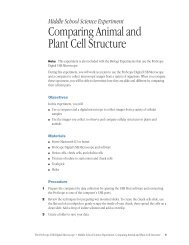
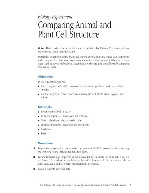
Biology Experiment<br />
<strong>Comparing</strong> <strong>Animal</strong> <strong>and</strong><br />
<strong>Plant</strong> <strong>Cell</strong> <strong>Structure</strong><br />
Note: This experiment is also included with the Middle School <strong>Science</strong> Experiments that use<br />
the ProScope Digital USB Microscope.<br />
During this experiment, you will work as a team to use the ProScope Digital USB Microscope<br />
<strong>and</strong> a computer to collect microscopic images from a variety of organisms. When you compare<br />
these specimens, you will be able to determine how they are alike <strong>and</strong> different by comparing<br />
their cellular parts.<br />
Objectives<br />
In this experiment, you will:<br />
m Use a computer <strong>and</strong> a digital microscope to collect images from a variety of cellular<br />
samples<br />
m Use the images you collect to observe <strong>and</strong> compare cellular structures in plants <strong>and</strong><br />
animals<br />
<strong>Materials</strong><br />
m Power Macintosh G3 or better<br />
m ProScope Digital USB Microscope <strong>and</strong> software<br />
m Onion cells, cheek cells, <strong>and</strong> elodea cells<br />
m Tincture of iodine to stain onion <strong>and</strong> cheek cells<br />
m Toothpick<br />
m Slides<br />
Procedure<br />
1 Prepare the computer for data collection by opening the USB Shot software <strong>and</strong> connecting<br />
the ProScope to one of the computer’s USB ports.<br />
2 Review the techniques for preparing wet mounted slides. To create the cheek cells slide, use<br />
the flat end of a toothpick to gently scrape the inside of your cheek, then spread the cells on a<br />
clean slide. Add a drop of iodine solution <strong>and</strong> add a coverslip.<br />
3 Create a folder to save your data.<br />
The ProScope USB Digital Microscope n Biology Experiment: <strong>Comparing</strong> <strong>Animal</strong> <strong>and</strong> <strong>Plant</strong> <strong>Cell</strong> <strong>Structure</strong> 1
4 Using the ProScope Digital USB Microscope, create a still image of each cell type: onion,<br />
cheek, <strong>and</strong> elodea. Make sure that you are saving the images in your data folder. (Adjust the<br />
microscope’s lens to increase the magnification.)<br />
5 Using a word-processing application, create a table like the one in the following “Data”<br />
section.<br />
6 Insert the images you selected in your table. Examine your samples <strong>and</strong> describe them <strong>and</strong><br />
identify the cell structures you observed in your table.<br />
Data<br />
Image<br />
Description of cell structures observed<br />
2 Biology Experiment: <strong>Comparing</strong> <strong>Animal</strong> <strong>and</strong> <strong>Plant</strong> <strong>Cell</strong> <strong>Structure</strong> n The ProScope USB Digital Microscope
Processing the data<br />
1. Describe how the onion <strong>and</strong> cheek cells were similar in observed parts. What parts did they<br />
have in common?<br />
2. Describe how the onion <strong>and</strong> elodea cells were similar in observed parts. What parts did<br />
they have in common?<br />
3. Describe how the onion <strong>and</strong> cheek cells were different in observed parts. What parts did<br />
they have in common?<br />
4. Describe how the onion <strong>and</strong> elodea cells were different in observed parts. What parts did<br />
they have in common?<br />
Extensions<br />
1. Why do you think the onion cells lacked chloroplasts?<br />
2. What structures do organisms that lack cell walls have to provide support?<br />
Sample results<br />
Image<br />
Description <strong>and</strong> cell structures observed<br />
Elodea 100X<br />
<strong>Cell</strong> wall, cytoplasm, <strong>and</strong> chloroplasts<br />
Elodea 200X<br />
<strong>Cell</strong> wall, cytoplasm, <strong>and</strong> chloroplasts<br />
Onion stained with iodine 200X<br />
<strong>Cell</strong> wall, cytoplasm, <strong>and</strong> nucleus<br />
Human cheek cells with iodine 100X<br />
<strong>Cell</strong> membrane, cytoplasm, <strong>and</strong> nucleus<br />
The ProScope USB Digital Microscope n Biology Experiment: <strong>Comparing</strong> <strong>Animal</strong> <strong>and</strong> <strong>Plant</strong> <strong>Cell</strong> <strong>Structure</strong> 3
Answers to questions<br />
Processing the data questions<br />
1. Both onion <strong>and</strong> cheek cells had a nucleus <strong>and</strong> both had cytoplasm.<br />
2. Both onion <strong>and</strong> elodea cells had a cell wall <strong>and</strong> both had cytoplasm.<br />
3. The onion cells had a cell wall but the cheek cells didn’t. Both had a nucleus <strong>and</strong><br />
cytoplasm.<br />
4. Both onion <strong>and</strong> elodea cells had a cell wall <strong>and</strong> cytoplasm but the onion lacked<br />
chloroplasts.<br />
Extension questions<br />
1. Onion is a storage organ for the onion plant. It is underground so it can’t do<br />
photosynthesis.<br />
2. They have a skeleton of some sort.<br />
Special thanks to the curriculum writer, Bruce Ahlborn, Technology Coordinator of<br />
Northbrook School District, Northbrook, IL.<br />
4 Biology Experiment: <strong>Comparing</strong> <strong>Animal</strong> <strong>and</strong> <strong>Plant</strong> <strong>Cell</strong> <strong>Structure</strong> n The ProScope USB Digital Microscope