Atypical Presentation of Ectopic Pregnancy in an Obese Woman: A ...
Atypical Presentation of Ectopic Pregnancy in an Obese Woman: A ...
Atypical Presentation of Ectopic Pregnancy in an Obese Woman: A ...

Create successful ePaper yourself
Turn your PDF publications into a flip-book with our unique Google optimized e-Paper software.
216<br />
J Emerg Crit Care Med. Vol. 21, No. 4, 2010<br />
<strong>Atypical</strong> <strong>Presentation</strong> <strong>of</strong> <strong>Ectopic</strong> <strong>Pregn<strong>an</strong>cy</strong> <strong>in</strong><br />
<strong>an</strong> <strong>Obese</strong> Wom<strong>an</strong>: A Case Report<br />
Teck-J<strong>in</strong> Y<strong>an</strong>g, Ho-Fu Hsiao, Hung-Hs<strong>in</strong> Mo<br />
A 37-year-old obese wom<strong>an</strong> (BMI=44.3) presented with disturbed consciousness, severe diarrhea <strong>an</strong>d<br />
tachypnea. Physical exam<strong>in</strong>ation revealed clear breath<strong>in</strong>g sounds <strong>an</strong>d a s<strong>of</strong>t abdomen. She was <strong>in</strong>itially<br />
treated as AGE <strong>an</strong>d later a case <strong>of</strong> pulmonary embolism was suspected due to a high level <strong>of</strong> D-dimer.<br />
However the cl<strong>in</strong>ical presentation <strong>an</strong>d the blood gas read<strong>in</strong>gs did not support a diagnosis <strong>of</strong> pulmonary<br />
embolism. Unstable hemodynamics developed with<strong>in</strong> one hour. Emergency abdom<strong>in</strong>al <strong>an</strong>d pelvic computed<br />
tomography showed that this was a case <strong>of</strong> <strong>in</strong>ternal bleed<strong>in</strong>g <strong>of</strong> unknown orig<strong>in</strong>. Subsequently, the patient<br />
was subjected to laparotomy <strong>an</strong>d a ruptured <strong>in</strong>tratubal pregn<strong>an</strong>cy was evacuated. She was discharged 5<br />
days after admission as well <strong>an</strong>d the recovery was still favorable at the 2 months follow-up.<br />
Key words: ectopic pregn<strong>an</strong>cy, pulmonary embolism, obesity<br />
Introduction<br />
<strong>Ectopic</strong> pregn<strong>an</strong>cy commonly occurs <strong>in</strong> women<br />
with impaired tubal function. The symptoms <strong>an</strong>d<br />
signs <strong>in</strong>clude abdom<strong>in</strong>al pa<strong>in</strong> with amenorrhea,<br />
pa<strong>in</strong> radiat<strong>in</strong>g to the shoulder, syncope, <strong>an</strong>d shock<br />
<strong>in</strong> 20% <strong>of</strong> patients. The diagnosis is difficult when<br />
there is <strong>an</strong> atypical presentation; however, late<br />
diagnosis <strong>of</strong> a ruptured ectopic pregn<strong>an</strong>cy c<strong>an</strong> be<br />
fatal due to massive <strong>in</strong>ternal bleed<strong>in</strong>g.<br />
Case Report<br />
A 37-year-old obese female (height: 160 cm, weight:<br />
116 Kg) presented with disturbed consciousness,<br />
shortness <strong>of</strong> breath <strong>an</strong>d severe diarrhea; she<br />
also had a past history <strong>of</strong> Cesare<strong>an</strong> section. She<br />
brought by ambul<strong>an</strong>ce to our hospital emergency<br />
department. Physical exam<strong>in</strong>ation revealed clear<br />
breath<strong>in</strong>g sounds <strong>an</strong>d a s<strong>of</strong>t abdomen with mild<br />
discomfort, a pulse <strong>of</strong> 131 beats per m<strong>in</strong>ute <strong>an</strong>d a<br />
blood pressure <strong>of</strong> 141/31 mmHg.<br />
Based on the history <strong>an</strong>d the cl<strong>in</strong>ical<br />
exam<strong>in</strong>ation, the patient was <strong>in</strong>itially diagnosed as<br />
AGE <strong>an</strong>d hav<strong>in</strong>g secondary hyperventilation. She was<br />
immediately resuscitated with I.V. fluids <strong>an</strong>d <strong>an</strong> O 2<br />
supply. CBC revealed a normal Hb (HB = 12.0 g/dL)<br />
<strong>an</strong>d a slightly elevated WBC (10630/uL). The other<br />
laboratory data showed <strong>an</strong> elevated blood glucose<br />
(280 mg/dL), a low pH <strong>of</strong> 7.278, a low bicarbonate<br />
level (HCO 3 = 17.4 mEq/L). Her FDP-D dimer<br />
measurement was checked at this po<strong>in</strong>t <strong>an</strong>d the<br />
result was 1140 ng/mL. Pulmonary embolism was<br />
then suspected as a differential diagnosis. However,<br />
the O 2 saturation was 100% while she was receiv<strong>in</strong>g<br />
3L/m<strong>in</strong> through a nasal c<strong>an</strong>ula <strong>an</strong>d acidosis with<br />
a low bicarbonate read<strong>in</strong>g is not a presentation<br />
<strong>of</strong> pulmonary embolism; therefore pulmonary<br />
embolism was not considered to likely.<br />
After <strong>in</strong>itial m<strong>an</strong>agement, the irritable mood<br />
Received: April 7, 2010 Accepted for publication: June 30, 2010<br />
From the Department <strong>of</strong> Emergency Department, Sijhih Cathay General Hospital<br />
Address repr<strong>in</strong>t requests <strong>an</strong>d correspondence: Dr. Teck-J<strong>in</strong> Y<strong>an</strong>g<br />
Department <strong>of</strong> Emergency Department, Sijhih Cathay General Hospital<br />
2 L<strong>an</strong>e 59, Ji<strong>an</strong>cheng Road, Sijhih City, Taipei County 221, Taiw<strong>an</strong> (R.O.C.)<br />
Tel: (02)26482121 ext 3910<br />
E-mail: y<strong>an</strong>gteckj<strong>in</strong>@hotmail.com
<strong>Atypical</strong> presentation <strong>of</strong> ectopic pregn<strong>an</strong>cy<br />
217<br />
progressed <strong>an</strong>d <strong>an</strong> unstable blood pressure was<br />
then noted (BP = 81/39 mmHg). A central venous<br />
catheter was <strong>in</strong>serted for rapid fluid <strong>in</strong>fusion <strong>an</strong>d<br />
<strong>an</strong> <strong>in</strong>otropic agent with dopam<strong>in</strong>e was also given.<br />
Ow<strong>in</strong>g to worsen<strong>in</strong>g abdom<strong>in</strong>al discomfort, bedside<br />
abdom<strong>in</strong>al sonography was performed <strong>an</strong>d moderate<br />
ascites was noted. There had been no ur<strong>in</strong>e output<br />
<strong>an</strong>d this was suspected to be due to hypovolemic<br />
shock. In these circumst<strong>an</strong>ces obta<strong>in</strong><strong>in</strong>g a ur<strong>in</strong>e<br />
sample for pregn<strong>an</strong>cy test<strong>in</strong>g was impossible <strong>an</strong>d<br />
<strong>an</strong> <strong>an</strong>alysis <strong>of</strong> her serum beta-HCG level was not<br />
available <strong>in</strong> less th<strong>an</strong> 2 hours. Her husb<strong>an</strong>d declared<br />
that her last menstrual period had been 4 weeks<br />
ago.<br />
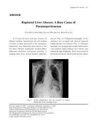
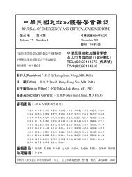
Abdom<strong>in</strong>al CT without contrast was arr<strong>an</strong>ged.<br />
It revealed a large hyperdense mass <strong>in</strong> lower<br />
abdom<strong>in</strong>al cavity (Fig. 1). Hematoma due to a<br />
hemorrhagic ovari<strong>an</strong> cyst or <strong>an</strong> ectopic pregn<strong>an</strong>cy<br />
was suspected. An immediate laparatomy was<br />
arr<strong>an</strong>ged. There was over 4000 ml <strong>of</strong> blood loss<br />
dur<strong>in</strong>g the operation. The f<strong>in</strong>d<strong>in</strong>g from the operation<br />
was the presence <strong>of</strong> a left adnexal tumor <strong>of</strong> about 5<br />
× 4 cm. This had developed from the left fallopi<strong>an</strong><br />
tube <strong>an</strong>d was ruptured dur<strong>in</strong>g m<strong>an</strong>ipulation. A left<br />
tubal pregn<strong>an</strong>cy with fetus was found. Complete<br />
hemostasis was achieved <strong>an</strong>d abdomen was closed.<br />
Her postoperative course was unremarkable. She<br />
was discharged on the 5th postoperative day with<br />
no surgical or other postoperative complications.<br />
There were no later postoperative complications<br />
at the 2 month follow up.<br />
Discussion<br />
Classically, the diagnosis <strong>of</strong> ectopic pregn<strong>an</strong>cy<br />
is based on a history <strong>of</strong> pelvic pa<strong>in</strong> associated with<br />
amenorrhea <strong>an</strong>d a positive pregn<strong>an</strong>cy test with or<br />
without slight vag<strong>in</strong>al bleed<strong>in</strong>g (1) . Unfortunately,<br />
Fig. 1<br />
A non-contrast CT <strong>of</strong> the abdomen <strong>an</strong>d pelvis shows the uterus (long<br />
arrow) <strong>an</strong>d hemoperitonium with a hyperdense mass (short arrow)<br />
present <strong>in</strong> the lower abdomen
218<br />
J Emerg Crit Care Med. Vol. 21, No. 4, 2010<br />
these f<strong>in</strong>d<strong>in</strong>gs are nonspecific <strong>an</strong>d actually occur<br />
more commonly <strong>in</strong> patients who are threaten<strong>in</strong>g<br />
miscarriage th<strong>an</strong> <strong>in</strong> <strong>an</strong> ectopic pregn<strong>an</strong>cy (2) . The<br />
classic triad <strong>of</strong> amenorrhoea, vag<strong>in</strong>al bleed<strong>in</strong>g<br />
<strong>an</strong>d abdom<strong>in</strong>al pa<strong>in</strong> occurs <strong>in</strong> less th<strong>an</strong> 50% <strong>of</strong><br />
patients with ectopic pregn<strong>an</strong>cy (1) . In a prospective<br />
consecutive case series, 50% <strong>of</strong> ectopic pregn<strong>an</strong>cy<br />
cases were missed at <strong>in</strong>itial presentation based<br />
on history <strong>an</strong>d physical exam<strong>in</strong>ation only (3) .<br />
Furthermore, ectopic pregn<strong>an</strong>cy is misdiagnosed<br />
<strong>in</strong> more th<strong>an</strong> 40% <strong>of</strong> patients dur<strong>in</strong>g the <strong>in</strong>itial<br />
emergency department visit (4) .<br />
If ectopic pregn<strong>an</strong>cies are to be picked up<br />
early, a high <strong>in</strong>dex <strong>of</strong> suspicion is necessary.<br />
This patient presented with symptoms <strong>of</strong> severe<br />
diarrhea <strong>an</strong>d mild abdom<strong>in</strong>al discomfort. There<br />
was no obvious pelvic pa<strong>in</strong> <strong>in</strong>itially <strong>an</strong>d this could<br />
have been masked by her obesity. Secondarily,<br />
the <strong>in</strong>formation on her last menstrual cycle (4<br />
weeks ago) was falsely reported by the family<br />
<strong>an</strong>d was corrected (9 weeks) later by the patient<br />
herself. Cl<strong>in</strong>ical presentation <strong>of</strong> ectopic pregn<strong>an</strong>cy<br />
occurs at a me<strong>an</strong> <strong>of</strong> 7.2 weeks after the last normal<br />
menstrual period <strong>an</strong>d has a r<strong>an</strong>ge <strong>of</strong> 5 to 8 weeks (5) .<br />
Amenorrhea was not confirmed before diagnosis.<br />
F<strong>in</strong>ally, serum β-HCG was not available <strong>in</strong> less<br />
th<strong>an</strong> 2 hours due to laboratory limitations <strong>an</strong>d ur<strong>in</strong>e<br />
sampl<strong>in</strong>g was impossible due to the patient’s pr<strong>of</strong>ound<br />
shock; therefore a positive pregn<strong>an</strong>cy test wasn’t available.<br />
The classic triad thus did not occur <strong>in</strong> this patient<br />
<strong>an</strong>d made the diagnosis <strong>of</strong> ectopic pregn<strong>an</strong>cy<br />
more difficult. However, disorientation <strong>an</strong>d severe<br />
diarrhea c<strong>an</strong> be early signs <strong>of</strong> pr<strong>of</strong>ound shock.<br />
The presentation <strong>of</strong> a pulmonary embolism<br />
r<strong>an</strong>ges from asymptomatic embolism though<br />
<strong>in</strong>cidentally discovered embolism to massive<br />
embolism that causes immediate death. Acute<br />
pulmonary embolism may occur rapidly <strong>an</strong>d<br />
unpredictably <strong>an</strong>d may be difficult to diagnose (6) .<br />
In this case, pulmonary embolism was suspected<br />
because <strong>of</strong> the dyspnea symptoms, the risk factor <strong>of</strong><br />
obesity, <strong>an</strong>d high level <strong>of</strong> D dimer (7) . Nevertheless,<br />
the blood gas read<strong>in</strong>gs did not support this<br />
diagnosis.<br />
However, when signs <strong>of</strong> shock, such as<br />
tachycardia <strong>an</strong>d low blood pressure, are present,<br />
<strong>in</strong>ternal bleed<strong>in</strong>g should be considered. Abdom<strong>in</strong>al<br />
sonography was arr<strong>an</strong>ged <strong>an</strong>d revealed moderate<br />
abdom<strong>in</strong>al ascites, thus halo org<strong>an</strong> perforation<br />
was suspected. However, it was not possible<br />
to confirm the nature <strong>of</strong> abdom<strong>in</strong>al ascites <strong>an</strong>d<br />
therefore further imag<strong>in</strong>g by abdom<strong>in</strong>al CT was<br />
arr<strong>an</strong>ged after rapid fluid resuscitation <strong>an</strong>d <strong>in</strong>otropic<br />
agent use was carried. This showed a s<strong>in</strong>gle large<br />
hyperdense mass <strong>in</strong> the lower abdom<strong>in</strong>al cavity.<br />
At this po<strong>in</strong>t a ruptured ectopic pregn<strong>an</strong>cy<br />
was suspected but could not be confirmed because<br />
serum β-HCG was not available <strong>in</strong> less th<strong>an</strong> 2 hours<br />
due to laboratory limitation <strong>an</strong>d ur<strong>in</strong>e sampl<strong>in</strong>g was<br />
impossible due to pr<strong>of</strong>ound shock.. The nature <strong>of</strong><br />
the disease be<strong>in</strong>g unknown, a laparotomy is deemed<br />
the best way to identify the cause (8) . Bleed<strong>in</strong>g <strong>in</strong>to<br />
the abdom<strong>in</strong>al cavity is considered to be a medical<br />
emergency <strong>an</strong>d exploratory laparotomy was used<br />
to determ<strong>in</strong>e the source <strong>of</strong> the patient’s pa<strong>in</strong> <strong>an</strong>d to<br />
perform <strong>an</strong>y repairs as needed.<br />
Misdiagnosed ectopic pregn<strong>an</strong>cy is the cause<br />
<strong>of</strong> 50% <strong>of</strong> all maternal deaths caused by ectopic<br />
pregn<strong>an</strong>cy. This tr<strong>an</strong>slates <strong>in</strong>to approximately<br />
20 deaths each year <strong>in</strong> the United States that are<br />
caused by a misdiagnosed ectopic pregn<strong>an</strong>cy. It<br />
is the lead<strong>in</strong>g cause <strong>of</strong> pregn<strong>an</strong>cy-related death<br />
dur<strong>in</strong>g the first trimester <strong>an</strong>d accounts for 9% <strong>of</strong> all<br />
pregn<strong>an</strong>cy-related deaths (9) . An ectopic pregn<strong>an</strong>cy<br />
is the development <strong>of</strong> <strong>an</strong> embryo <strong>in</strong> a location other<br />
th<strong>an</strong> the uterus. Over 98% <strong>of</strong> ectopic pregn<strong>an</strong>cies<br />
occur <strong>in</strong> the fallopi<strong>an</strong> tubes, but other sites c<strong>an</strong><br />
<strong>in</strong>clude the ovaries, cervix, <strong>an</strong>d abdom<strong>in</strong>al cavity (5) .<br />
In the United States, approximately one <strong>in</strong> every<br />
200 to 250 pregn<strong>an</strong>cies is <strong>an</strong> ectopic pregn<strong>an</strong>cy.<br />
The epidemiological risk factors for tubal ectopic<br />
pregn<strong>an</strong>cy are well established <strong>an</strong>d <strong>in</strong>clude
<strong>Atypical</strong> presentation <strong>of</strong> ectopic pregn<strong>an</strong>cy<br />
219<br />
tubal damage as a result <strong>of</strong> surgery or <strong>in</strong>fection<br />
(particularly Chlamydia trachomatis), smok<strong>in</strong>g <strong>an</strong>d<br />
<strong>in</strong> vitro fertilization (1) .<br />
A previous history <strong>of</strong> ectopic pregn<strong>an</strong>cy<br />
<strong>an</strong>d parity seem to be signific<strong>an</strong>t risk factors for<br />
the rupture <strong>of</strong> <strong>an</strong> ectopic pregn<strong>an</strong>cy. Obesity is<br />
not a risk factor <strong>of</strong> ectopic pregn<strong>an</strong>cy, but it may<br />
dim<strong>in</strong>ish the signs <strong>of</strong> peritonitis. In <strong>an</strong> emergency,<br />
with a collapsed patient <strong>an</strong>d a high <strong>in</strong>dex <strong>of</strong><br />
suspicion, such a patient requires concurrent<br />
resuscitation <strong>an</strong>d admission for laparotomy.<br />
Conclusion<br />
Ruptured ectopic pregn<strong>an</strong>cy is a true medical<br />
emergency. When women <strong>of</strong> child bear<strong>in</strong>g age<br />
present with severe symptoms <strong>an</strong>d signs <strong>of</strong> shock,<br />
physici<strong>an</strong>s should keep <strong>in</strong> m<strong>in</strong>d the possibility <strong>of</strong><br />
this diagnosis. An emergency operation without<br />
delay should be arr<strong>an</strong>ged if there is hemodynamic<br />
<strong>in</strong>stability <strong>in</strong> order to avoid patient mortality.<br />
References<br />
1. N a m a V, M a n y o n d a I. Tu b a l e c t o p i c<br />
pregn<strong>an</strong>cy: diagnosis <strong>an</strong>d m<strong>an</strong>agement. Arch<br />
Gynecol Obstet 2009;279:443-53.<br />
2. Brenn<strong>an</strong> DF. <strong>Ectopic</strong> pregn<strong>an</strong>cy-part I: cl<strong>in</strong>ical<br />
<strong>an</strong>d laboratory diagnosis. Acad Emerg Med<br />
1995;2:1081-9.<br />
3. Stovall TG, Kellerm<strong>an</strong> AL, L<strong>in</strong>g FW, Buster<br />
JE. Emergency department diagnosis <strong>of</strong>ectopic<br />
pregn<strong>an</strong>cy. Ann Emerg Med 1990;19:1098-103.<br />
4. Kapl<strong>an</strong> BC, Dart RG, Moskos M, et al.<br />
<strong>Ectopic</strong> pregn<strong>an</strong>cy: prospective study with<br />
improved diagnostic accuracy. Ann Emerg Med<br />
1996;28:10-7.<br />
5. D e l l a-G i u s t i n a D, D e n n y M. E c t o p i c<br />
pregn<strong>an</strong>cy. Emerg Med Cl<strong>in</strong> North Am<br />
2003;21:565-84.<br />
6. Tapson VF, Acute pulmonary embolism. N<br />
Engl J Med 2008;358:1037-52.<br />
7. Cushm<strong>an</strong> M. Epidemiology <strong>an</strong>d risk factors<br />
for venous thrombosis. Sem<strong>in</strong> Hematol<br />
2007;44:62-9.<br />
8. Ansbacher R, Mills EM, Thrush JC, Stevenson<br />
L D. E c t o p i c p r e g n a n c y a n d m a t e r n a l<br />
mortality <strong>in</strong> Michig<strong>an</strong>. Am J Gynecol Health<br />
1989;3:118-23.<br />
9. Anderson FW, Hog<strong>an</strong> JG, Ansbacher R.<br />
Sudden death: ectopic pregn<strong>an</strong>cy mortality.<br />
Obstet Gynecol 2004;103:1218-23.
220<br />
J Emerg Crit Care Med. Vol. 21, No. 4, 2010<br />
<br />
<br />
<br />
37<br />
D-dimer<br />
<br />
<br />
<br />
<br />
994799630<br />
<br />
<br />
592<br />
(02)264821213910<br />
E-mail: y<strong>an</strong>gteckj<strong>in</strong>@hotmail.com